
JoVE 비디오를 활용하시려면 도서관을 통한 기관 구독이 필요합니다. 전체 비디오를 보시려면 로그인하거나 무료 트라이얼을 시작하세요.
Method Article
예는 폐 전이 및 그들의 미세 환경의 라이브 영상을 생체 내
요약
우리는 쥐에 형광 기자를 활용, 폐 전이에서 종양 세포 기질 상호 작용의 생체 라이브 영상에 대한 비교적 간단한 방법을 설명합니다. 스피닝 디스크 공 촛점 현미경을 사용하여,이 기술은 최소 4 시간 동안 생균의 시각화를 가능하게하고 다른 염증성 폐 상태를 학습하도록 구성 될 수있다.
초록
전이 암 관련 이환율과 사망률의 주요 원인이다. 전이는 다단계 과정과 그 복잡성으로 인해, 전이성 보급과 성장을 지배하는 정확한 세포 및 분자 프로세스는 여전히 애매하다. 라이브 영상은 세포 및 미세 환경의 동적 및 공간 상호 작용을 시각화 할 수 있습니다. 고형 종양은 일반적으로 폐에 전이. 그러나, 폐의 해부학 적 위치는 생체 내에 영상에 도전을 포즈. 이 프로토콜은, 종양 세포 및 폐 전이 내에서 주변 기질 간의 동적 상호 작용의 실시간 생체 외 이미징을위한 비교적 간단하고 빠른 방법을 제공한다. 이 방법을 사용하여, 그 미세 암 세포 및 간질 세포와 암세포의 운동과 상호 작용은 몇 시간 동안 실시간으로 시각화 될 수있다. 형질 형광 리포터 생쥐를 사용하여 형광 세포주 주사 형광 표지분자 및 / 또는 항체, 폐 미세 여러 구성 요소는 이러한 혈관 면역 세포로서 가시화 될 수있다. 상이한 세포 유형 이미지를 신속, 4 색 화상 취득 장기 연속 촬영을 허용 회전 디스크 공 촛점 현미경을 사용하고있다. 여러 위치와 초점면을 통해 수집 된 이미지에서 컴파일 시간 경과 영화는 적어도 4 시간 동안 라이브 전이성 및 면역 세포 사이의 상호 작용을 보여줍니다. 이 기술은 또한 화학 요법 또는 치료 대상을 시험 할 수있다. 또한, 본 방법은 폐 미세 환경에 영향을 미칠 수있는 다른 폐 관련된 병리의 연구에 적용 할 수있다.
서문
The deadliest aspect of cancer is metastasis, which accounts for more than 90% of cancer-related morbidity and mortality1. Metastasis is a multistep process and due to its complexity, the exact cellular and molecular mechanisms that govern metastatic dissemination and growth are still elusive. To metastasize, tumor cells in the primary tumor must detach from their neighboring cells and basement membrane, cross through the extracellular matrix, intravasate, travel via blood or lymphatic vessels, extravasate at the secondary site, and finally, survive and establish secondary tumors. In addition to the properties of the tumor cells, the contribution from the microenvironment, which includes the adjacent stroma along with the normal counterparts of the cancer cells, is crucial for the seeding and establishment of metastatic lesions2.
Traditional methods to study metastatic seeding and growth examine static states, as tissues are excised and sectioned for histology. These data only generate a snapshot of this highly dynamic process. Although some useful information can be gained from these studies, the complicated process by which tumor and stromal cells interact during metastatic formation cannot be adequately assessed by these methods. Furthermore, it is not possible to gain insights into tumor or stromal cell migration patterns, which are important in establishing a colony at the distant site. In order to effectively study the metastatic process, it is essential to visualize various interactions between cancer cells and their microenvironment in a continuous manner and at real time.
The lung is a common site for metastases from solid tumors as breast, colorectal, pancreatic cancer, melanoma and sarcoma3. Intravital imaging was previously used to study cell-cell interaction in various primary tumor and metastatic models4,5. Methods of lung imaging in mice, including intravital imaging, lung section imaging, and an ex vivo pulmonary metastasis assay have been published6–9. Intravital imaging of mouse lungs utilizes a thoracic suction window to stabilize the lungs6. This method is used for time-lapse imaging of the lung microcirculation and alveolar spaces. The anatomical location of the lungs poses a challenge to intravital imaging. In order to access the lungs, the chest cavity must be opened which leads to loss of negative pressure and collapsed lungs. This method only allows the visualization of a small part of the lungs and is technically demanding; an unnecessary complication in studies that examine processes that are independent of blood flow. Moreover, this method also requires gating out movement caused by breathing. This is done either by collecting images between breaths or during post image acquisition analyses10. The alternative ex vivo lung section imaging provides stability and depth, and also prepares lung parenchyma for immunostaining7. However, the lengthy sectioning process leads to an extensive delay between the time of animal sacrifice and the start of the imaging session. Moreover, the process of sectioning a mouse lung causes considerable amount of cell death8, thus interfering with the quality and quantity of imaging samples and perhaps needlessly altering tumor-stroma interactions. In order to technically bridge between the methods of intravital imaging and lung section imaging, while exploiting the advantages of the two techniques, a relatively fast and easy method for ex vivo lung imaging was developed. This method was achieved by imaging of non-sectioned whole lung lobes. Using this method, the motility of cancer cells as well as interactions between cancer cells and stromal cells in their microenvironment can be visualized in real time for several hours.
프로토콜
설명하는 모든 절차는 지역 기관 동물 관리 및 사용위원회 (IACUC)의 사전 승인을 포함한 척추 동물의 사용에 대한 지침과 규정에 따라 수행해야합니다.
예를위한 폐 전이의 1 세대는 라이브 영상을 생체 내 (형질 전환 또는 꼬리 정맥 주입)
주 : 폐전이 유전자 조작 마우스 모델을 이용하여 암 세포의 정맥 주사에 의해 생성 될 수있다.
- 예를 들면, 유전자 변형 기자 마우스에 유전자 조작 종양 마우스 모델을 교차하여 이미징을위한 폐 전이를 생성, ACTB - ECFP에 유방암 마우스 모델, 마우스 유방 종양 바이러스 긴 터미널 반복-폴리오 중간에 T 항원 (MMTV-PyMT) (11)를 교차 마우스 모델 (12).
참고 : ACTB - ECFP 모델은 β-행위에서 시안 형광 단백질 (ECFP)를 강화 표현한다프로모터의 모든 세포는 블루, CFP 채널 형광되도록. 그러나 암세포에 의해 지금까지 가장 두드러진이며 현미경 ECFP 양성 세포 대량으로 나타난다. MMTV-PyMT 마우스 모델 유방 종양 성장, 특히 폐, 주변으로 암세포의 보급과 연관되는 진행성 질환을 개발한다. FVB / n를 배경에 MMTV-PyMT 마우스에서 미세 전이 나이 10~11주 주위에 관찰 할 수있다. 일반적으로 13 세의 약 14주에서 macrometastases 이러한 진행.
또는 - 일차 전지 또는 동계 세포주를 사용하여 실험 전이를 생성합니다. IV 주입 (14) 다음에 시험관 조작 차 종양 세포 또는 세포주 (예., 전달)에 사용한다.
- 요약하면,이 프로토콜에서 형광 리포터 생쥐 (ACTB-ECFP)로 이루어지는 군으로부터 선택된다 또는 야생형 마우스에 녹색 형광 단백질 (GFP) 발현 (+) MMTV-PyMT 세포주를 주입. 그때,녹색 GFP 채널을 이용하여 VO-PyMT 전지 (15)로 지칭 이들 세포를 시각화.
참고 : 원래의 VO-PyMT 세포주는 내쉬빌, TN의 밴더빌트 정형 외과에서 유래되었다. VO는 밴더빌트 정형 외과를 의미합니다. - 주입 후 수 시간 바로 위로 암세포의 혈관 외 유출을 관측 (200 μL)에 10 6 세포 주입 다음; 주사 후 1-3주 사이의 미세 전이를 관찰하고 주입 15 일 이후 macrometastases 3 주 검출한다.
주 : 적은 세포 전이성 성장 주사까지의 시간을 연장 주입 될 수있다.
- 요약하면,이 프로토콜에서 형광 리포터 생쥐 (ACTB-ECFP)로 이루어지는 군으로부터 선택된다 또는 야생형 마우스에 녹색 형광 단백질 (GFP) 발현 (+) MMTV-PyMT 세포주를 주입. 그때,녹색 GFP 채널을 이용하여 VO-PyMT 전지 (15)로 지칭 이들 세포를 시각화.
전이성 미세 환경에 대한 관심의 구성 요소 2. 라벨 (형질 전환 및 / 또는 주사 가능한)
주 : 라벨은 트랜스 제닉 마우스에 의해 및 / 또는 각종 주사제에 의해 달성 될 수있다. 다양한 세포 유형의 표시에 대해 서로 다른 형광 색상을 사용하십시오.
- 형질 전환 마우스를 사용하여 전이성 미세 환경의 레이블 구성 요소. 전술 한 마우스 종양 모델을 교차 관심 간질 세포 ECFP없는 형광 단백질, 예를 들면., C-FMS-EGFP로 표지 된 형질 전환 마우스 모델에 (예., MMTV-PyMT는 ACTB-ECFP을 X) 4,16.
주 : CFP 채널에서 암세포의 시각화 외에도, 이는 GFP 채널 4에서 골수 세포의 가시화를 가능하게한다.
AND / OR - 형질 전환 형광 기자 마우스 또는 (비 형광) 야생형 마우스에 주사제를 사용하여 전이성 미세 환경의 다양한 구성 요소 레이블.
참고 : 여러 화합물은 전이성 미세 환경, 예를 들어, AF647 - 복합 GR-1 항체가 호중구 레이블을 여기에 사용되며, 일부 단핵구 (13)와 다른 분자량 덱스 트란이 사용되는 다양한 구성 요소 레이블을 주입 할 수있다폐 모세 혈관에 레이블을합니다. 이 주사제의 제조를위한 4 단계를 참조하십시오.
해부하기 전에 재료 3. 준비
- 2 % 아가로 오스
- 아가로 오스 0.2 g을 달아 10ml를 1 × PBS에 추가 할 수 있습니다. 아가로 오스를 분해하는 용액을 가열한다. 아가로 오스는 실온에서 응고 때문에 인플레이션에 사용할 때까지 37 ° C의 물을 욕조에 그것을 유지합니다.
- CO (2) 및 온도 제어기
- 가습 실에서 DDH 2 O를 확인합니다. 필요한 경우 리필. 온도 스테이지 판 홀더 (기후 실)로 구성 플레이트를 삽입합니다. CO 2 컨트롤러의 전원을 켜고 5 % CO 2로 설정합니다. 공기 흐름 속도가 0.4 N1 개의 / 분으로 설정되어 있는지 확인합니다.
- 공기와 CO 2 밸브를 엽니 다. 온도 조절기의 전원을 켭니다. 기후 챔버의 온도와 뚜껑을 37 ° C로 설정되어 있는지 확인합니다.
- CO 2 미터 공기 압력을 해제합니다. CO 2가 증가 확인, 전자quilibration 30 분까지 걸릴 수 있습니다.
- 회전 디스크 공 초점 현미경
참고 : 현미경 설정의 세부 사항 이전에 4,17을 설명 하였다.- 레이저 (488 nm의 여기 및 고체 405 nm의, 561 nm 내지 640 nm의 레이저에 대한 아르곤 레이저)를 켭니다. 현미경, 카메라, 회전 디스크 제어부 AOTF, 레이저 제어 유닛과 카메라 제어부 켜기.
- 현미경 셔터를 열고 현미경을 실행하는 컴퓨터를 켜고 소프트웨어를 엽니 다.
- 도구 및 해부 플랫폼의 준비.
- 뜨거운 비드 살균기를 켜고는 250 ° C에 도달 할 수 있습니다. 물과 비누로 수술 가위와 집게의 청소 2 쌍. 적어도 30 초 동안 도구를 소독. 도구를 식혀 보자. 해부 플랫폼으로 폴리스티렌 뚜껑을 사용하십시오. 실험실 담그는의 조각으로 그것을 커버.
주사 4. 준비
참고 : 반감기와 선호하는 응답에 따라 분사 형광 표지 항체 및 / 또는 형광 분자 중 동물의 희생 또는 그 이전 일 몇 시간 직전에.
- 이미지 GR1 양성 호중구와 단핵구에, 후드 멸균 PBS 100 ㎕에 스톡 AF647 - 복합 GR-1 항체의 7 μL (1 ㎎ / ㎖)을 주사기를 준비합니다. 주사기에 27 G ½ 바늘을 놓습니다.
- 화상 폐 모세 혈관 (100) 중 70 kD의 로다 민 접합 된 덱스 트란 (4 ㎎ / ㎖)을 10 μL kD의 AF647 접합 된 덱스 트란 (4 ㎎ / ㎖)과 함께 두 번째 및 세 번째 주사기를 준비한다. 주사기에 27 G ½ 바늘을 놓습니다.
- 이전에 폐 '절제에 AF647 - 복합 항체 솔루션 IV 5 시간을 주입한다.
- IV 직전 폐 '절제 하나 또는 둘 덱스 트란 용액을 주입한다.
예를위한 폐 5. 준비 라이브 영상을 생체 내
참고 : 폐 내의 면역 세포의 불필요한 문제를 피하기 위해 가능한 멸균 및주의 일을보십시오.
- IACUC 승인 동물 프로토콜, 예. 허용 마취의 치명적인 과다 복용으로 마우스 복강 내 (IP)을 주입, 2.5 % AVERTIN 1 ㎖. 호흡을 중지하고 유해 자극 (뒷발 핀치)에 완전히 비 반응 할 마우스 기다립니다.
주 : 경추 탈구 이산화탄소 안락사는 유해한 폐 세포 생존에 영향을 미칠 수 있으므로 피해야한다. - 해부 보드에 마우스를 고정하고 70 % 에탄올로 소독 마우스.
- 먼저 복막을 통해 비슷한 절개 한 다음 피부를 통해 가로 상복부 절개를 만들기 위해 수술 가위를 사용합니다. , 수직으로 절개 보드를 잡고 하행 대동맥을 절단하지 흉강의 복부에 혈액 풀 다운 때문에.
- 에스진공을 해제 다이어프램 작은 개구 닙. 다이어프램을 절제 및 폐 시각 액세스를 얻기 위해 10, 12 늑골을 따라 잘라.
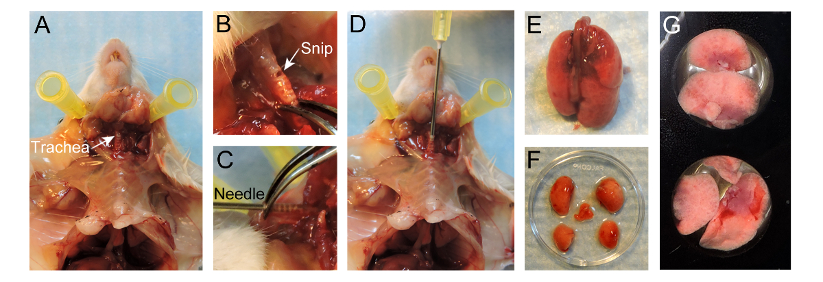
- 흉곽을 통해 기관에 피부를 잘라하지만 그대로 흉곽을 떠나 수술 가위를 사용합니다. 흉곽으로부터 피부를 분리합니다. 기관 자체 (그림 1A)을 손상되지 않도록주의하면서 주변의 결합 조직을 제거하여기도를 노출.
- 작은 구멍을 수 (그림 1B)와 후두에 가까운 연골 고리에 노출 된 기관 평행 직경 약 1mm를 싹둑. 기관을 통해 완전히 절단하지 않도록주의하십시오.
- 20 G 바늘을 가지고 부드럽게 모든 카운터 힘 (그림 1D)이없는 기관에 바늘 4-5mm를 삽입합니다. 바늘의 끝은 기관 (그림 1C)를 통해 볼 수 있어야합니다. 기관에서 바늘을 안정시키기 위해 집게를 사용합니다. 대안 적으로, 봉합사 ARO 묶여있다장소에 바늘을 유지하는 기관 핀클.
주 : 너무 깊이 삽입함으로써, 카리나는 충격되거나 폐의 한쪽 만이 팽창 될 수있다. - (항온조에서 직접 촬영) 37 ° C 2 % 낮은 용융 온도 아가로 오스의 400 μL와 주사기를 입력합니다. 해부 보드가 서 있는지 확인하고 천천히 폐에 바늘을 통해 따뜻한 아가로 오스를 주입, 폐를 팽창 ~ 400 μl를 사용합니다.
참고 : 흉곽 내에서 팽창 폐를보세요. 이 파열되므로 폐를 과다 팽창하지 마십시오. - 폐이 팽창되면, 주사기를 분리하고 누설하는 아가 방지 기관 내부 바늘을 유지 흉곽 ~ ⅔ 충전.
- 설정 고화 폐 내부 아가 있도록 팽창 폐 위에 20 ° C PBS 약 50 mL로 붓는다. 천천히 방지하기 위해 집게와 기관을 바늘을 제거하고 닫습니다 비 응고 아가로 오스 누출.
- 흉골 절개술을 수행하여 폐 노출이어서, 폐를 절제. 완전히 기관을 통해 절단하는 동안 폐 절제술의 경우, 기관에 개최합니다. 조심스럽게 폐는 마우스 (그림 1E)에서 분리 될 때까지 흉강에서 폐를 당기면서 결합 조직과 식도를 절단,기도를 당깁니다.
- 과도한 혈액을 씻어 따뜻한 RPMI-1640에 폐를 담가 부드럽게 폐문 (그림 1 층)에서 로브의 주요 줄기 기관지를 잘라 가위와 집게를 사용하여 로브를 분리합니다.
- 24- 웰 플레이트 촬상 (도 1G)의 웰에 촬상면을 최대화하기 위해 아래 평면에, 돌출부를 배치했다. 로브의 상단에 37 ° C RPMI-1640의 100 μl를 추가합니다. 부동 것을 방지하기 위해 로브의 상단에 여러 15mm 원형 현미경 커버 슬라이드를 놓습니다.
- EVAP에서 RPMI-1640 미디어를 방지하기 위해 주변의 우물에 따뜻한 PBS를 붓고orating. 평형 기후 챔버로 24 웰 플레이트를 삽입하고 공기와 5 % CO 2와 37 ° C에서 폐 엽 (叶)을 유지한다. 공 초점 현미경의 무대에서 기후 챔버를 삽입합니다.
주의 : 다른 가스 혼합물 (예를 들어, N이 5 % O 2, 5 % CO 2는 저산소증 / 낮은 산소의 조건으로 세포를 검사하는 동작)도 고려 될 수있다.

라이브 이미징을위한 폐의 준비를위한 그림 1. 프로토콜. 마우스의 준비 후 기관의 (A) 노출. 연골 고리에 노출 기관지 평행 제 (B) 작은 한조각. (C) 20 G 바늘 기관에 4-5mm 삽입. (D) 아가 폐로 400 ㎕의 2 % 저 융점 온도 점적. (E) Inflated 폐 마우스에서 분리. (F) 로브는 인플레이션 후 분리. (G) 24 웰 이미징 플레이트의 웰에 위치 로브. 이 그림의 더 큰 버전을 보려면 여기를 클릭하십시오.
6. 수집 및 이미지 분석
주 : 이미지는 다양한 소프트웨어 프로그램에 의해 지원되는 디스크 공 초점 현미경을 방사 다양한 얻을 수있다. Imaris 영화 편집 및 분석에 사용되는 동안이 프로토콜에서 시판 회전 디스크 공 초점 현미경으로 주문 제작 회전 디스크 공 초점 현미경 선으로 하나 μManager은 이미지 수집에 사용됩니다.
- μManager을 사용하여 이미지를 획득. μManager 소프트웨어를 사용하여 화상 취득 단계 프로토콜에 의해 상세한 단계 이전에 (18)를 설명한다.
또는 - 이미지 획득이러한 선으로서 사용하는 이미지 분석 소프트웨어 (도 S1 참조).
- '찾기'탭을 클릭하고 '광경'도구 (그림 S1A, 빨간색 상자)에서 (10 배 또는 20 배) 목표를 선택합니다. 접안 렌즈 (그림 S1A, 파란색 상자)를 통해 CFP 채널을보고 - 'DAPI 눈'그 후, 클릭합니다. 수동 현미경을 사용하여 샘플을 현지화. 조직 Off'after 모든 시야의 중심 '을 클릭합니다.
- 이미지 획득을위한 모든 매개 변수를 설정하는 '수집'탭을 클릭합니다.
- '채널'도구에서 '+'버튼 (그림 S1B, 빨간색 상자)을 클릭합니다. 팝업 메뉴가 나타납니다하고 '염료 데이터베이스'(그림 S1B)의 샘플에 존재하는 염료 (들)를 검색합니다. 염료를 선택하고 '추가'를 클릭합니다.
참고 :이 프로그램은 모든 필터가 최적화되도록 설정됩니다. 염료는 삭제 될 수 있습니다선택은 휴지통 버튼 (그림 S1B, 노란색 상자)를 클릭 하였다. - '획득 모드'메뉴에서, 5 × 5에 '비닝 (Binning)'로 설정합니다. 선택하는 채널 메뉴에서 ECFP을 더블 클릭합니다. 샘플 화상 취득을위한 파라미터를 설정하는 동안 표백되지 않도록 20 % 레이저 파워를 낮출.
- '실험 관리자'섹션에서 '타일'확인란을 선택하고 타일 도구는 '다차원 취득'도구 그룹 (그림 S1C)에 나타납니다. 카메라에서 라이브 영상을 볼 수 있습니다 '고급 설정'버튼을 클릭합니다. 실험에 4 ~ 6 위치를 추가하려면 '위치'섹션에서 '추가'버튼을 클릭합니다. 위치를 삭제하려면 해당 위치를 선택하고 휴지통 버튼을 클릭합니다.
- '획득 매개 변수'도구 그룹에서 '앱솔루트을'집중 전략 '도구를 열고 선택전자는 드롭 다운 목록에서 Z-위치를 '수정되었습니다.
- '실험 관리자'섹션과 Z-스택 도구의 Z 스택 상자를 확인은 '다차원 취득'도구 그룹 (그림 S1D)에 나타납니다. '위치'섹션를 눌러 '라이브'의 위치 중 하나를 더블 클릭합니다. 수동으로 제 1 세트의 촬상 영역의 마지막 위치를 설정한다. 4 μm의 간격을 설정합니다.
참고 :이 프로그램은 선택된 범위와 간격 조각의 수를 결정합니다. 이상적으로, 5-7 조각 충분한 시각화와 빠른 영상 획득이 가능하도록 편리합니다. - '실험 관리자'섹션에서 '시계열'확인란을 선택합니다. 설정 원하는 '시간'과 '다차원 취득'도구 그룹 (그림 S1E)에 나타난 '시계열'도구 '간격'시간.
- 'Acquisi에서기 모드 2 × 2로 '비닝 (Binning)'메뉴 설정 '. 채널 메뉴에서 형광을 두 번 클릭을 선택하고 100 %로 레이저 파워를 증가시킵니다. 를 눌러 '라이브'와 '노출 시간'을 조정합니다. 모든 형광에 대해이 작업을 반복합니다.
- '사용 자동 저장'확인란을 선택합니다. 파일의 이름으로 폴더 및 유형을 선택합니다. 모든 획득 된 영상은 자동으로이 폴더에 저장됩니다.
- 이미지 수집을 시작하는 '실험 관리자'섹션에서 '시작 실험'을 클릭합니다.
- 이미지 획득 후 Imaris 소프트웨어의 원시 데이터를 컴파일한다. 파일을 .ims하는 이미지 변환 및 조정을 할 수있다. 파일의 변환, 조정을하고 Imaris를 사용하여 저장하는 영화 단계 프로토콜에 의해 자세한 단계는 이전에 18 설명한다.
- 동영상을 저장할 때, 두 번째 (FPS) 당 5 프레임 '프레임 레이트'를 설정합니다.
결과
스피닝 디스크 공 초점 현미경, 다양한 마우스 모델 시스템 및 주사제를 사용하여, 미세 전이가 가시화하고 시간을 추적 할 수있다. MMTV-PyMT를 사용하여; ACTB - ECFP; C-FMS-EGFP 트리플 유전자 변형 마우스 모델, 다른 세포 성분이 찬란 (그림 2A, 영화 1) 레이블이 표시됩니다. 폐 실질의 일반적인 구조는 모든 셀 β 액틴 프로모터 하에서 발현 ECFP 때문에 CFP 채널에서 가시?...
토론
이 원고는 전이의 마우스 모델에서 폐 전이의 생체 라이브 영상에 대한 자세한 방법을 설명합니다. 이 촬상 프로토콜 폐 미세 환경 내에서 동적 공간 종양 세포 - 기질 상호 작용의 직접적인 시각화를 제공한다. 적어도 4 시간 동안 폐전이 신뢰성있게 이미징 비교적 쉽고 빠른 방법이다. 이 실험에서 얻은 동영상 세포 운동성 및 셀룰러 상호 동적 프로세스를 추적 할 수있다.
공개
The authors have no conflicts of interest to disclose. All animal experiments were conducted in accordance with IACUC approved protocols, UCSF.
감사의 말
We thank Nguyen H. Nguyen for her technical help and Audrey O’Neill for support with the Zeiss Cell Observer spinning-disk confocal microscope. This work was supported by a Department of Defense postdoctoral fellowship (W81XWH-11-01-0139) and the Weizmann Institute of Science-National Postdoctoral Award Program for Advancing Women in Science (to V.P.).
자료
| Name | Company | Catalog Number | Comments |
| MMTV-PyMT/FVB mice | Jackson Laboratory | 2374 | Female mice |
| ACTB-ECFP/FVB mice | UCSF Werb lab | Female mice | |
| c-fms-EGFP/FVB mice | UCSF Werb lab | Female mice | |
| FVB mice | Jackson Laboratory | 1800 | Female mice |
| GFP+ VO-PyMT cells | UCSF Werb lab | ||
| 70,000 kDa Dextran, rhodamine-conjugated | Invitrogen | D1818 | Dilute to 4mg/ml in 1 x PBS and store at -20 °C. Use 0.4 mg per animal. |
| 10,000 kDa Dextran, Alexa Fluor 647 conjugated | Invitrogen | D22914 | Dilute to 4mg/ml in 1 x PBS and store at -20 °C. Use 0.4 mg per animal. |
| Anti-mouse Gr-1 antibody Alexa Fluor 647 | UCSF Monoclonal antibody core | Stock 1mg/ml. Use 7 ug per animal. | |
| Anesthetic | Anesthesia approved by IACUC, used for anesthesia and/or euthanesia | ||
| 1X PBS | UCSF cell culture facility | ||
| PBS, USP sterile | Amresco INC | K813-500ML | Ultra pure grade for i.v. injection |
| Styrofoam platform | Will be used as dissection board | ||
| Fine scissors sharp | Fine Science Tools | 14060-11 | |
| Forceps | Roboz Surgical Store | RS-5135 | |
| Hot bead sterilizer | Fine Science Tools | 18000-45 | Turn ON 30min before use |
| Air | UCSF | ||
| Oxygen | UCSF | ||
| Carbon dioxide | UCSF | ||
| 1 mL syringe without needle | BD | 309659 | |
| 27 G x 1/2 needle | BD | 305109 | for i.v. injection |
| 20 G x 1 needle, short bevel | BD | 305178 | |
| Low-melting-temperature agarose | Lonza | 50111 | To make 10 ml of solution, weigh 0.2 g of agarose, add to 10 ml 1 x PBS, and heat to dissolve. Agarose will solidify at room temperature, so maintain in a 37 °C water bath until used for inflation. |
| RPMI-1640 medium without phenol red | Life Technologies | 11835-030 | |
| 24 well Imaging plate | E&K scientific | EK-42892 | |
| Glass cover slides, 15 mm | Fisher Scientific | 22-031-144 | |
| Digital CO2 and temperature controller | Okolab | DGTCO2BX | http://www.oko-lab.com |
| Climate chamber | Okolab | http://www.oko-lab.com | |
| Cell Observer spinning disk confocal microscope | Zeiss | ||
| Zen software | Zeiss | ||
| Inverted microscope | Carl Zeiss Inc | Zeiss Axiovert 200M | |
| ICCD camera | Stanford Photonics | XR-Mega-10EX S-30 | |
| Spinning disk confocal scan-head | Yokogawa Corporation | CSU-10b | |
| Imaris | Bitplane | ||
| mManager | Vale lab, UCSF | Open-source software |
참고문헌
- Chaffer, C. L., Weinberg, R. A. A perspective on cancer cell metastasis. Science. 331 (6024), 1559-1564 (2011).
- Plaks, V., Kong, N., Werb, Z. The cancer stem cell niche: how essential is the niche in regulating stemness of tumor cells. Cell stem cell. 16 (3), 225-238 (2015).
- Nguyen, D. X., Bos, P. D., Massague, J. Metastasis: from dissemination to organ-specific colonization. Nat Rev Cancer. 9 (4), 274-284 (2009).
- Egeblad, M., Ewald, A. J., et al. Visualizing stromal cell dynamics in different tumor microenvironments by spinning disk confocal microscopy. Dis Model Mech. 1 (2-3), 155-167 (2008).
- Ellenbroek, S. I. J., van Rheenen, J. Imaging hallmarks of cancer in living mice. Nat Rev Cancer. 14 (6), 406-418 (2014).
- Looney, M. R., Thornton, E. E., Sen, D., Lamm, W. J., Glenny, R. W., Krummel, M. F. Stabilized imaging of immune surveillance in the mouse lung. Nat Methods. 8 (1), 91-96 (2011).
- Thornton, E. E., Krummel, M. F., Looney, M. R. Live imaging of the lung. Cur Protoc Cytom. , (2012).
- Thornton, E. E., Looney, M. R., et al. Spatiotemporally separated antigen uptake by alveolar dendritic cells and airway presentation to T cells in the lung. J Exp Med. 209 (6), 1183-1199 (2012).
- Mendoza, A., Hong, S. -. H., et al. Modeling metastasis biology and therapy in real time in the mouse lung. J Clin Invest. 120 (8), 2979-2988 (2010).
- Lelkes, E., Headley, M. B., Thornton, E. E., Looney, M. R., Krummel, M. F. The spatiotemporal cellular dynamics of lung immunity. Trends Immunol. 35 (8), 379-386 (2014).
- Guy, C. T., Cardiff, R. D., Muller, W. J. Induction of mammary tumors by expression of polyomavirus middle T oncogene: a transgenic mouse model for metastatic disease. Mol Cell Biol. 12 (3), 954-961 (1992).
- Hadjantonakis, A. -. K., Macmaster, S., Nagy, A. Embryonic stem cells and mice expressing different GFP variants for multiple non-invasive reporter usage within a single animal. BMC Biotechnol. 2, (2002).
- Casbon, A. -. J., Reynaud, D., et al. Invasive breast cancer reprograms early myeloid differentiation in the bone marrow to generate immunosuppressive neutrophils. Proc Natl Acad Sci USA. 112 (6), 566-575 (2015).
- Donovan, J., Brown, P. Parenteral injections. Curr Protoc Immunol. , (2006).
- Halpern, J., Lynch, C. C., et al. The application of a murine bone bioreactor as a model of tumor: bone interaction. Clin Exp Metastas. 23 (7-8), 345-356 (2006).
- Sasmono, R. T., Oceandy, D., et al. A macrophage colony-stimulating factor receptor-green fluorescent protein transgene is expressed throughout the mononuclear phagocyte system of the mouse. Blood. 101 (3), 1155-1163 (2003).
- Ewald, A. J., Werb, Z., Egeblad, M. Dynamic, long-term in vivo imaging of tumor-stroma interactions in mouse models of breast cancer using spinning-disk confocal microscopy. Cold Spring Harb Protoc. (2), (2011).
- Bonnans, C., Lohela, M., Werb, Z. Real-time imaging of myeloid cells dynamics in ApcMin/+ intestinal tumors by spinning disk confocal microscopy. J Vis Exp. (92), (2014).
- Nakasone, E. S., Askautrud, H. A., et al. Imaging tumor-stroma interactions during chemotherapy reveals contributions of the microenvironment to resistance. Cancer cell. 21 (4), (2012).
- Cheng, N., Lambert, D. L. Mammary transplantation of stromal cells and carcinoma cells in C57BL/6J mice. J Vis Exp. (54), (2011).
- Al-Mehdi, A. B., Tozawa, K., Fisher, A. B., Shientag, L., Lee, A., Muschel, R. J. Intravascular origin of metastasis from the proliferation of endothelium-attached tumor cells: a new model for metastasis. Nat Med. 6 (1), 100-102 (2000).
- Wong, C. W., Song, C., et al. Intravascular location of breast cancer cells after spontaneous metastasis to the lung. Am J Pathol. 161 (3), 749-753 (2002).
- Liang, C. -. C., Park, A. Y., Guan, J. -. L. In vitro scratch assay: a convenient and inexpensive method for analysis of cell migration in vitro. Nat protoc. 2 (2), 329-333 (2007).
- Nelson, K., Bobba, C., Ghadiali, S., Hayes, D. J., Black, S. M., Whitson, B. A. Animal models of ex vivo lung perfusion as a platform for transplantation research. World J Exp Med. 4 (2), (2014).
- Magness, S. T., Bataller, R., Yang, L., Brenner, D. A. A dual reporter gene transgenic mouse demonstrates heterogeneity in hepatic fibrogenic cell populations. Hepatology. 40 (5), 1151-1159 (2004).
- Motoike, T., Loughna, S., et al. Universal GFP reporter for the study of vascular development. Genesis. 28 (2), (2000).
- Srivastava, M. K., Andersson, A., et al. Myeloid suppressor cells and immune modulation in lung cancer. Immunotherapy. 4 (3), (2012).
- Craig, A., Mai, J., Cai, S., Jeyaseelan, S. Neutrophil recruitment to the lungs during bacterial pneumonia. Infect Immun. 77 (2), 568-575 (2009).
- Kreisel, D., Nava, R. G., et al. In vivo two-photon imaging reveals monocyte-dependent neutrophil extravasation during pulmonary inflammation. Proc Natl Acad Sci USA. 107 (42), 18073-18078 (2010).
재인쇄 및 허가
JoVE'article의 텍스트 или 그림을 다시 사용하시려면 허가 살펴보기
허가 살펴보기더 많은 기사 탐색
This article has been published
Video Coming Soon
Copyright © 2025 MyJoVE Corporation. 판권 소유