
Longitudinal Evaluation of Mouse Hind Limb Bone Loss After Spinal Cord Injury using Novel, in vivo, Methodology
December 7th, 2011
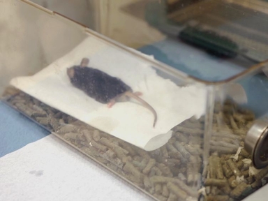
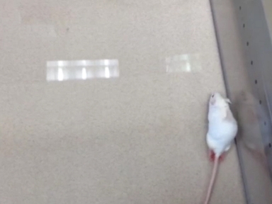
•A longitudinal examination of bone loss in the femurs and tibiae of adult mice was performed following spinal cord injury using sequential low-dose X-ray scans. Tibia bone loss was detected throughout the study, while bone loss in the femur was not detected until 40 days post injury.
Tags
Related Videos
Acute and Chronic Tactile Sensory Testing after Spinal Cord Injury in Rats

A Contusion Model of Severe Spinal Cord Injury in Rats

A Novel Vertebral Stabilization Method for Producing Contusive Spinal Cord Injury
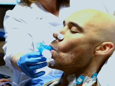
Evaluation of Respiratory Muscle Activation Using Respiratory Motor Control Assessment (RMCA) in Individuals with Chronic Spinal Cord Injury
Controlled Cortical Impact Model for Traumatic Brain Injury
A Radio-telemetric System to Monitor Cardiovascular Function in Rats with Spinal Cord Transection and Embryonic Neural Stem Cell Grafts
Calibrated Forceps Model of Spinal Cord Compression Injury
Contrast Enhanced Ultrasound Imaging for Assessment of Spinal Cord Blood Flow in Experimental Spinal Cord Injury

Orthotopic Hind Limb Transplantation in the Mouse

A Neonatal Mouse Spinal Cord Compression Injury Model
Copyright © 2024 MyJoVE Corporation. 판권 소유