
Electrophysiological Method for Recording Intracellular Voltage Responses of Drosophila Photoreceptors and Interneurons to Light Stimuli In Vivo
June 19th, 2016
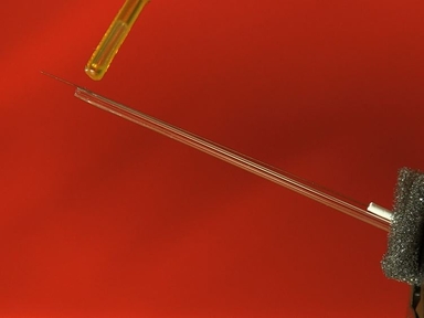
•Sharp microelectrodes enable accurate electrophysiological characterization of photoreceptor and visual interneuron output in living Drosophila. Here we show how to use this method to record high-quality voltage responses of individual cells to controlled light stimulation. This method is ideal for studying neural information processing in insect compound eyes.
Related Videos

Dual Electrophysiological Recordings of Synaptically-evoked Astroglial and Neuronal Responses in Acute Hippocampal Slices

A Method for Systematic Electrochemical and Electrophysiological Evaluation of Neural Recording Electrodes

Electrophysiological Recording From Drosophila Labellar Taste Sensilla
A Guide to In vivo Single-unit Recording from Optogenetically Identified Cortical Inhibitory Interneurons

Analyzing Synaptic Modulation of Drosophila melanogaster Photoreceptors after Exposure to Prolonged Light

Electrophysiological Method for Whole-cell Voltage Clamp Recordings from Drosophila Photoreceptors

Electrophysiological Recording from Drosophila Trichoid Sensilla in Response to Odorants of Low Volatility

Ex Vivo Calcium Imaging for Visualizing Brain Responses to Endocrine Signaling in Drosophila

Electrophysiological Recording of The Central Nervous System Activity of Third-Instar Drosophila Melanogaster

Preparing Adult Drosophila melanogaster for Whole Brain Imaging during Behavior and Stimuli Responses
Copyright © 2024 MyJoVE Corporation. 판권 소유