É necessária uma assinatura da JoVE para visualizar este conteúdo. Faça login ou comece sua avaliação gratuita.
Q Suture Technique: A Suturing Technique to Reduce Gap Formation and Increase Tensile Strength in Tendon Repair
Neste Artigo
Overview
In this video, we demonstrate suturing techniques — core suture and Q suture — to repair the foot flexor tendon in an ex vivo porcine model. Q sutures enhance the strength of the core suture in tendon repair and decrease gapping at the tendon repair site.
Protocolo
All procedures involving animal models have been reviewed by the local institutional animal care committee and the JoVE veterinary review board.
1. Tendon repair using core suture and Q suture
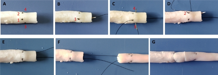
- Mark the anterior surface of one of the tendon stumps with 2 points that are 10 mm from the cut tendon end, with each point locating one-fourth of the way from the left (point 1) and the right (point 2), respectively, in the medial-lateral direction (Figure 1A).
- Mark each of the left (point 3) and right (point 4) lateral surfaces of the tendon with one point that is 8 mm from the cut tendon end and locate in the middle in the anterior-posterior direction (Figure 1A). Determine all lengths with a Vernier calliper (rated accuracy of 0.02 mm).
- Repair the tendon with 4-0 suture. Insert the needle into the cut surface of one tendon stump from the point that is in the middle in the anterior-posterior direction and one-fourth of the way from the left in the medial-lateral direction (Figure 1B). Pass the needle longitudinally through the tendon and withdraw the needle on the anterior surface of the tendon, exiting from point 1 (Figure 1B).
- Re-insert the needle obliquely from point 3 and pass it transversely toward point 4, creating a small loop at the lateral surface of the tendon (Figure 1C). Pull out the suture and re-insert the needle obliquely from point 2 and pass it longitudinally toward the cut end (Figure 1D,E).
- Insert the needle into the cut end of the other tendon stump and repair it with the same construct, forming a symmetrical repair (Figure 1F).
- Tighten the suture with 10% shortening of the tendon segment within the core suture. Tie the tendon ends together with 3 to 4 knots and complete the 2-strand core suture (Figure 1G).
- Repeat the operation once to complete the 4-strand core suture. Do not cut off the first core suture when performing the second core suture.
- Insert the same needle into the tendon anterior surface 2 mm away from the joined tendon end and pass through the full thickness of the tendon stump (Figure 2A).
- Withdraw the needle on the posterior surface of the tendon and re-insert the needle into the posterior surface of the tendon 2 mm away from the other side of the joined tendon end (Figure 2B).
- Pull out the suture from the anterior surface of the tendon and tie 3 knots to complete 1 Q suture (Figure 2C). Repeat the procedure to complete the second Q suture (Figure 2D).
- In the 2-strand and 4-strand core suture plus running group, add a running epitendinous suture of 9 to 10 stitches to the tendon ends using 6-0 suture. Keep a similar purchase of 1.5 mm and depth of 1 mm (Figure 2E,F,G).
- Keep the repaired tendon moist by wet gauzes before biomechanical testing.
Resultados

Figure 1: 2-strand core suture in tendon repair.
(A) Surface of tendon stump was marked with point 1, 2, 3, and 4. (B-E) Core suture in one tendon stump was completed. (F) The entire core suture was completed. (G) Suture was tightened, and knots were tied.
Divulgações
Materiais
| Name | Company | Catalog Number | Comments |
| 4-0 suture | Ethicon, Somerville, NJ | Ethilon 1667 | |
| 6-0 suture | Ethicon, Somerville, NJ | Ethilon 689 |
This article has been published
Video Coming Soon
Source: Mao, W. F., et al., Using Q Suture to Enhance Resistance to Gap Formation and Tensile Strength of Repaired Flexor Tendons. J. Vis. Exp. (2020).
Copyright © 2025 MyJoVE Corporation. Todos os direitos reservados