A subscription to JoVE is required to view this content. Sign in or start your free trial.
Chick Chorioallantoic Membrane Harvest: A Method to Isolate Tumor Graft-bearing Chorioallantoic Membrane from Chicken Egg
In This Article
Overview
This video demonstrates the isolation of tumor-bearing chorioallantoic membrane from the chicken embryo. The harvested CAM can be used for further downstream applications.
Protocol
All procedures involving animal models have been reviewed by the local institutional animal care committee and the JoVE veterinary review board.
1. Harvesting the CAM
- Prepare 6-well plates with 4% PFA (paraformaldehyde) pH 7.0; one well per egg, 2 mL per well.
- Bring the eggs to a laboratory bench. Using a needle attached to a syringe to perforate the film dressing, drop around 300 µL of PFA over the CAM to slightly stiffen the CAM, thus facilitating the harvesting process. Repeat this for all eggs.
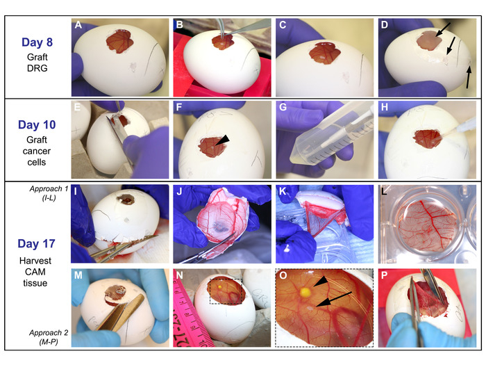
- With a scissor, remove the upper half of the eggshell (where the operating window is located) with the CAM attached to it (Figure 1I). Reduce the size of this half to approximately 3 cm in diameter. Grasp the CAM with fine forceps and detach it from the eggshell while placing it into PFA.
Results

Figure 1: Grafting of DRG, cells, and harvesting of CAM: On day 8: A. CAM easily observed after paraffin wax membrane removal. B-C. With fine forceps, DRG is placed onto the CAM. D. Egg is covered with film dressing and put in the incubator; arrows point to the openings that are covered. On day 10: E-F. Film dressing is...
Disclosures
Materials
| Name | Company | Catalog Number | Comments |
| PFA (paraformaldehyde solution) | Sigma-Aldrich | # P6148-1KG | Dilute in water to make a 4% PFA solution |
| Fine surgical straight sharp scissor | Fine Science Tools (FST) | #14060-09 | Used to harvest the CAM tissue on day 17 |
| Fertilized Lohmann White Leghorn eggs | Fertilized eggs at early fertilization days, preferably on first day postfertilization. Eggs used in this protocol are from Michigan State University Poultry Farm | ||
| Extra fine Graefe forceps, straight | Fine Science Tools (FST) | # 11150-10 | Used to graft DRG onto the CAM on day 8 and to harvest CAM tissue on day 17 |
This article has been published
Video Coming Soon
Source: Schmidt, L. B. et al. The Chick Chorioallantoic Membrane In Vivo Model to Assess Perineural Invasion in Head and Neck Cancer. J. Vis. Exp. (2019)
Copyright © 2025 MyJoVE Corporation. All rights reserved