A subscription to JoVE is required to view this content. Sign in or start your free trial.
Inferior Lacrimal Gland Removal From Rabbit Model: A Surgical Procedure to Remove the Larger Inferior Tear Gland From Rabbit Eye Orbit
In This Article
Overview
In this video, we describe a surgical procedure to remove the large inferior lacrimal gland from a rabbit model to study the effect on tear production in the animal.
Protocol
All procedures involving animal models have been reviewed by the local institutional animal care committee and the JoVE veterinary review board.
1. Resect the ILG
- Allow at least 5 min for the local anesthetic to take effect in the rabbit.
- Incise the skin, the depressor muscle of the inferior palpebra, the zygomaticolabial part of the zygomatic muscle, and orbicularis muscle with the Colorado microdissection needle and make the skin incisions along the surgical markings. Settings can vary based on clinical response and typically are between 10 to 15 units for both cut and coagulation.
- Maintain hemostasis with the monopolar cautery.
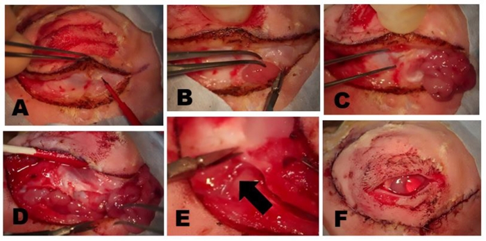
- As the incision is carried deeper through the skin marking, look for the sheen of a fascial plane over the zygomatic bone or superficial part of the masseter muscle. At this point, maintain the tissue plane and carry it superiorly toward the orbital rim using the Colorado needle for cutting (Figure 1A).
NOTE: For the purpose of identifying the ILG, it is easiest to perform this part of the dissection over the head of the ILG which is typically inferior to the anterior limbus of the eye. - After identifying and incising the capsule surrounding the ILG, identify the tan tissue of the ILG. Only the anterior portion of the ILG head will be visible (Figure 1B). However, the head can be followed medially as it passes beneath the zygomatic arch and transitions into the tail (Figure 1C).
- Use tenotomy scissors to cut the orbital septum along the inferior rim exposing the more posterior portion of the ILG tail. Once the tissue plane is identified, extend the dissection posteriorly along the entire incision line (Figure 1D).
NOTE: The duct of the ILG passes through the lower fibrous connective tissues to enter the inferior conjunctival space in the temporal aspect of the lid. At the posterior rim, the tail of the ILG can have varying anatomic configurations. Sometimes it terminates inferior to the posterior (lateral) canthus, while in other dissections it extends more superiorly around the temporal orbit. - Use extreme care to prevent inadvertent damage to the blood supply, which the ILG receives from branches of the carotid artery. The blood supply can be seen during this part of the dissection (Figure 1E).
- In cases where the tail terminates under the posterior (lateral) canthus, it may be necessary to bisect the temporal portion of the frontoscutular muscle to expose the tail of the ILG, which lies along the zygomatic bone.
- After the entire ILG has been isolated and exposed, remove it. Due to its large size, it is often preferable to cut the gland in half with scissors and remove the head separately from the tail.
- Proceed very cautiously when removing the head of the ILG as it lies immediately adjacent to a large venous sinus in the orbit. Although bleeding from this structure during surgical resections has not occurred, have ample hemostatic aids present to mitigate this risk.
- After removal of all gland tissue, close the deep connective tissue plane with multiple interrupted 5-0 ethylene terephthalate sutures. Close the superficial muscles and skin with a running 6-0 polyglactin 910 suture (Figure 1F) using 0.3 tissue forceps and a needle driver.
Results

Figure 1: Removal of the ILG. (A) The skin and superficial muscle are incised until the fascial plane overlying the zygomatic bone or superficial part of the masseter muscle is reached. The head of the ILG usually is clearly evident as a small bulge located under the anterior limbus. (B) The fibrous capsule of the ILG is incised with scissors exposing the ILG. On...
Disclosures
Materials
| Name | Company | Catalog Number | Comments |
| Acepromazine, Aceproinj | Henry Schein Animal Health, Dublin, OH | NDC11695-0079-8 | 0.1ml/kg subcutaneously injection for rabbit sedation |
| Anesthesia vaporizer | VetEquip, Pleasanton, CA | Item # 911103 | |
| Animal restraining bag | Henry Schein Animal Health, Dublin, OH | Jorvet J0170 | Use appropriately sized bag |
| Cautery unit, high-temperature, battery-powered | Medline Industries Inc, Northfield, IL | REF ESCT001 | Keep on hand in case of bleeding |
| Clipper, Wahl Mini Arco | Henry Schein Animal Health, Dublin, OH | No. 022573 | Cordless shears for fur removal |
| Colorado needle | Stryker Craniomaxillofacial, Kalamazoo, MI | N103A | Use with electrosurgical unit to make incisions |
| Electrosurgical unit with monopolar cautery plate | Valleylab, Boulder, CO | Force FXc | Use with electrosurgical unit to make incisions |
| Forceps, curved dressing | Bausch and Lomb (Storz), Bridgewater, NJ | Storz E1406 | delicate serrated dressing forceps |
| Forceps, 0.3 | Bausch and Lomb (Storz), Bridgewater, NJ | ET6319 | For removal of nictating membrane |
| Forceps, Bishop Harmon | Bausch and Lomb (Storz), Bridgewater, NJ | E1500-C | Use toothed forceps for dacryoadenectomy |
| Hair remover lotion, Nair | Widely available | Softening Baby oil | Dipilitory cream for sensitive skin |
| Isoflurane | Henry Schein Animal Health, Dublin, OH | 29405 | Possible alternative sedation |
| Laryngeal mask airway | Docsinnovent Ltd, London, UK | Vgel R3 | |
| Lid speculum, wire | Bausch and Lomb (Storz), Bridgewater, NJ | Barraquer SUH01 | For removal of nictating membrane |
| Scissors, Vannas | McKesson Medical-Surgical, San Francisco, CA | Miltex 2-130 | Capsulotomy scissors for dacryoadenectomy |
| Sedation gas mask | DRE Veterinary, Louisville, KY | #1381 | Possible alternative sedation |
| Surgical marking pen | Medical Action Industries, Arden, ND | REF 115 | |
| Sutures, 5-0 Mersilene | Ethicon US, LLC | Ethylene terephthalate sutures, used for deep connective tissue closure | |
| Rabbit, New Zealand White | Charles River Labs, Waltham, MA (NZW) | 2-3 kg | Research animals |
| Sutures, Vicryl 6-0 | Ethicon US, LLC | Polyglactin 910 sutures, used for superficial muscle and skin closure | |
| Syringe, 1 cc | BD, Franklin Lakes, NJ | Ref 309659 | For injection of lidocaine/ epinephrine |
| Tissue forceps, 0.8mm Graefe | Roboz Surgical Store, Gaithersburg, MD | RS-5150 | Curved Weck forceps |
This article has been published
Video Coming Soon
Source: Honkanen, R. A., et al. Establishment of a Severe Dry Eye Model Using Complete Dacryoadenectomy in Rabbits. J. Vis. Exp. (2020).
Copyright © 2025 MyJoVE Corporation. All rights reserved