A subscription to JoVE is required to view this content. Sign in or start your free trial.
Extracellular Vesicle Uptake Assay: A Method to Visualize and Quantify Cellular Uptake of Extracellular Vesicles Using 3D Fluorescence Imaging Technique
In This Article
Overview
In this video, we demonstrate an assay to visualize and quantify the cellular uptake of extracellular vesicles or EVs. This assay utilizes 3D fluorescent imaging using a confocal microscope and allows for distinguishing internalized EVs from ones adhered to the cell surface.
Protocol
1. EV isolation and on-chip immuno-fluorescent EV labeling
- Collection of cell culture media (CCM) and pre-processing of CCM for EV isolation
- Seed PC3 cells at 30% confluency in a 75 cm2 cell culture flask. Allow control cells to grow to 90% confluency (~48 h) in standard media and cell-line-specific supplements.
NOTE: To prevent EV-containing components from affecting cellular uptake (i.e., fetal bovine serum), use exosome-depleted media and supplements. - Harvest the CCM.
- Centrifuge the CCM at 1000 x g for 10 min at room temperature (RT) to pellet any unattached cells and large debris harvested with the media. Transfer the supernatant to a new conical tube.
- In the new tube, centrifuge the supernatant at 10,000 x g for 20 min at 4 °C to pellet smaller debris and apoptotic bodies remaining in the media. Some larger EVs will pellet. Transfer the supernatant to a new tube.
- Filter the supernatant through a 0.45 µm hydrophilic Polyvinylidene fluoride (PVDF) membrane syringe filter.
NOTE: If not immediately processing CCM for EV isolation, store the pre-processed CCM at -80 °C until isolation is performed. If frozen, limit freeze-thaw cycles to one.
- Seed PC3 cells at 30% confluency in a 75 cm2 cell culture flask. Allow control cells to grow to 90% confluency (~48 h) in standard media and cell-line-specific supplements.
- EV isolation from CCM using a nano-filtration based microfluidic device
- If frozen, completely thaw CCM and vortex for 30 s before step 1.2.2.
- Inject 1 mL of pre-processed CCM (step 1.1) into the sample chamber of the nano-filtration-based microfluidic device (see Table of Materials).
NOTE: Follow the standard operating procedure for the nano-filtration-based microfluidic device. - Spin at 3000 rpm for 10 min in the bench-top spinning machine (see Table of Materials) to operate the microfluidic device.
NOTE: If CCM remains on the sample chamber following the initial run, perform additional spins until all CCM has emptied from the sample chamber. - Remove the fluid from the waste chamber by pipetting and repeat steps 1.2.1, 1.2.2, and 1.2.3 twice.
NOTE: In total, 3 mL of CCM will be processed for EV isolation. - Inject 1 mL phosphate-buffered saline (PBS) into the sample chamber to wash the isolated EVs. Spin in the bench-top spinning machine for operating the microfluidic device as mentioned in step 1.2.3. Locate the pure EVs on the membrane of the device.
NOTE: The quality of EVs isolated from the nano-filtration based microfluidic device, specifically, was confirmed and compared to the conventional UC method by transmission electron microscopy (TEM), scanning electron microscope (SEM), nanoparticle tracking analysis (NTA), structured illumination microscopy, enzyme-linked immunosorbent assay and real-time PCR in the previous research.
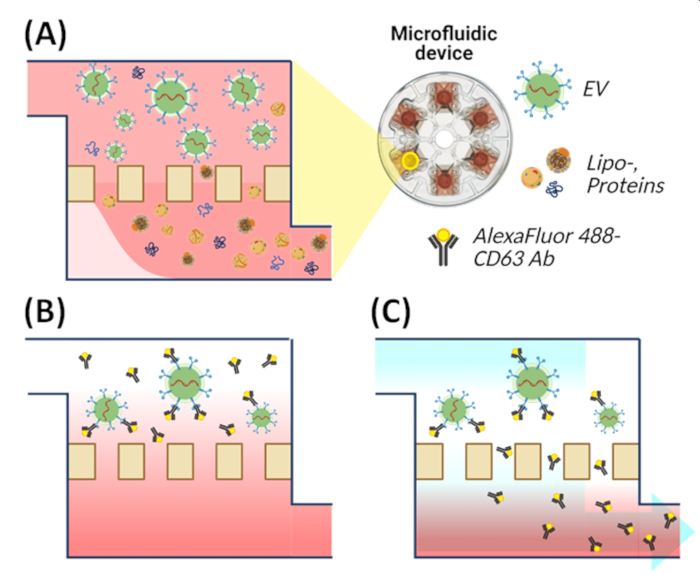
- Immunofluorescent labeling of EV using nano-filtration based microfluidic device (Figure 1)
- Select an EV-specific antibody according to the purpose of the assay (see Table of Materials).
NOTE: Certain antibodies may interfere with ligand binding sites specific to EV-uptake pathways (i.e., endocytosis). - Inject 1 µg/mL of the EV-specific antibody into the elution hole of the device containing 100 µL of isolated EVs.
- Incubate for 1 h in the dark at RT on a plate shaker to ensure the even distribution of the antibody across the sample.
- Attach an adhesive tape to the elution hole. Inject 1 mL PBS into the sample chamber to wash out any residual antibodies.
- Spin the device at 3000 rpm until the sample chamber is empty. Remove any fluid from the waste chamber by pipetting. Inject 1 mL PBS into the sample chamber.
NOTE: Fluorescently labeled EVs will be located in the membrane chamber. - Pipette the fluorescently labeled EVs (Figure 2) from the membrane chamber to an amber tube. Block from light until use.
- Select an EV-specific antibody according to the purpose of the assay (see Table of Materials).
2. Incubation of the cells with fluorescently labeled EVs for the EV-uptake assay
- Target cell seeding and culture on the cell-culture compatible dishes.
- Seed 1 x 10⁴ PC3 cells into the microslide 8-well plate (9.4 x 10.7 mm for each well) with 0.2 mL of media or 4 x 10⁴ PC3 cells into a 35 mm dish with 1 mL of media. Plate the cells into a cell-culture compatible dish consisting of a thin coverslip (thickness: 0.18 mm).
NOTE: The thin coverslip minimizes the adverse scattering of light. - Allow cells to adhere overnight in optimal cell-culture conditions (37 °C, 5% CO2 concentration, 90% humidity).
- Wash adhered cells twice with exosome-depleted media (described in step 1.1.1).
- Seed 1 x 10⁴ PC3 cells into the microslide 8-well plate (9.4 x 10.7 mm for each well) with 0.2 mL of media or 4 x 10⁴ PC3 cells into a 35 mm dish with 1 mL of media. Plate the cells into a cell-culture compatible dish consisting of a thin coverslip (thickness: 0.18 mm).
- Cell incubation with fluorescently labeled EVs.
- Measure the concentration of fluorescently labeled EVs (step 1.3.5) by nanoparticle tracking analysis (NTA, Figure 3). Determine the optimal concentration of fluorescently labeled EVs to be added to the cultured cells (step 2.1.2.).
- Dilute the fluorescently labeled EVs with exosome-depleted media to match the desired concentration measured in step 2.2.1. (i.e., 7.80 x 10⁹ EVs (in NTA value) in 200 µL of exosome-depleted media.)
- Add the diluted EVs (Step 2.2.2) to the adhered target cells prepared at 2.1.2. Incubate for experimental time (i.e., 4, 8, or 12 h).
- Wash cells thrice with exosome-free media to remove any non-internalized EVs.
NOTE: Optional: Cells can be fixated following wash. - Label the cytoplasm of the adhered cells with 1 µg/mL of CMTMR ((5-(and-6)-(((4-chloromethyl)benzoyl)amino) tetramethylrhodamine) (see Table of Materials) and incubate in optimal cell-culture conditions (37 °C, 5% CO2 concentration, 90% humidity).
NOTE: Cell area dyes should fluoresce separately from labeled EVs to aid in determining the spatial location (internalized or superficial) of the spiked EVs during the EV uptake assay. - Wash labeled cells twice with exosome-depleted media to remove the residual dye. Add fresh exosome-depleted media to the cells in preparation for live-cell confocal imaging.
3. Confocal microscopy
- To perform live-cell imaging, utilize an on-stage incubator to maintain optimal cell-culture conditions (37 °C, 5% CO2 concentration, 90% humidity).
- Place the prepared cells in the on-stage incubator.
- Set the imaging parameters based on control samples.
NOTE: Suggested control samples include: Fluorescently labeled EVs only, fluorescently labeled cells, unlabeled EVs, and unlabeled cells. - Determine the depth of the target cells and the range of stacking size in the z-direction to acquire 3D confocal images.
NOTE: The thickness of a Z-stack is 1 µm. The confocal 3D image acquisition lasted 2 min 34 s (each Z-plane image acquisition took approximately 8 s; a total of twenty Z-stack images). - Set image acquisition to multiple z-stacked images of both cell-specific dye (i.e., red) and EV-specific dye (i.e., green) simultaneously (Figure 4 and Figure 5A).
4. Image processing
- Utilize automatic image-processing software to analyze the raw z-stacked confocal images and determine the EV uptake by cells (see Table of Materials).
- Set thresholding parameters to the fluorescent signal of the cells and EV-specific dyes. Build the virtual surfaces of cells (Figure 5A,B).
- To build the virtual surfaces of cells, click the button Add new Surfaces.
- Select Shortest Distance Calculation as "Algorithm Settings" to use the provided algorithm by the software, then click Next: Source Channel.
- Select Channel 2 - CMTMR as "Source Channel" in this experiment.
- Select Smooth and put the appropriate value into "Surfaces Detail" for surface smoothing.
NOTE: 0.57 µm in this experiment since 1 pixel represents 0.57 µm in raw imaging data. - Select Absolute Intensity as "Thresholding."
- To automatically threshold the fluorescent image by the provided algorithm, click Threshold (Absolute Intensity): The value is automatically set.
- Select Enable as "Split touching Objects (Region Growing)" and put the value of estimated cell size into "Seed Points Diameter," 10.0 µm in this experiment. Then click Next: Filter Seed Points.
- To configure the virtual cell surfaces, click + Add button, then select Quality as "Filter Type." Threshold the appropriate value (210 in this experiment) for the low limit by a visual inspection and the maximum value (1485) for the upper limit, then click the Finish button.
NOTE: A visual inspection means that a researcher can discriminate the cellular area from a raw fluorescent image. - Next, to build the virtual dots of EVs, click the button Add new Spots.
- Select Different Spot Sizes (Region Growing) and Shortest Distance Calculation as "Algorithm Settings," then click Next: Source Channel.
- Select Channel 1 - Alexa Fluor 488 as "Source Channel" in this experiment.
- Put the appropriate value into "Estimated XY Diameter" for the spot detection, 1 µm in this experiment. Then, click Next: Filter Spots.
- To configure the virtual EV dots, click + Add button, select Quality as "Filter Type," and set "Lower Threshold" by a visual inspection, 100 in this experiment. Then, click the Next: Spot Region Type button.
- Select Absolute Intensity as "Spot Regions Type," then click Next: Spot Regions.
- To threshold, the region of EV dots, put the appropriate value into "Region Threshold" by a visual inspection, 100 as "Region Threshold" in this experiment.
NOTE: A visual inspection means that a researcher can discriminate the EVs area from a raw fluorescent image. - Select Region Volume as "Diameter from," then click Finish.
- Use the software's provided algorithms to split the grouped spots inside the built surface at step 4.2 (Figure 5C, i-iv).
- Click the built Spots, then go into Filters.
- Click + Add button, then select Shortest Distance to Surfaces Surfaces = Surface 1 as Filter Type, then click Duplicate Selection to new Spots button. The lowest threshold (-7.0 in this experiment) for the low limit and the appropriate value (-0.5) for the upper limit.
NOTE: Set the upper limit with the estimated radius of Spots. In this experiment, the estimated diameter of Spots,i.e., EV dots, was set to 1 µm in step 4.2.11.; thus, the upper limit can be 0.5.
- Automatic count of EVs inside the cells
NOTE: The software will automatically count the number of EVs inside the cells, indicating the number internalized by the target cells.- Click the built Spots 1 selection [Shortest Distance to Surfaces Surfaces = Surfaces 1 between -7.00 and -0.500].
- Go to the Statistics, and export the value from "Total Number of Spots"
NOTE: The software's provided algorithms will automatically calculate the number and volume of cells.
- Determine the yield of EV uptake per incubation period based on the above-calculated values (Figure 6).
- To obtain the number of cells, click the built Surfaces 1, then go to the Statistics, and export the value of "Total Number of Surfaces" from Overall.
- Go to the Detailed into Statistics to export the Volume from Detailed.
Access restricted. Please log in or start a trial to view this content.
Results

Figure 1: Schematic illustration of the EV isolation and on-chip labeling using a nano-filtration-based microfluidic device. (A) EVs isolation from CCM. (B) On-chip Immunofluorescent labeling of EVs. (C) Removal of unbound antibodies.

Figure 2: Imaging of fluorescently labeled EVs. The fluorescently labeled (anti-CD63-Alexa Fluor 488) EVs were detected using the confocal microscope (40x objective). (A) Positive sample (anti-CD63-Alexa Fluor 488 labeled EVs). (B) Negative control 1 for the EV labeling (EVs with 2nd antibody (Alexa Fluor 488) only, without 1st antibody). (C) Negative control 2 (EVs with the mouse (MS) IgG antibody and 2nd antibody (Alexa Fluor 488)).

Figure 3: NTA measurement of anti-CD63-Alex Fluor 488 labeled EVs.

Figure 4: Imaging of the internalized EVs into cells in a 2D image. (A) The fluorescently labeled (anti-CD63-Alexa Fluor 488, green) EVs and the cells (CMTMR, red) were detected by using the confocal microscope (20x objective) after the incubation. (B) A separate image of the fluorescently labeled EVs only. (C) A separate image of the fluorescently labeled cells only. The excitation/emission laser wavelengths for CMTMR and Alexa Fluor 488 are 560.6/595 (±50) nm and 487.8/525 (±50) nm. Laser power settings are 3.0 % for CMTMR and 10.0 % for Alexa Fluor 488.

Figure 5: Quantification of the internalized EVs by the post-imaging process. (A) Raw confocal image obtained from the EV-uptake assay. (B) Virtual rendering of the EVs as a dot (green) and the cells as a surface (red) by using the image-processing software. (C, i-iv) Discrimination of the internalized EVs (yellow dots) and non-internalized EVs (green dots, white arrow) using the software provided algorithm.

Figure 6: The amount of EV uptake as a function of incubation time. (A) The number of internalized EVs per cell. (B) The number of internalized EVs per cell volume. The number of internalized EVs was increased depending on the incubation time.
Access restricted. Please log in or start a trial to view this content.
Disclosures
Materials
| Name | Company | Catalog Number | Comments |
| Alexa Fluor 488 anti-human CD63 Antibody | Biolegend | 353038 | Fluorescent dye conjugated EV-specific antibody |
| CellTracker Orange CMTMR Dye | Thermo Fisher Scientific | C2927 | Live cell (cytoplasm) fluoresent labeling reagent |
| CFI Apo Lambda S 40XC WI | Nikon | MRD77400 | Objective for confocal imaging, NA=1.25 |
| CFI Plan Apo VC 20X | Nikon | MRD70200 | Objective for confocal imaging, NA=0.75 |
| Exodisc | Labspinner Inc. | EX-D1001 | A nano-filtration based microfluidic device for EV isolation |
| ExoDiscovery | Labspinner Inc. | EX-R1001 | Operation device for Exodisc |
| Exosome-depleted FBS | Thermo Fisher Scientific | A2720801 | Nutrient of cell culture media for PC3 cell line derived EV collection |
| Fetal bovine serum (FBS) | VWR | 1500-500 | Nutrient for cell cultivation |
| Goat Anti-Mouse IgG H&L preadsorbed | abcam | ab7063 | Mouse IgG antibody for negatvie control of EV labeling |
| Ibidi USA U DISH μ-Dish 35 mm | Ibidi | 81156 | Culture dish for confocal imaging |
| Imaris 9.7.1 | Oxford Instruments | 9.7.1 | Post-image processing software |
| Incubator System+ CO2/O2/N2 gas mixer | Live Cell Instrument | TU-O-20 | Incubator system for live cell imaging |
| Nikon A1 HD25 / A1R HD25 camera | Nikon | NA | Camera for confocal imaging |
| Nikon Eclipse Ti microscope | Nikon | NA | Inverted microscope for confocal imaging |
| NIS-Elements AR 4.50.00 | Nikon | 4.50.00 | Image processing software for Nikon microscope |
| NTA, NanoSight NS500 | Malvern Panalytical | NS500 | Measurement device for EV concentration |
| OriginPro 2020 | OriginLab | 9.7.0.185 | Graphing software |
| Penicillin-Streptomycin | Thermo Fisher Scientific | 15140122 | Antibiotics for cell cultivation |
| RPMI 1640 | Thermo Fisher Scientific | 21875034 | Cell culture media for PC3 cell line cultivation |
| SYTO RNASelect Green Fluorescent cell Stain - 5 mM Solution in DMSO | Thermo Fisher Scientific | S32703 | RNA staining fluorescent dye for the EV labeling |
References
Access restricted. Please log in or start a trial to view this content.
This article has been published
Video Coming Soon
Source: Kim, C. J., et al., Extracellular Vesicle Uptake Assay via Confocal Microscope Imaging Analysis. J. Vis. Exp. (2022).
Copyright © 2025 MyJoVE Corporation. All rights reserved