
A subscription to JoVE is required to view this content. Sign in or start your free trial.
Method Article
Oct4GiP Reporter Assay to Study Genes that Regulate Mouse Embryonic Stem Cell Maintenance and Self-renewal
In This Article
Summary
We describe a fluorescence reporter assay to quickly identify and characterize genes that regulate mouse embryonic stem cell maintenance and self-renewal.
Abstract
Pluripotency and self-renewal are two defining characteristics of embryonic stem cells (ES cells). Understanding the underlying molecular mechanism will greatly facilitate the use of ES cells for developmental biology studies, disease modeling, drug discovery, and regenerative medicine (reviewed in 1,2).
To expedite the identification and characterization of novel regulators of ES cell maintenance and self-renewal, we developed a fluorescence reporter-based assay to quantitatively measure the self-renewal status in mouse ES cells using the Oct4GiP cells 3. The Oct4GiP cells express the green fluorescent protein (GFP) under the control of the Oct4 gene promoter region 4,5. Oct4 is required for ES cell self-renewal, and is highly expressed in ES cells and quickly down-regulated during differentiation 6,7. As a result, GFP expression and fluorescence in the reporter cells correlates faithfully with the ES cell identity 5, and fluorescence-activated cell sorting (FACS) analysis can be used to closely monitor the self-renewal status of the cells at the single cell level 3,8.
Coupled with RNAi, the Oct4GiP reporter assay can be used to quickly identify and study regulators of ES cell maintenance and self-renewal 3,8. Compared to other methods for assaying self-renewal, it is more convenient, sensitive, quantitative, and of lower cost. It can be carried out in 96- or 384-well plates for large-scale studies such as high-throughput screens or genetic epistasis analysis. Finally, by using other lineage-specific reporter ES cell lines, the assay we describe here can also be modified to study fate specification during ES cell differentiation.
Protocol
1. Oct4GiP Mouse ES Cell Maintenance
Oct4GiP cells were kindly provided by Dr. Austin Smith. They were derived from the 129/Ola mice carrying an Oct4-GFPiresPac transgene 4,5 . They are maintained in gelatin-coated tissue culture plates in the ESGRO complete plus clonal grade medium (Millipore), or in the M15 medium: DMEM (Invitrogen) supplemented with 15% FBS, 1000 U/ml ESGRO (Millipore), 1x Non-essential amino acids (Invitrogen), 1x EmbryoMax Nucleasides (Millipore), and 10 μM β-mercaptoethanol.
- Coat plates with 0.1% gelatin (Sigma) at room temperature for 30 minutes (0.1 ml gelatin solution/cm2).
- Plate Oct4GiP cells in gelatin-coated plates at ~2 x 10⁴/cm2.
- Split the cells every two days with 0.05% trypsin.
2. siRNA Transfection in Oct4GiP Cells
The Oct4GiP reporter assay is most conveniently carried out in gelatin-coated flat bottom 96- or 384-well plates (Corning or BD) in the M15 medium. The overall procedure is outlined in Figure 1.
- siRNA-lipid complexes assembly:
- For transfection in 96-well plates, assemble siRNA-lipid complexes in U-bottom 96-well plates.
- In each well, mix 10 μl OptiMEM (Invitrogen) and 0.3 μl Lipofectamine 2000 (Invitrogen) and incubate at room temperature for 5 min.
- Add 5 pmol siRNA to the lipid-OptiMEM mixture and incubate for another 15 minutes. The siRNA : lipid ratio is 100 pmol : 6ul.
- Transfer the siRNA-lipid complexes in OptiMEM from the U-bottom plate to the gelatin-coated flat-bottom plate with a multi-channel pipette.
- For transfection in 384-well plates, prepare the siRNA-lipid complexes using 0.1 μl Lipofectamine 2000 and 2 pmol siRNA in 10 μl OptiMEM.
Note: The final siRNA concentration in the transfections is 50 nM. Carry out 3-4 biological replicates for each siRNA transfection. Set up master mixtures of the siRNA-lipid complexes in the U-bottom plate and aliquot into the flat-bottom gelatin-coated plate.
- Oct4GiP cell plating and transfection:
- For transfection in 96-well plates, collect the Oct4GiP cells and resuspend them in fresh M15 medium to 9 x 10⁴ cells/ml.
- Add 100 μl of the cell suspension to each well using a multi-channel pipette.
- Mix the cell suspension with the pre-aliquoted siRNA-lipids complexes in the well by pipeting up and down 3-5 times. Change tips for different siRNA transfections.
- For Transfection in 384-well plates, resuspend Oct4GiP cells to 6 x 10⁴ cells/ml and aliquot 30 μl cell suspension to each well.
Note: The optimal cell plating density is usually 3 x 10⁵ cells/cm2, but it may require further optimization for siRNAs that dramatically affect cell growth or viability. Low plating density will lead to poor cell survival during transfection. High plating density will lead to high background due to high cell confluence induced differentiation. If necessary, test plating density between 2-4 x 10⁵ cells/cm2.
- Medium change:
- Remove old medium using multi-channel aspirator (Corning) or vacuum wand (VP-Scientific). Be careful not to scratch the cells at the bottom of the plates.
- Feed the cells with fresh ES cell medium (100 μl for 96-well plate and 30 μl for 384-well plate) using multi-channel pipette every day.
3. FACS Analysis
- Cell dissociation:
- Four days after transfection, remove medium using multi-channel aspirator (Corning) or vacuum wand (VP-Scientific).
- For 96-well plates, rinse cells once with 100μl PBS.
- Add 25 μl 0.25% trypsin to each well using a multi-channel pipette.
- Incubate at room temperature for 5minutes with occasional agitation, and visually inspect to ensure complete detachment of the cells.
- Add 90 μl of PBS with 10% FBS to inactivate the trypsin and dissociate cells into a single cell suspension by repeated pipetting.
- For 384-well plates, disassociate cells with 10 μl trypsin and quench with 30 μl PBS with 10% FBS.
- FACS analysis:
- Analyze the GFP fluorescence on the BD LSRII FACS analyzer equipped with the HTS unit or other similarly equipped FACS analyzer (such as Accuri or Intellicyt).
- Use the high-throughput mode on the HTS and analyze 10 μl of cell suspension. Set the count threshold to 1.0 x 10⁴ cells.
- Adjust PMT voltage and threshold to capture the cells in the forward vs. side-scatter plot. Gate for the live cell population in the forward vs. side-scatter plot.
- Create a histogram plot for the GFP channel. Set the gate in the GFP channel so that ~10% of the cells appear to be GFP-negative in the mock or control-siRNA transfected cells.
- Determine % GFP-negative cells from each treatment: % Differentiated cells = % GFP-negative cells. Compare the % Differentiated cells between experimental- and control-siRNA transfections using 2-tailed t-test, and score the positive hits with p-values < 0.01.
4. Representative Results
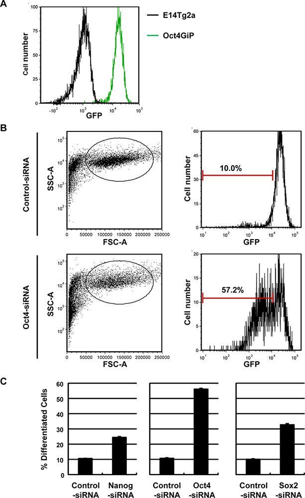
Oct4, Nanog, and Sox2 are three genes that play critical roles in the maintenance of ES cell self-renewal 6,7,9-11. Figure 2 shows that the Oct4GiP reporter assay can readily detect the differentiation caused by silencing these factors in the Oct4GiP ES cells.
Figure 2A shows the Oct4GiP ES cells are GFP-positive when maintained as ES cells. Figure 2B shows the forward vs. side scatter plot and the histogram of the GFP channel of the Oct4GiP cells transfected with the control- or Oct4-siRNAs. It is necessary to gate for live cells in the forward vs. side scatter plot, as the dead cells and debris are GFP-negative and will increase background. On the other hand, doublet discrimination to exclude possible cell clumps is not always needed. In the control-siRNA transfected cells, the vast majority of the cells should be GFP-positive. If obvious GFP-negative populations are present in the control-siRNA transfected wells, the starting Oct4GiP cells or the transfection procedure may have been compromised and the result may not be interpretable.
Figure 2C shows the bar graph of % Differentiated cells (% Differentiated cells = % GFP-negative cells) from control-, Oct4-, Nanog-, and Sox2-siRNA transfected cells.

Figure 1. Outline of the Oct4GiP reporter assay.

Figure 2. Oct4GiP reporter assay can detect ES cell differentiation caused by Nanog, Oct4, or Sox2 silencing. A) E14Tg2a (wild-type, black line) and Oct4GiP (green line) cells were analyzed by FACS. Histogram of the GFP-channel shows that Oct4GiP cells are GFP-positive. B) Oct4GiP cells were transfected in 96-well plates with the Control- or Oct4-siRNA and FACS analyzed 4 days after transfection. Forward vs. side scatter plots and histograms of the GFP-channel of the transfected cells are shown. C) Oct4GiP cells were transfected with the Control-, Nanog-, Oct4-, or Sox2-siRNA. The % Differentiated Cells was determined from the % GFP-negative cells 4 days after transfection, and was plotted as mean +/- standard error of the mean (n = 4). Click here to view larger figure.
Access restricted. Please log in or start a trial to view this content.
Discussion
The Oct4GiP reporter assay we describe above can quantitatively measure the extent of self-renewal vs. differentiation. Compared to other available methods, such as the morphology-based 12 and proliferation/viability-based assays, it offers higher sensitivity and throughput, as well as a more direct measurement of the ES cell state. It is therefore well suited for large-scale screens and genetic epistasis analysis. Indeed, we and others have successfully used the Oct4GiP reporter assay for genome-wide RNAi scr...
Access restricted. Please log in or start a trial to view this content.
Disclosures
No conflicts of interest declared.
Acknowledgements
We thank Brad Lackford for reading and editing the manuscript. This research was supported by the National Institute of Environmental Health Sciences, National Institutes of Health Intramural Research Program Z01ES102745 (to G. H.).
Access restricted. Please log in or start a trial to view this content.
Materials
| Name | Company | Catalog Number | Comments |
| ESGRO complete plus clonal grade medium | EMD Millipore | SF001-500P | |
| DMEM (High glucose 1X) | Invitrogen | 11965 | |
| 0.25% Trypsin-EDTA | Invitrogen | 25200 | |
| Lipofectamine 2000 | Invitrogen | 1001817 | |
| OPTI-MEM(reduce serum medium) | Invitrogen | 31985 | |
| ESGRO mLIF (107 units/1ml) | EMD Millipore | DAM1776540 | |
| MEM NEAA (Non-Essential Amino Acids) | Invitrogen | 11140 | |
| 100x Nucleosides for ES cell | EMD Millipore | 10620-1 | |
| 2-mercapt–thanol | Sigma-Aldrich | M7522-100ml | |
| ES-qualified fetal bovine serum | Invitrogen | 10437 | |
| Nanog siRNA | Invitrogen | MSS231181 | |
| Oct4 siRNA | Dharmacon | D-046256-02 | |
| Sox2 siRNA | Dharmacon | M-058489-01 | |
| Control siRNA: siRNA duplex targeting firefly luciferase (5’-CGTACGCGGAATACTTCGA) synthesized by Dharamcon. | |||
References
- Keller, G. Embryonic stem cell differentiation: emergence of a new era in biology and medicine. Genes Dev. 19, 1129-1155 (2005).
- Murry, C. E., Keller, G. Differentiation of embryonic stem cells to clinically relevant populations: lessons from embryonic development. Cell. 132, 661-680 (2008).
- Hu, G. A genome-wide RNAi screen identifies a new transcriptional module required for self-renewal. Genes Dev. 23, 837-848 (2009).
- Ying, Q. L., Nichols, J., Evans, E. P., Smith, A. G. Changing potency by spontaneous fusion. Nature. 416, 545-548 (2002).
- Ying, Q. L., Smith, A. G. Defined conditions for neural commitment and differentiation. Methods Enzymol. 365, 327-341 (2003).
- Nichols, J. Formation of pluripotent stem cells in the mammalian embryo depends on the POU transcription factor Oct4. Cell. 95, 379-391 (1998).
- Niwa, H., Miyazaki, J., Smith, A. G. Quantitative expression of Oct-3/4 defines differentiation, dedifferentiation or self-renewal of ES cells. Nat. Genet. 24, 372-376 (2000).
- Ding, L. A genome-scale RNAi screen for Oct4 modulators defines a role of the Paf1 complex for embryonic stem cell identity. Cell Stem Cell. 4, 403-415 (2009).
- Chambers, I. Functional expression cloning of Nanog, a pluripotency sustaining factor in embryonic stem cells. Cell. 113, 643-655 (2003).
- Masui, S. Pluripotency governed by Sox2 via regulation of Oct3/4 expression in mouse embryonic stem cells. Nat. Cell Biol. 9, 625-635 (2007).
- Mitsui, K. The homeoprotein Nanog is required for maintenance of pluripotency in mouse epiblast and ES cells. Cell. 113, 631-642 (2003).
- Fazzio, T. G., Huff, J. T., Panning, B. An RNAi screen of chromatin proteins identifies Tip60-p400 as a regulator of embryonic stem cell identity. Cell. 134, 162-174 (2008).
- Pease, S., Braghetta, P., Gearing, D., Grail, D., Williams, R. L. Isolation of embryonic stem (ES) cells in media supplemented with recombinant leukemia inhibitory factor. 141, 344-352 (1990).
- Young, R. A. Control of the embryonic stem cell state. Cell. 144, 940-954 (2011).
- Chambers, I. Nanog safeguards pluripotency and mediates germline development. Nature. 450, 1230-1234 (2007).
- Maherali, N. Directly reprogrammed fibroblasts show global epigenetic remodeling and widespread tissue contribution. Cell Stem Cell. 1, 55-70 (2007).
- Rodda, D. J. Transcriptional regulation of nanog by OCT4 and SOX2. J. Biol. Chem. 280, 24731-24737 (2005).
- Schaniel, C. Delivery of short hairpin RNAs--triggers of gene silencing--into mouse embryonic stem cells. Nat. Methods. 3, 397-400 (2006).
- Toyooka, Y., Shimosato, D., Murakami, K., Takahashi, K., Niwa, H. Identification and characterization of subpopulations in undifferentiated ES cell culture. Development. 135, 909-918 (2008).
Access restricted. Please log in or start a trial to view this content.
Reprints and Permissions
Request permission to reuse the text or figures of this JoVE article
Request PermissionExplore More Articles
This article has been published
Video Coming Soon
Copyright © 2025 MyJoVE Corporation. All rights reserved