
Method Article
Best Current Practice for Obtaining High Quality EEG Data During Simultaneous fMRI
In This Article
Summary
Simultaneous electroencephalography (EEG) and functional Magnetic Resonance imaging (fMRI) is a powerful neuroimaging tool. However, the inside of an MRI scanner forms a difficult environment for EEG data recording and safety must be considered whenever operating EEG equipment inside a scanner. Here, we present an optimised EEG-fMRI data acquisition protocol.
Abstract
Simultaneous EEG-fMRI allows the excellent temporal resolution of EEG to be combined with the high spatial accuracy of fMRI. The data from these two modalities can be combined in a number of ways, but all rely on the acquisition of high quality EEG and fMRI data. EEG data acquired during simultaneous fMRI are affected by several artifacts, including the gradient artefact (due to the changing magnetic field gradients required for fMRI), the pulse artefact (linked to the cardiac cycle) and movement artifacts (resulting from movements in the strong magnetic field of the scanner, and muscle activity). Post-processing methods for successfully correcting the gradient and pulse artifacts require a number of criteria to be satisfied during data acquisition. Minimizing head motion during EEG-fMRI is also imperative for limiting the generation of artifacts.
Interactions between the radio frequency (RF) pulses required for MRI and the EEG hardware may occur and can cause heating. This is only a significant risk if safety guidelines are not satisfied. Hardware design and set-up, as well as careful selection of which MR sequences are run with the EEG hardware present must therefore be considered.
The above issues highlight the importance of the choice of the experimental protocol employed when performing a simultaneous EEG-fMRI experiment. Based on previous research we describe an optimal experimental set-up. This provides high quality EEG data during simultaneous fMRI when using commercial EEG and fMRI systems, with safety risks to the subject minimized. We demonstrate this set-up in an EEG-fMRI experiment using a simple visual stimulus. However, much more complex stimuli can be used. Here we show the EEG-fMRI set-up using a Brain Products GmbH (Gilching, Germany) MRplus, 32 channel EEG system in conjunction with a Philips Achieva (Best, Netherlands) 3T MR scanner, although many of the techniques are transferable to other systems.
Introduction
Simultaneous electroencephalography (EEG) and functional Magnetic Resonance imaging (fMRI) allows the excellent temporal resolution of EEG to be combined with the high spatial accuracy of fMRI. There are a number of ways in which the data from these two modalities can be combined 1, but all rely on the acquisition of high quality EEG and fMRI data. To date, simultaneous EEG-fMRI has been used to study the correlation between oscillatory rhythms (measured with EEG) and blood oxygenation responses (using blood oxygenation level dependent (BOLD) fMRI) e.g. 2,3. It has also been used to study whether the characteristics of the evoked signal can explain the variance in the BOLD signal on a trial-by- trial basis4,5. In clinical studies the main use of the technique has been to investigate the foci of interictal epileptic discharges, which can help in surgical planning and are currently difficult to localize non-invasively6,7. To achieve the fusion of EEG and fMRI data which is desired, it is essential to have high quality data from both modalities. However, EEG data acquired during simultaneous fMRI are affected by several artifacts, including the gradient artefact (due to the changing magnetic fields required for fMRI), the pulse artefact (linked to the cardiac cycle) and movement artifacts (resulting from movements in the strong magnetic field of the scanner, as well as muscle activity). These artifacts are significantly larger than the neuronal activity of interest and therefore reduction (at source) and correction of the artifacts (via post-processing) are both needed to enable successful implementation of simultaneous EEG-fMRI.
The post-processing methods currently available for correcting the gradient and pulse artifacts require a number of criteria to be satisfied during data acquisition in order to produce high quality EEG data. Over the previous decade the optimal experimental set-up for recording high quality data has evolved as our understanding of the causes of the artifacts 8-10 has improved and we have learnt how to modify experimental methods so as to reduce the artifacts at source 11,12 and to improve the performance of post-processing correction algorithms. These developments include improving the sampling of the gradient waveforms through synchronization of scanner clocks 13,14 and use of a vectocardiogram15,16 to provide a cleaner cardiac trace than the traditional ECG. The vectocardiogram trace is derived from four electrodes placed on the chest with a stringent low-pass filter employed14-16. As a result the trace is relatively unaffected by gradient artifacts and is insensitive to the blood flow artifact making R-peak detection easier. However, the facility to record a vectocardiogram is not available on all MRI scanners and therefore will only be mentioned briefly in this study. The importance of the minimization of artifacts and stringent cleaning of data has been highlighted by the recent demonstration that motion artifacts recorded in the EEG data can correlate with BOLD activity unrelated to the task of interest, producing spurious results if extreme care is not taken throughout the experimental process 17.
The method presented here represents the current optimal approach for obtaining high quality EEG and fMRI data simultaneously using MR hardware and pulse sequences which are widely available, along with commercially supplied EEG equipment. Implementation of the suggested acquisition method, in conjunction with the use of appropriate post-processing methods, will yield EEG and fMRI data that can be used to answer a number of important neuroscience questions.
Protocol
1. Preparing the Experimental Setup
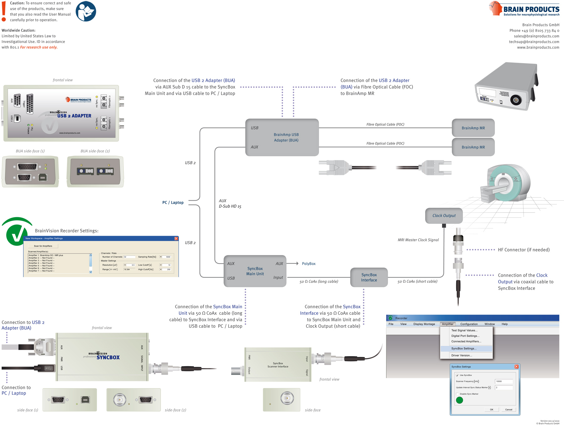
- Prior to the subject's arrival set up the EEG equipment in the control room where the scanner operator will sit. Connect the laptop computer to the EEG hardware as shown in Figure 1. Note: all triggers from peripheral devices and the MR scanner must have durations of more than 200 μsec to be detected by the EEG system.
- Set up the stimulus computer; in this study, we use a visual stimulus; markers are read into the BrainVision Recorder at the beginning and end of each stimulation period.
- Ensure the workspace for recording the data is set to the highest temporal resolution available and correct filter settings. For the majority of studies AC-coupling with a filter of 0.016-250 Hz is optimal although DC-coupling or a higher (1 kHz) low-pass filter may be required if ultra low or high frequency neuronal signals are of interest, respectively.
- Check the markers from the scanner and stimulus presentation to confirm that they are being recorded by the EEG system correctly. Turn on the synchronization of the scanner and EEG clocks using the BrainVision Recorder control panel. Then check if synchronization is successful; if the set up is correct the green icon and "Sync On" dot will appear.
- Set up the MR scanner in the conventional way; here we're using the body transmit RF coil and a 32 channel head receive RF coil. When possible, it is best to use a head-sized transmit coil to minimize the risk of RF heating of the EEG cap and associated cables. However on most scanners, the head transmit coil cannot be used in conjunction with a multi-element receiver coil, which leads to a sub-optimal set up for acquisition of fMRI data (in particular parallel imaging speed-up is not possible). We are using this specific head receive coil because it incorporates an access port which allows the cables from the EEG cap to run along a straight path out of the scanner.
- Ensure the MR sequences to be run are set up. The fMRI sequence must use a slice TR which is a multiple of the EEG clock period (200 μsec). If using a Philips MR system the Philips Timing calculator can be used to determine the possible slice and TR combinations.
- Make one final check that all equipment is recording as expected.
2. Subject Arrival
- Ask the subject to arrive with clean hair and wearing comfortable, non-metallic clothing.
- Explain to the subject the purpose of the experiment and what will happen.
- Ask the subject to fill out forms that are used to establish that there are no contra-indications for MR scanning and that the subject consents to participation in the experiment. Check the forms before proceeding. In this study the approval of the local ethics committee has been obtained and all subjects gave informed consent.
- Measure the head-circumference and select the appropriately sized cap (i.e. the smallest available cap that is larger than the head size). Place the cap on the head starting at the front of the head and pulling backwards. Position the cap correctly, such that Cz electrode is sited half way between the nasion and inion and also centered left-right.
- Connect the electrodes to the head by: moving hair out of the way, applying alcohol and then Abralyte gel. Attach the ECG electrode to the base of the back using a similar method to that used for the cap electrodes. This electrode is used to measure the heartbeat. The positioning at the base of the back is recommended to maximize the signal to noise of the R-peak in the ECG trace as well as for subject comfort.
- Work on the contacts so as to reduce the impedances of electrodes on the head to less than 10 kΩ (excluding the resistance of the internal resistors in each electrode) . ECG and EOG resistance may be higher as the signals are stronger and good connections can be hard to achieve, but must be kept below 50kΩ.
- Check the EEG data quality is satisfactory by visually inspecting the data on the monitor screen.
3. Recording Outside of the MR Scanner
(Optional: Only Required if You Wish to Compare EEG Data Quality from Inside and Outside the MR Scanner)
- Set up the presentation apparatus and EEG equipment outside the scanner (in a location where the magnetic field is low). Ensure that the setup is as similar as possible to that used inside the MR scanner (in particular the subject should be supine and a similar process of stimulus presentation should be used).
- Perform the experiment and record the data in a similar way to that used inside the scanner (see Section 4).
4. Setting Subject up Inside the MR Scanner
- Ask the subject to be seated whilst you set up the EEG equipment in the MR scanner room.
- Take the amplifier into the shielded room and place it on a table at the back of the scanner. Attach the amplifier to a long fiber optic cable. Pass the fiber optic cable through the waveguide and attach it to the BrainAmp USB adaptor in the control room (Figure 1).
- Register the patient in the MR scanner patient data-base.
- Take the subject into the room and ask them to lie on the scanner bed.
- Give the subject earplugs, head-phones and call button, and ensure that they are comfortable.
- Put the head coil over the subject's head. The EEG cables must leave the head coil along the shortest path possible. Now pad the subject's head to minimize head movement.
- Move the subject into the scanner bore, ensuring that electrodes Fp1 and Fp2 are at isocentre of the MR scanner in the z-direction. This is normally achieved by aligning these two electrodes with the light that is used to position the subject before they enter the bore.
- Attach the EEG cap to the amplifier at the rear of the scanner. Ensure that there are no wire loops in the EEG leads (as these can lead to RF heating and also cause larger EEG artifacts to be induced) and that the cabling is isolated from MR scanner vibrations as much as possible; here we use a cantilevered beam to achieve this isolation.
5. Recording Inside the Scanner
- Talk to the subject from the console room to confirm that they can hear the scanner operator and are OK.
- A second experimenter starts the EEG monitoring, checking for noisy electrodes in the traces, as well as for the green "Sync On" dot at the bottom of the screen.
- The clear effect of the cryo-pumps on the recording can be seen (see Figure 2). Therefore, switch off these pumps during data acquisition, following manufacturer guidelines.
- Ask the subject to move their head by a small amount. The importance of keeping the head still can be seen from the large voltages in the EEG recording that result from small head movements.
- Test the recording of neuronal activity by asking the subject to open and close their eyes. Look for occipital alpha activity. This will test whether you are measuring physiological signals rather than noise. If an alpha signal cannot be seen on a subject (which occurs in some subjects) it is possible to test for neuronal activity by performing a short run of the experimental paradigm without the MR scanner running and to look for the averaged evoked potential.
- The pulse artifact can clearly be seen in the raw data (see Figure 2) particularly on electrodes over the temples. Use the ECG trace to correct this artifact in real time using RecView (or in post processing software packages).
- As soon as each MRI scan begins the gradients will cause large artifacts in the EEG data.
- When the fMRI experiment is ready to start, - with the stimulus presentation system in a ready state - then begin saving the EEG data by following the steps shown.
- Now start the experiment, checking that the markers from the stimulus presentation and the MR scanner can be seen in BrainVision Recorder. Here the stimulus consists of a full-field radial checkerboard at 100% contrast. The reversal rate is 2 Hz such that an evoked response will occur every 500 msec and a marker is placed in the EEG file at each image reversal.
- The EEG data quality will appear to be very poor, but it can be cleaned up, either on-line in RecView or during post processing. In order for the gradient artefact correction to work without compromising the recording of neuronal signals, the stimulus presentation must not be locked to the TR and the frequency of stimulus repetition must not be equal to the slice repetition frequency.
- Gradient artefact correction must be carried out prior to pulse artefact correction (see Figures 3 and 4). Data may then be segmented according to stimulus presentation and analyzed with numerous techniques; the simplest of which is averaging for investigation of evoked responses (see Figure 6).
6. Debriefing the Subject
- Once the scanning is complete, take the subject out of the scanner and help them to take off the EEG cap.
- Allow them to wash their hair.
- They are now free to leave.
7. Clearing up at the End of Experiment
- Pack up the EEG equipment as required by your laboratory. If the MR manufacturer requires it, make sure that the synchronization hardware is unplugged at the end of each session and not left attached to the scanner electronics.
- Finally, the EEG cap must be cleaned. To do this, soak the cap in water (normally for approximately 5 min) or a water and disinfectant mixture (the disinfectant should be selected according relevant pathogen spectrum and disinfectant recommendation of the cap manufacturer. Exposure time and disinfectant concentration must follow the guidelines of the disinfectant manufacturer.). Then use a toothbrush to clean away residual gel. It is very important to clean the cap fully to ensure the proper performance of the cap when subsequently used.
8. Analysis
- Here, real time EEG analysis has been demonstrated, however it is also possible and normally desirable to post-process the EEG data. This can be done in a number of analysis packages such as Brain Products Analyzer 2 or EEGLAB.
- Gradient and pulse artifact correction can be performed using a variety of methods such as: average artifact subtraction18,19 (commonly used for gradient correction and often used for pulse artifact correction), independent component analysis20,21 or optimal basis sets22 (for pulse artifact correction).
- Data may then be analyzed in the time or frequency domain to look at evoked responses and on-going oscillatory activity.
- Here, we recorded the ECG trace using the Brain Products system so as to obtain information necessary for pulse artifact correction. In the standard setup the ECG trace is recorded by means of a dedicated electrode placed on the back of the subject. In our lab we use also a non-standard solution, which employs a vectrocardiogram to generate the cardiac trace (this solution is available only with the Philips physiological monitoring equipment). We have found this can be useful if a clean trace cannot be obtained using the conventional ECG set-up.
Results
Figure 3 shows the signal quality to be expected when no artifact correction has been performed. It is clear that any neuronal activity is obscured. Figure 3C shows that the gradient artifact occurs at distinct frequencies which are harmonics of the frequency of slice acquisition in the fMRI sequence, spanning the entire frequency range of the recording. Figure 4 shows the pulse artifact which is revealed once the gradient artifact has been removed using the post-processing method of average artifact subtraction in Analyzer 2 (version 2.0.2). It is clear that there is considerable spatial variation of this artifact and that O1, one of the channels of interest for this visual experiment, displays a particularly large pulse artifact. This artifact has a lower frequency than the gradient artifact (mainly below 10 Hz - Figure 4C) and is linked to the cardiac activity. Figure 5 shows the EEG data quality that can be achieved after gradient and pulse artifact correction; here the pulse artifact was corrected using average artifact subtraction in Analyzer 2 and the R-peaks of the cardiac waveform were detected from the ECG trace. It is clear that the amplitude of the remaining signals are far smaller and therefore neuronal signals are no longer obscured, as shown by the evoked responses obtained in Figures 6 and 7. Figure 6 shows a typical evoked response produced by averaging across all 300 stimuli. However, the variability of this response across blocks can be seen in Figure 7 and it is this natural and unpredictable variation in neuronal responses which may be used to interrogate correlations between the BOLD and EEG responses when simultaneous recordings have been performed.

Figure 1. A schematic diagram of the set-up of the EEG equipment and the connections required between hardware, as described in the protocol. Click here to view larger figure.

Figure 2. Fourier transform of the signal collected on a subject lying still with the cryo-pumps on (red) and off (black) for a representative channel (P7).

Figure 3. Ten seconds of raw EEG data recorded during concurrent MRI on 16 different channels (A); focusing on 5 seconds of data from Oz (B); with the associated Fourier transform (C). Click here to view larger figure.

Figure 4. Ten seconds of EEG data recorded on 16 different channels during concurrent MRI shown after gradient artefact correction using AAS on 16 different channels (A); focusing on 5 seconds of data from Oz (B); with the associated Fourier transform (C). Click here to view larger figure.

Figure 5. Ten seconds of EEG data recorded on16 different channels during concurrent fMRI, shown after gradient and pulse artefact correction using AAS (A); focusing on 5 seconds of data from Oz (B); with the associated Fourier transform (C). Click here to view larger figure.

Figure 6. Average EEG evoked response (300 averages) for channels 01 and 02 (left) and associated topographic map for the P120 (right).

Figure 7. Variation of evoked response across blocks for channel O1 (responses have been averaged within 30 sec blocks).
Discussion
General Advice Since the physical layout of all scanner rooms is different we recognize that you may not be able to position your EEG amplifiers outside the bore of the magnet. In this case a good compromise is to place the amplifiers on a thick rubber pad so as to decouple them from the scanner vibrations as much possible. If you find that the gradient artifact correction is not working well, then check the times between volume or slice markers, as it is likely in this case that the TR that has been input to the MR console is not precisely the TR that is being generated. In this case you will need to contact the relevant MR scanner manufacturer for further assistance.
The most important steps in the process of EEG data acquisition during simultaneous fMRI are those taken to ensure that all external noise sources have been minimized (e.g. cyrocooler pumps and vibration of the EEG equipment). To allow optimal gradient artifact correction it is important to ensure that the EEG and MR scanner clocks are synchronized, the slice TR is a multiple of the scanner clock period and that the subject is optimally positioned. To ensure optimal pulse artifact correction many techniques require a clean cardiac trace from which R-peaks can be detected, we suggest that this can be best achieved using a VCG, although it is also possible with a well-positioned ECG lead. If using the ECG then it is recommended to place this at the base of the back to maximize the signal to noise ratio of the R-peak with the added benefit of this being an easier site to access than a position near the heart23. Positioning the ECG lead on the chest results in motion artifacts due to respiration being added to the trace from this lead as well as causing the gradient artifact to vary over time. This can result in the trace saturating and/or gradient artifact correction not working due to template variability and therefore is not recommended.
General Discussion EEG-fMRI is a powerful tool for studying brain function, as the high temporal resolution of EEG can be combined with the high spatial resolution of fMRI. To date, a number of studies have used this multi-modal approach to gain a better understanding of brain function. EEG-fMRI has been applied to healthy volunteers in order to investigate the correlation between oscillatory rhythms (measured with EEG) and blood oxygenation responses (using BOLD fMRI) e.g. 2,3. It has also been used to study whether characteristics of the evoked signal can explain the variance in the BOLD signal on a trial-by- trial basis4,5. In clinical studies the main use of the technique has been to investigate the foci of interictal epileptic discharges which are inherently difficult to localize non-invasively6,7. These examples show the power of this multi-modal imaging tool. However, to enable the study of such phenomena, it is important to have access to the best possible quality of EEG and MRI data. To achieve this inside the MR scanner it is important to have the best experimental set-up and also to choose the most appropriate analysis methods. The optimal analysis methods will to some extent depend upon the research question of interest, as will the correction methods used for removal of artifacts. For example the size and number of movements that have occurred during the recording will determine the most effective combination of algorithms for removing the gradient artifact. However, the optimal experimental set-up of the EEG and fMRI hardware is relatively independent of particular research questions. The guidelines outlined here are therefore of general value and can be followed in experiments using different EEG and MR scanner hardware than we used.
Here we have demonstrated the acquisition methods which should be followed to acquire high quality EEG and fMRI data. We used a visual stimulus based on a previously employed stimulus paradigm 24. However, the same techniques for data acquisition can be applied regardless of the paradigm used to stimulate the brain activity of interest. When choosing your paradigm it should be noted that the quality of the EEG data that can be achieved when recording inside the MR environment with the techniques currently available to users (and described here) still place some limitations on the brain activity which may be studied: there are particular difficulties in recording EEG activity in low (<5 Hz) and high frequency (>80 Hz) bands where residual pulse and gradient artifacts may reside. Additionally, care must be taken when choosing the paradigm so that the possibility of subject movement related to the task is minimized. This is a problem because motion artifacts in the EEG data are often difficult to correct and small artifacts can be difficult to identify clearly, although they still may dominate neuronal signals. These motion artifacts can cause spurious but plausible correlations with the fMRI data17.
Post-processing methods for simultaneous EEG-fMRI are numerous and as such their discussion is beyond the scope of this work. As previously mentioned the gradient and pulse artifact can be removed using a number of techniques which include average artifact subtraction18,19, independent component analysis20,21, optimal basis sets22 and beamformers25. Often a combination of these methods may be employed23 and the performance of the methods is dependent upon factors such as the magnetic field strength and the paradigm used. The optimal post-processing methods for a specific study will also depend on the signals to extract from the data, whether these are oscillatory rhythms or evoked potentials may have an influence on the post-processing methods employed.
Whilst there is considerable on-going research targeting improved data acquisition and analysis methods for simultaneous EEG-fMRI, it is already possible, using the techniques described here, to answer important neuroscience questions which require the combination of the high spatial resolution of fMRI and the excellent temporal resolution of EEG.
Disclosures
The production of this article was sponsored by Brain Products GmbH. Pierluigi Castellone is an employee of Brain Products GmbH, which manufactures some instruments and software used in this article.
Acknowledgements
We would like to thank Brain Products GmbH for providing their equipment, expertise and help in producing this work. We would also like to thank Glyn Spencer, University of Nottingham, in assisting with the production of the video. We also thank Engineering and Physical Science Research Council (EPSRC), EP/J006823/1 and University of Nottingham for funding this research.
Materials
| Name | Company | Catalog Number | Comments |
| 3T MR scanner | Here we use a Philips Achieva but any MR scanner should work. | ||
| BrainVision Recorder | Brain Products GmbH | BP-00010 | 1st License item |
| BrainVision RecView | Brain Products GmbH | BP-00051 | basis module |
| BrainAmp MR plus | Brain Products GmbH | BP-01840 | single amplifier |
| BrainAmp USB Adapter | Brain Products GmbH | BP-02041 | BUA64 |
| SyncBox | Brain Products GmbH | BP-02675 | SyncBox complete |
| Fibre Optic cables and USB connectors | Brain Products GmbH | BP-02300 (FOC5) BP-02310 (FOC20) BP-02042 USB2 Cable) | These come with the above listed equipment. |
| BrainCap MR | EASYCAP GmbH | BP-03000-MR | 32 channel EEG cap for use in MR |
| Abralyte 2000 conductive Gel | Brain Products GmbH | FMS-060219 | Conductive and abrasive gel to connect electrodes to scalp |
| Isopropyl Alcohol BP | Brain Products GmbH | FMS-060224 | To be applied before Abralyte Gel. Isopropylalcohol 70% (60 ml)-for degreasing the skin |
| Cotton tipped swab | Brain Products GmbH | FMS-060234 | For application of Abralyte and Isopropyl Alcohol. Cotton Swabs Non-sterile, 100 pieces |
References
- Kilner, J. M., Mattout, J., Henson, R., Friston, K. J. Hemodynamic correlates of EEG: A heuristic. Neuroimage. 28, 280-286 (2005).
- Goldman, R. I., Stern, J. M., Engel, J., Cohen, M. S. Simultaneous EEG and fMRI of the alpha rhythm. Neuroreport. 13, 2487-2492 (2002).
- Laufs, H. Endogenous Brain Oscillations and Related Networks Detected by Surface EEG-Combined fMRI. Human Brain Mapping. 29, 762-769 (2008).
- Debener, S., Ullsperger, M., Siegel, M., Engel, A. K. Single-trial EEG-fMRI reveals the dynamics of cognitive function. Trends in Cog. Sci. 10, 558-563 (2006).
- Eichele, T., et al. Assessing the spatiotemporal evolution of neuronal activation with single-trial event-related potential and functional MRI. Proc. Natl. Acad. Sci. U.S.A. 102, 17789-17803 (2005).
- Lemieux, L. Electroencephalography-correlated functional MR imaging studies of epileptic activity. Neuroimaging Clinics of North America. 14, 487(2004).
- Grouiller, F., et al. With or without spikes: localization of focal epileptic activity by simultaneous electroencephalography and functional magnetic resonance imaging. Brain. 134, 2867-2886 (2011).
- Debener, S., Mullinger, K. J., Niazy, R. K., Bowtell, R. W. Properties of the ballistocardiogram artefact as revealed by EEG recordings at 1.5, 3 and 7 Tesla static magnetic field strength. Int. J. of Psychophys. 67, 189-199 (2008).
- Yan, W. X., Mullinger, K. J., Brookes, M. J., Bowtell, R. W. Understanding Gradient Artefacts in Simultaneous EEG/fMRI. Neuroimage. 46, 459-471 (2008).
- Yan, W. X., Mullinger, K. J., Geirsdottir, G. B., Bowtell, R. W. Physical modelling of pulse artefact sources in simultaneous EEG/fMRI. Human Brain Mapping. 31, 604-620 (2010).
- Mullinger, K. J., Brookes, M. J., Stevenson, C. M., Morgan, P. S., Bowtell, R. W. Exploring the feasibility of simultaneous EEG/fMRI at 7 T. Magnetic Resonance Imaging. 26, 607-616 (2008).
- Mullinger, K. J., Yan, W. X., Bowtell, R. W. Reducing the Gradient Artefact in Simultaneous EEG-fMRI by Adjusting the Subject's Axial Position. NeuroImage. 54, 1942-1950 (2011).
- Mandelkow, H., Halder, P., Boesiger, P., Brandeis, D. Synchronization facilitates removal of MRI artefacts from concurrent EEG recordings and increases usable bandwidth. Neuroimage. 32, 1120-1126 (2006).
- Mullinger, K. J., Morgan, P. S., Bowtell, R. W. Improved Artefact Correction for Combined Electroencephalography/Functional MRI by means of Synchronization and use of VCG Recordings. Journal of Magnetic Resonance Imaging. 27, 607-616 (2008).
- Chia, J. M., Fischer, S. E., Wickline, S. A., Lorenz, C. H. Performance of QRS detection for cardiac magnetic resonance imaging with a novel vectorcardiographic triggering method. Journal of Magnetic Resonance Imaging. 12, 678-688 (2000).
- Fischer, S. E., Wickline, S. A., Lorenz, C. H. Novel Real-Time R-Wave Detection Algorithm Based on the Vectorcardiogram for Accurate Gated Magnetic Resonance Acquisitions. Magnetic Resonance In Medicine. 42, 361-370 (1999).
- Jansen, M., et al. Motion-related artefacts in EEG predict neuronally plausible patterns of activation in fMRI data. Neuroimage. 59, 261-270 (2012).
- Allen, P. J., Josephs, O., Turner, R. A Method for removing Imaging Artifact from Continuous EEG Recorded during Functional MRI. Neuroimage. 12, 230-239 (2000).
- Allen, P. J., Poizzi, G., Krakow, K., Fish, D. R., Lemieux, L. Identification of EEG Events in the MR Scanner: The Problem of Pulse Artifact and a Method for Its Subtraction. Neuroimage. 8, 229-239 (1998).
- Briselli, E., et al. An independent component ballistocardiogram analysis-based approach on artifact removing. Magnetic Resoance Imaging. 24, 393-400 (2006).
- Mantini, D., et al. Complete artifact removal for EEG recorded during continuous fMRI using independent component analysis. Neuroimage. 34, 598-607 (2007).
- Naizy, R. K., Bechmann, C. F., Iannetti, G. D., Brady, J. M., Smith, S. M. Removal of fMRI environment artifacts from EEG data using optimal basis sets. Neuroimage. 28, 720-737 (2005).
- Eichele, T., Moosmann, M., Wu, L., Gutberlet, I., Debener, S. Simultaneous EEG and fMRI: recording, analysis and application. Ullsperger, M., Debener, S. 1, Oxford University Press. (2010).
- Sandmann, P., et al. Visual activation of auditory cortex reflects maladaptive plasticity in cochlear implant users. Brain. 135, 555-568 (2012).
- Brookes, M. J., Mullinger, K. J., Stevenson, C. M., Morris, P. G., Bowtell, R. W. Simultaneous EEG source localisation and artifact rejection during concurrent fMRI by means of spatial filtering. NeuroImage. 40, 1090-1104 (2008).
Reprints and Permissions
Request permission to reuse the text or figures of this JoVE article
Request PermissionThis article has been published
Video Coming Soon
Copyright © 2025 MyJoVE Corporation. All rights reserved