A subscription to JoVE is required to view this content. Sign in or start your free trial.
Method Article
Live-Cell Imaging of the Life Cycle of Bacterial Predator Bdellovibrio bacteriovorus using Time-Lapse Fluorescence Microscopy
In This Article
Summary
Presented here is a protocol that describes monitoring of the complete life cycle of predatory bacterium Bdellovibrio bacteriovorus using time-lapse fluorescence microscopy in combination with an agarose pad and cell-imaging dishes.
Abstract
Bdellovibrio bacteriovorus is a small gram-negative, obligate predatory bacterium that kills other gram-negative bacteria, including harmful pathogens. Therefore, it is considered a living antibiotic. To apply B. bacteriovorus as a living antibiotic, it is first necessary to understand the major stages of its complex life cycle, particularly its proliferation inside prey. So far, it has been challenging to monitor successive stages of the predatory life cycle in real-time. Presented here is a comprehensive protocol for real-time imaging of the complete life cycle of B. bacteriovorus, especially during its growth inside the host. For this purpose, a system consisting of an agarose pad is used in combination with cell-imaging dishes, in which the predatory cells can move freely beneath the agarose pad while immobilized prey cells are able to form bdelloplasts. The application of a strain producing a fluorescently tagged β-subunit of DNA polymerase III further allows chromosome replication to be monitored during the reproduction phase of the B. bacteriovorus life cycle.
Introduction
Bdellovibrio bacteriovorus is a small (0.3–0.5 µm by 0.5–1.4 µm) gram-negative bacterium that preys on other gram-negative bacteria, including harmful pathogens such as Klebsiella pneumoniae, Pseudomonas aeruginosa, and Shigella flexneri1,2,3. Since B. bacteriovorus kills pathogens, it is considered a potential living antibiotic that can be applied to combat bacterial infections, particularly those caused by multidrug-resistant strains.
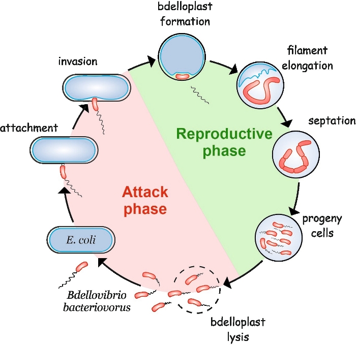
B. bacteriovorus exhibits a peculiar life cycle consisting of two phases: a free-living non-replicative attack phase and an intracellular reproductive phase (Figure 1). In the free-living phase, this highly motile bacterium, which moves at speeds of up to 160 µm/s, searches for its prey. After attaching to the prey’s outer membrane, it enters the periplasm4,5. During the interperiplasmic reproductive phase, B. bacteriovorus uses a plethora of hydrolytic enzymes to degrade the host’s macromolecules and reuse them for its own growth. Soon after invading the periplasm, the host cell dies and bloats into a spherical structure called a bdelloplast, inside which the predatory cell elongates and replicates its chromosomes. The replication process starts at the replication origin (oriC)6 and proceeds until several copies of the chromosome have been completely synthesized7. Interestingly, replication of each chromosome is not followed by cell division. Instead, the predator elongates to form a long, multinucleoid and filamentous cell. Upon nutrient depletion, the filament undergoes synchronous septation and progeny cells are released from the bdelloplast8.
Before B. bacteriovorus can be used as a living antibiotic against bacterial infections, it is crucial to understand the major stages of its life cycle, particularly those related to its proliferation inside the prey. Live-cell imaging of B. bacteriovorus has been challenging, due to the various morphological forms of the predator and its prey during the complex life cycle. So far, the interactions between B. bacteriovorus and its host cell have been mainly studied by electron microscopy and snap-shot analysis2,9,10, both of which have limitations, especially when they are used to monitor successive stages of the predatory life cycle. These methods provide high-resolution images of B. bacteriovorus cells and enable observation of a small predator during the attack or growth phase. However, they do not allow tracking of single B. bacteriovorus cells throughout both life cycle phases.
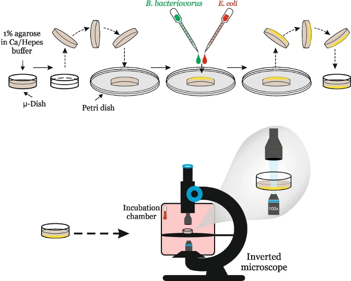
Presented here is a comprehensive protocol for using time-lapse fluorescence microscopy (TLFM) to monitor the complete life cycle of B. bacteriovorus. A system consisting of an agarose pad is used in combination with a cell-imaging dish, in which the predatory cells can move freely beneath the agarose pad while the immobilized prey cells are able to form bdelloplasts (Figure 2). This set-up is prepared based on specific strains of both E. coli and B. bacteriovorus, but the protocol may be easily altered to fit a user’s individual strains (e.g., carrying different selection markers, proteins fused with different fluorophores, etc.).
In this case, to visualize B. bacteriovorus during the attack phase, a specific strain (HD100 DnaN-mNeonGreen/PilZ-mCherry) was constructed that expresses a fluorescently tagged version of the cytoplasmic protein, PilZ (available in our laboratory upon request)7. This strain additionally produces DnaN (the β-sliding clamp), a subunit of DNA polymerase III holoenzyme, fused with a fluorescent protein. This enables ongoing DNA replication to be monitored inside the predatory cells as they grow within bdelloplasts.
Although the described protocol and software used for image acquisition refer to an inverted microscope provided by a specific manufacturer (see Table of Materials), this technique may be adjusted for any inverted microscope equipped with an environmental chamber or other external heating holder and capable of time-lapse imaging. For data analysis, users may choose any available software compatible with the individual output formats.

Figure 1: B. bacteriovorus life cycle in E. coli as a host cell. During the attack phase, a free-swimming B. bacteriovorus cell searches for and attaches to a host E. coli cell. After the invasion, the predatory cell becomes localized in the prey’s periplasm, changing the host cell’s shape and forming a bdelloplast. The reproductive phase starts with bdelloplast formation. The predatory cell digests the prey cell and reuses simple compounds to build its own structures. B. bacteriovorus grows as a long single filament inside the host’s periplasm. When the prey cell’s resources are exhausted, the B. bacteriovorus filament synchronously septates and forms progeny cells. After the progeny cells develop their flagella, they lyse the bdelloplast. Please click here to view a larger version of this figure.
Access restricted. Please log in or start a trial to view this content.
Protocol
1. Preparation of B. bacteriovorus lysate for microscopic analysis
- In a 250 mL flask, set up the coculture by combining 1 mL of fresh 24 h B. bacteriovorus culture (e.g., HD100 DnaN-mNeonGreen/PilZ-mCherry) and 3 mL of overnight prey culture (e.g., E. coli S17-1 pZMR100) with 50 mL of Ca-HEPES buffer (25 mM HEPES, 2 mM CaCl2 [pH = 7.6]) supplemented with antibiotics when needed (here, 50 µg/mL kanamycin).
- Incubate the coculture for 24 h at 30 °C with shaking at 200 rpm.
- Check that the prey cells are fully lysed by liquid mounted phase-contrast microscopy. At this point, numerous motile attack phase B. bacteriovorus cells should be present, and no E. coli cell or bdelloplast should be visible.
- Filter the B. bacteriovorus culture through a 0.45 µm pore size filter to remove any remaining host cells. Predatory cells (0.3–0.5 µm x 0.5–1.4 µm) freely penetrates 0.45 µm pores, whereas host cells are retained on the filter surface.
- Spin down the filtrate for 20 min at 2469 x g and 30°C in a 50 mL conical tube to collect predatory cells. Resuspend the B. bacteriovorus pellet in 3 mL of Ca-HEPES buffer (to a final OD600 of approximately 0.2) and incubate at 30 °C and 200 rpm for 30 min.
2. Preparation of host cells for B. bacteriovorus invasion
- Use a single colony of the host strain (E. coli S17-1 pZMR100 is recommended due to various cell sizes resulting in the formation of unequally sized bdelloplasts) to inoculate 10 mL of YT medium (0.8% Bacto tryptone, 0.5% yeast extract, 0.5% NaCl [pH = 7.5]) that has been supplemented with antibiotics, if needed (here, 50 µg/mL kanamycin). Culture cells overnight at 37 °C and 180 rpm.
- Transfer 2 mL of overnight E. coli culture (OD600 of ~3.0) to a 2 mL test tube and centrifuge at room temperature (RT) and 2469 x g for 5 min. Resuspend the pellet in 200 µL of Ca-HEPES buffer.
3. Set-up of the microscope (see Table of Materials)
- Pre-warm a microscopic chamber with a 100x oil immersion objective to 30 °C for at least 1 h before use. This step is required to prevent errors in maintaining focus during the experiment.
- Turn on both the microscope and microscopy automation. Initialize the control software and select the appropriate filters to acquire brightfield/DIC/phase-contrast and fluorescence images, as needed (here: BF images, GFP/mCherry polychromic filter, and emission/excitation for mCherry and GFP).
4. Assembly of layouts for time-lapse fluorescence microscopy
- Mix 200 mg of low fluorescent molecular grade agarose with 20 mL of Ca-HEPES buffer (1% final agarose concentration) and dissolve the agarose in a microwave. The batch can be reused several times after it solidifies, so simply repeat the heating step.
- Pour 3 mL of melted agarose into a 35 mm glass-bottom dish (Table of Materials). Allow the agarose to solidify.
- Using a laboratory micro-spatula, carefully remove the agarose pad from the 35 mm dish without disturbing the center pole of the agarose pad. Flip the pad bottom side facing up and place it in a Petri dish cover (Figure 2).
- Put 5 µL of concentrated E. coli suspension on top of the flipped pad and spread cells in the center pole using an inoculation loop. Add 5 µL of concentrated B. bacteriovorus suspension (OD600 of ~0.2) onto the E. coli-coated surface. Do not spread the cells.
- Quickly return the agarose gel to the 35 mm glass-bottom dish (top side facing down), then cover with a lid. If necessary, remove any air bubbles by gently pressing down the agar against the plate.

Figure 2: Schematic depiction of the experimental workflow. The agarose pad is removed from the 35 mm dish and flipped so that the bottom side faces upwards. Fresh suspensions of predatory cells and overnight culture of prey cells are placed on the flipped pad to coat the “bottom” side. The agarose pad is then flipped to its original orientation and placed back in the 35 mm dish, which is mounted onto the inverted microscope stage in the microscope chamber. Please click here to view a larger version of this figure.
5. Conduction of time-lapse fluorescence microscopy
- Place the 35 mm dish in the Petri dish holder such that it will not move during the course of the experiment. Place one drop of immersion oil (1.518 refractive index) onto the objective as well as the bottom of the 35 mm dish.
- Mount the holder onto the stage of the inverted microscope in the microscope chamber.
- Set up the options in the microscope control software to collect multiple images at multiple stage positions over time.
- Set the magnification (100x) and polychromic lens (GFP/mCherry). Using an eye piece and brightfield (BF), find the focal plane and open the point list manager.
- Select at least 10 positions of interest by moving the stage and storing the coordinates for each position in the microscope software by clicking the Mark point button. Avoid positions located close to each other to prevent photobleaching and phototoxicity. Turn the camera valve from the eyepiece to the monitor and calibrate each point by setting the focal plane and clicking the Replace point button in the point list manager.
- Open the experiment designer and set all experimental parameters in each tab as follows:
- Unmark the Z-stacking box.
- Choose fluorescent channels and set the optimal illumination settings. Here, the following settings were used: for the mCherry channel, mCherry filter sets (EX575/25; EM625/45), 50% intensity, and 200 ms exposure time; for the GFP channel, GFP filter set (EX475/28; EM525/48), 50% intensity, and 80 ms; and for the BF images, POL channel, 5% intensity, and 50 ms.
- Select intervals between acquiring images and the total time of the experiment. Here, 5 min intervals were performed over the 10 h period of the experiment.
- Enable focus maintenance for the chosen positions (for the system used here, by marking the Maintain focus with UltimateFocus check box).
- Select the data folder in which the image files should be automatically saved.
- Recheck all settings in the microscope software control, then start the time-lapse experiment.
- After the first hour of the experiment, check that all stage positions are still in focus. Adjust the focus during time intervals between image acquisition, if needed.
- When the time-lapse experiment is finished, remove the Petri dish and utilize the 35 mm dish with an agarose pad according to the biosafety protocol.
NOTE: All substeps after step 5.2 are specific for DeltaVision Elite users. Researchers using other systems need to adjust the settings according to individual systems.
6. Processing of time-lapse images and generation of movies using Fiji software
- Transfer experimental data to a computer loaded with the Fiji software. Launch the Fiji program and open an image using File | Open or by simply dragging and dropping the image file into Fiji.
- In Bio-Formats Import Options, choose View stack with Hyperstack in the stack viewing options, Click on Composite color mode in the color options, and mark Autoscale.
- By scrolling through the image stacks, assess whether the cells and bdelloplasts remained in focus over the course of the experiment. Check whether green fluorescent spots from DnaN-mNeonGreen are present within B. bacteriovorus cells inside bdelloplasts throughout the growth phase, and ensure that all stages of the predatory life cycle can be visualized.
- To analyze a particular B. bacteriovorus cell/bdelloplast, mark it using the selection tools in the Fiji menu (Rectangle, Oval, Polygon, or Freehand). Duplicate the chosen region using Image | Duplicate or by using Ctrl + Shift + D.
- Adjust the brightness and contrast for each fluorescence channel as follows:
- To choose a single channel, open Channel tools by clicking Image | Color | Channels tools or Ctrl + Shift + Z, and select the channel of interest (Channel 1: mCherry, Channel 2: mNeonGreen, Channel 3: brightfield).
- Adjust the brightness and contrast of images in the fluorescence channel by opening the B&C menu in Image | Adjust | Brightness/Contrast or by clicking Ctrl + Shift + C.
- Add scale bar using Analyze | Tools | Scale bar. Add the time stamper to the images by clicking Images | Stacks | Time Stamper.
- To save modified images, go to File | Save As and choose TIFF to save as an image sequence, AVI to produce a movie, or PNG to save a single image of the current frame.
Access restricted. Please log in or start a trial to view this content.
Results
The described TLFM-based system allows individual cells of B. bacteriovorus to be tracked in time (Figure 3, Movie 1) and provides valuable information about each stage of the complex predatory life cycle. The PilZ-mCherry fusion enables the entire predatory cell to be labeled in the attack phase as well as early stage of the growth phase (Figure 3). The transition from the attack to replicative phase was visualized not only by the host...
Access restricted. Please log in or start a trial to view this content.
Discussion
Due to the increased interest in using B. bacteriovorus as a living antibiotic, new tools for observing the predatory life cycle, particularly predator-pathogen interactions, are needed. The presented protocol is used to track the entire B. bacteriovorus life cycle, especially during its growth inside the host, in real-time. Moreover, the application of a strain producing fluorescently tagged beta clamp of DNA polymerase III holoenzyme enabled monitoring of chromosome replication progression throughout ...
Access restricted. Please log in or start a trial to view this content.
Disclosures
The authors have nothing to disclose.
Acknowledgements
This study was supported by the National Science Centre grant Opus 2018/29/B/NZ6/00539 to J.Z.C.
Access restricted. Please log in or start a trial to view this content.
Materials
| Name | Company | Catalog Number | Comments |
| Centrifuge | MPW MED. INSTRUMENTS | MPW-260R | Rotor ref. 12183 |
| CertifiedMolecular Biology Agarose | BIO-RAD | 161-3100 | low fluorescence agarose for agarose pad |
| Fiji | ImageJ | https://imagej.net/Fiji | Open source image processing package |
| Glass Bottom Dish 35 mm | ibidi | 81218-200 | uncoated glass |
| Microscope | GE | DeltaVision Elite | Microtiter Stage, ultimate focus laser module, DV Elite CoolSnap HQ2 Camera, SSI assembly FP DV, kit obj. Oly 100x oil 1.4 NA, prism Nomarski 100x LWD DIC, ENV ctrl IX71 uTiter opaQ 240 V |
| Minisart Filter 0.45 µm | Sartorius | 16555----------K | Cellulose Acetate, Sterile, Luer Lock Outlet |
| Start SoftWoRx | GE | Manufacturer-supplied imaging software |
References
- Shatzkes, K., et al. Predatory Bacteria Attenuate Klebsiella pneumoniae Burden in Rat Lungs. mBio. 7 (6), (2016).
- Iebba, V., et al. Bdellovibrio bacteriovorus directly attacks Pseudomonas aeruginosa and Staphylococcus aureus Cystic fibrosis isolates. Frontiers in Microbiology. 5, (2014).
- Willis, A. R., et al. Injections of Predatory Bacteria Work Alongside Host Immune Cells to Treat Shigella Infection in Zebrafish Larvae. Current biology: CB. 26 (24), 3343-3351 (2016).
- Lambert, C., et al. Characterizing the flagellar filament and the role of motility in bacterial prey-penetration by Bdellovibrio bacteriovorus. Molecular Microbiology. 60 (2), 274-286 (2006).
- Lambert, C., et al. A Predatory Patchwork: Membrane and Surface Structures of Bdellovibrio bacteriovorus. Advances in Microbial Physiology. 54, 313-361 (2008).
- Makowski, Ł, et al. Initiation of Chromosomal Replication in Predatory Bacterium Bdellovibrio bacteriovorus. Frontiers in Microbiology. 7, 1898(2016).
- Makowski, Ł, et al. Dynamics of Chromosome Replication and Its Relationship to Predatory Attack Lifestyles in Bdellovibrio bacteriovorus. Applied and Environmental Microbiology. 85 (14), (2019).
- Fenton, A. K., Kanna, M., Woods, R. D., Aizawa, S. I., Sockett, R. E. Shadowing the actions of a predator: backlit fluorescent microscopy reveals synchronous nonbinary septation of predatory Bdellovibrio inside prey and exit through discrete bdelloplast pores. Journal of Bacteriology. 192 (24), 6329-6335 (2010).
- Kuru, E., et al. Fluorescent D-amino-acids reveal bi-cellular cell wall modifications important for Bdellovibrio bacteriovorus predation. Nature Microbiology. 2 (12), 1648-1657 (2017).
- Dashiff, A., Junka, R. A., Libera, M., Kadouri, D. E. Predation of human pathogens by the predatory bacteria Micavibrio aeruginosavorus and Bdellovibrio bacteriovorus. Journal of Applied Microbiology. 110 (2), 431-444 (2011).
Access restricted. Please log in or start a trial to view this content.
Reprints and Permissions
Request permission to reuse the text or figures of this JoVE article
Request PermissionExplore More Articles
This article has been published
Video Coming Soon
Copyright © 2025 MyJoVE Corporation. All rights reserved