Bu içeriği görüntülemek için JoVE aboneliği gereklidir. Oturum açın veya ücretsiz deneme sürümünü başlatın.
Harvesting DRG from Rat Spine: A Procedure to Extract Dorsal Root Ganglion from the Spine of a Rat Model
Overview
In this video, we demonstrate the extraction of dorsal root ganglion or DRG from a harvested rat spine for downstream applications.
Protokol
All procedures involving animal models have been reviewed by the local institutional animal care committee and the JoVE veterinary review board.
1. Harvest and Preparation of DRGs (estimated timing: 2 h, day 8)
NOTE: Experiments with mice and rats require approval from the IACUC. In some countries, the use of chicken eggs also requires approval.
- Get six to seven week-old Sprague Dawley rats (~200 g in weight) to extract DRGs.
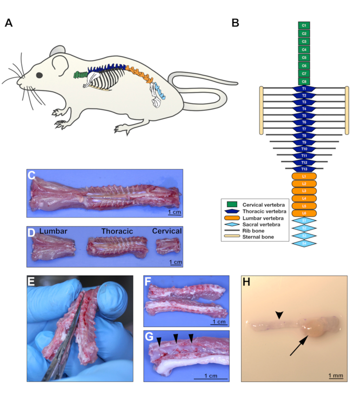
NOTE: One rat should yield ~40 cervical and thoracic DRGs. Mouse DRG also integrates in the CAM; however, the conditions for this species need to be optimized independently. - In a laminar flow cabinet, harvest DRGs from cervical and thoracic regions following the protocol for mouse DRG extraction published elsewhere. Follow Figure 1 for orientation on how to harvest DRGs.
- Euthanize the rat by cardiac puncture after administration of Ketamine/ Xylazine injected intraperitoneally. Clean the rat skin with 70% ethanol and remove the rat spine using a scissor. Do not perform cervical dislocation because this would damage the cervical DRGs.
- Separate the cervical, thoracic, and lumbar regions with the same scissor, following the schematic anatomic representation and gross images provided in the Figure 1A-D. Place the spine sections in a 10 cm culture dish with 1X PBS to keep tissues wet.
- With a delicate bone scissor, open the vertebral bones in the dorsal and ventral aspects, separating the spine in two lateral halves (Figure 1E-F). Place the tissue sections in a clean 10 cm dish with fresh 1X PBS.
- Using forceps, gently detach the spinal cord from the vertebral bones to visualize the DRGs (Figure 1G).
- With fine forceps held underneath each DRG, grasp it and pull it out from the bone cavity in which it is lodged. Do not hold the DRG directly because this will cause tissue damage. Do not trim the axon bundles from the DRG (Figure 1H).
NOTE: Avoid using lumbar DRGs since these have reduced integration in the CAM. For DRG region location, follow the schematic illustration and gross anatomic images in Figure 1A-D.
- Immediately after harvesting, place each DRG into DMEM culture medium supplemented with 2% Penicillin/Streptomycin (Pen/Strep) and 10% heat-inactivated Fetal Bovine Serum (FBS) to help prevent bacterial contamination of the DRGs. Group all DRGs in the same 6 cm culture dish with 4 mL of culture medium.
- After harvesting all DRGs, transfer them to a new culture dish with DMEM culture medium supplemented with 2% Pen/Strep plus 10% FBS and containing 1.25 µg/mL of red fluorescent dye. Incubate for 1 h in the cell culture incubator.
Sonuçlar

Figure 1: DRG extraction on day 8. A. Rat schematic illustrating the anatomical location of the spine. B. Diagram of the rat vertebrae configuration showing different body regions; green for cervical, dark blue...
Açıklamalar
Malzemeler
| Name | Company | Catalog Number | Comments |
| PBS (1x) pH 7.4 | Gibco | # 10010-023 | Phosphate Buffered Saline |
| Sprague Dawley rats (females) | Charles River laboratories | Strain code: 001 | 6-7 weeks old (190-210g in weight) |
| Dumont # 5 fine forceps | Fine Science Tools (FST) | # 11254-20 | Used to harvest DRG |
| Extra fine Graefe forceps, curved | Fine Science Tools (FST) | # 11151-10 | Used to graft DRG onto the CAM on day 8 and to harvest CAM tissue on day 17 |
| Extra fine Graefe forceps, straight | Fine Science Tools (FST) | # 11150-10 | Used to graft DRG onto the CAM on day 8 and to harvest CAM tissue on day 17 |
| DMEM (1x) | Gibco | # 11965-092 | Dulbeecco`s Modified Eagle Medium |
| CellTracker Red CMTPX fluorescent dye | Life Technologies | # C34552 | Reconstitute 50µg in 40µL of DMSO and stock at -20oC. Use 1µL of stock solution/mL of culture medium |
| Pen/Strep | Gibco | # 15140-122 | 10,000 Units/mL Penicilin, 10,000 µg/mL Streptomycin |
| HI FBS | Gibco | # 10082-147 | Heat-inactivated Fetal Bovine Serum |
| DMSO | Fisher Bioreagents | # BP231-100 | Dimethyl Sulfoxide |
This article has been published
Video Coming Soon
Source: Schmidt, L. B. et al. The Chick Chorioallantoic Membrane In Vivo Model to Assess Perineural Invasion in Head and Neck Cancer. J. Vis. Exp. (2019)
JoVE Hakkında
Telif Hakkı © 2020 MyJove Corporation. Tüm hakları saklıdır