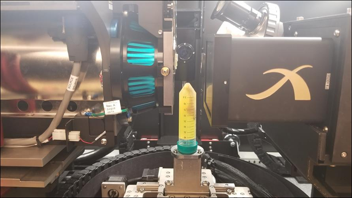
Micro-CT Imaging of a Mouse Spinal Cord
Genel Bakış
Source: Peiman Shahbeigi-Roodposhti and Sina Shahbazmohamadi, Biomedical Engineering Department, University of Connecticut, Storrs, Connecticut
It's a little-known fact that the discovery and (inadvertent) use of X-rays garnered the first ever Nobel Prize in Physics. The famous X-ray image of Dr. Röntgen's wife's hand from 1895 that sent shock waves through the scientific community looks like most modern day 2D medical X-ray images. Though it is not the newest technology, X-ray absorption imaging is an indispensable tool and can be found in the world's top R&D and university labs, hospitals, airports, among other places. Arguably the most advanced uses of X-ray absorption imaging involve attaining information like the kind found in a 2D medical X-ray but realized in 3D through a computed tomography (CT or micro-CT). By taking a series of 2D X-ray projections, advanced software is capable of reconstructing data to form a 3D volume. The 3D information can, and most likely will include information from the inside of the probed object without having to be cut open. Here, a micro-CT scan will be obtained, and the major factors impacting image quality will be discussed.
Prosedür
1. Mounting a Sample (Bone)
- For examining a network of bones, like a spine, suspend the structure in an agarose gel and allow to cure in a very thin-walled plastic tube (Figure 2). The thinness of the tube is very important, greatly affecting the signal throughput and overall image quality. This in turn affects your ability to resolve features. The transmission value of the tube should be as close to 100% as possible.
- Mount the tube on sample stage with tape or by making a custom stand, ultimately e
Sonuçlar
The following images give an overview of results that can be obtained from using micro-CT with the above stated procedure. Qualitative measurements on varying absorption can be directly noted based on these images. Quantitative data such as material porosity, feature size and distribution, etc. would require additional image processing in a different software.

Figure 3: 3D volume of mouse spinal cord (
Başvuru ve Özet
This experiment examined the many factors that should be considered when using micro-CT, particularly for a biological sample. This project was designed to help the investigator understand how their decisions will impact the data that micro-CT can provide. As demonstrated there are many dependent and sensitive parameters that should be considered including: mounting, X-ray energy, exposure time, source and detector positioning, number of projections, and total scan angular displacement. This exercise is meant as an intro
Referanslar
- http://www.spectroscopyonline.com/tutorial-attenuation-X-rays-matter [cited 1 November 2017]
- http://hyperphysics.phy-astr.gsu.edu/hbase/quantum/xrayc.html [cited 1 November 2017]
- A.G. Rao, V.P. Deshmukh, L. L. Lavery, H. Bale, "3D investigation of the microstructural modification in hypereutetic aluminum silicon (Al-30Si) alloy." Microscopy and Analysis 2017 [cited 1 November 2017].
Atla...
Bu koleksiyondaki videolar:

Now Playing
Micro-CT Imaging of a Mouse Spinal Cord
Biomedical Engineering
8.0K Görüntüleme Sayısı

Imaging Biological Samples with Optical and Confocal Microscopy
Biomedical Engineering
35.7K Görüntüleme Sayısı

SEM Imaging of Biological Samples
Biomedical Engineering
23.6K Görüntüleme Sayısı

Biodistribution of Nano-drug Carriers: Applications of SEM
Biomedical Engineering
9.3K Görüntüleme Sayısı

High-frequency Ultrasound Imaging of the Abdominal Aorta
Biomedical Engineering
14.4K Görüntüleme Sayısı

Quantitative Strain Mapping of an Abdominal Aortic Aneurysm
Biomedical Engineering
4.6K Görüntüleme Sayısı

Photoacoustic Tomography to Image Blood and Lipids in the Infrarenal Aorta
Biomedical Engineering
5.7K Görüntüleme Sayısı

Cardiac Magnetic Resonance Imaging
Biomedical Engineering
14.7K Görüntüleme Sayısı

Computational Fluid Dynamics Simulations of Blood Flow in a Cerebral Aneurysm
Biomedical Engineering
11.7K Görüntüleme Sayısı

Near-infrared Fluorescence Imaging of Abdominal Aortic Aneurysms
Biomedical Engineering
8.2K Görüntüleme Sayısı

Noninvasive Blood Pressure Measurement Techniques
Biomedical Engineering
11.9K Görüntüleme Sayısı

Acquisition and Analysis of an ECG (electrocardiography) Signal
Biomedical Engineering
105.0K Görüntüleme Sayısı

Tensile Strength of Resorbable Biomaterials
Biomedical Engineering
7.5K Görüntüleme Sayısı

Visualization of Knee Joint Degeneration after Non-invasive ACL Injury in Rats
Biomedical Engineering
8.2K Görüntüleme Sayısı

Combined SPECT and CT Imaging to Visualize Cardiac Functionality
Biomedical Engineering
11.0K Görüntüleme Sayısı
JoVE Hakkında
Telif Hakkı © 2020 MyJove Corporation. Tüm hakları saklıdır
