
Peripheral Vascular Exam Using a Continuous Wave Doppler
Overview
Source: Joseph Donroe, MD, Internal Medicine and Pediatrics, Yale School of Medicine, New Haven, CT
Peripheral vascular disease (PVD) is a common condition affecting older adults and includes disease of the peripheral arteries and veins. While the history and physical exam offer clues to its diagnosis, Doppler ultrasound has become a routine part of the bedside vascular examination. The video titled "The Peripheral Vascular Exam" gave a detailed review of the physical examination of the peripheral arterial and venous systems. This video specifically reviews the bedside assessment of peripheral arterial disease (PAD) and chronic venous insufficiency using a handheld continuous wave Doppler.
The handheld Doppler (HHD) is a simple instrument that utilizes continuous transmission and reception of ultrasound (also referred to as continuous wave Doppler) to detect changes in blood velocity as it courses through a vessel. The Doppler probe contains a transmitting element that emits ultrasound and a receiving element that detects ultrasound waves (Figure 1). The emitted ultrasound is reflected off of moving blood and back to the probe at a frequency directly related to the velocity of blood flow. The reflected signal is detected and transduced to an audible sound with a frequency directly related to that of the received Doppler signal (thus, faster blood flow produces a higher frequency sound).

Figure 1. Generation of a Doppler signal. The handheld Doppler emits an ultrasound signal, which is then reflected back by moving blood, and finally received by the Doppler probe.
The HHD is easily used in the office or hospital setting to detect pulses, screen for PAD using the ankle brachial pressure index (ABPI), and localize venous insufficiency. This video reviews these procedures; however, it is not intended to be a comprehensive review of non-invasive vascular testing.
Procedure
1. Preparation
- Obtain a blood pressure cuff, an HHD machine, Doppler gel, and skin marker.
- Wash hands prior to examining the patient.
- Begin with the patient in a gown, lying comfortably supine on the exam table.
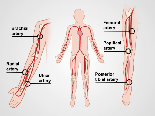
Figure 2. The major arteries of the upper and lower extremities.
2. Lower Extremity Arterial Asses
Application and Summary
A careful history and physical exam are important for anyone suspected of peripheral vascular disease based on symptoms or risk factors. The HHD has become part of the routine bedside vascular examination and should be used to complement the physical exam, if PVD is suspected. It is not a technically difficult tool to use, and the maneuvers described in the video can be performed by general physicians. Just like for the physical exam, knowledge of the vascular anatomy is critical to the success of the HHD exam.

Skip to...
Videos from this collection:

Now Playing
Peripheral Vascular Exam Using a Continuous Wave Doppler
Physical Examinations I
38.4K Views

General Approach to the Physical Exam
Physical Examinations I
115.7K Views

Observation and Inspection
Physical Examinations I
93.1K Views

Palpation
Physical Examinations I
82.7K Views

Percussion
Physical Examinations I
99.6K Views

Auscultation
Physical Examinations I
60.3K Views

Proper Adjustment of Patient Attire during the Physical Exam
Physical Examinations I
83.2K Views

Blood Pressure Measurement
Physical Examinations I
107.4K Views

Measuring Vital Signs
Physical Examinations I
113.9K Views

Respiratory Exam I: Inspection and Palpation
Physical Examinations I
156.6K Views

Respiratory Exam II: Percussion and Auscultation
Physical Examinations I
212.1K Views

Cardiac Exam I: Inspection and Palpation
Physical Examinations I
175.8K Views

Cardiac Exam II: Auscultation
Physical Examinations I
139.4K Views

Cardiac Exam III: Abnormal Heart Sounds
Physical Examinations I
91.4K Views

Peripheral Vascular Exam
Physical Examinations I
67.9K Views
Copyright © 2025 MyJoVE Corporation. All rights reserved