
Cardiac Magnetic Resonance Imaging
Overview
Source: Frederick W. Damen and Craig J. Goergen, Weldon School of Biomedical Engineering, Purdue University, West Lafayette, Indiana
In this video, high field, small-bore magnetic resonance imaging (MRI) with physiological monitoring is demonstrated to acquire gated cine loops of the murine cardiovascular system. This procedure provides a basis for assessing left-ventricular function, visualizing vascular networks, and quantifying motion of organs due to respiration. Comparable small animal cardiovascular imaging modalities include high-frequency ultrasound and micro-computed tomography (CT); however, each modality is associated with trade-offs that should be considered. While ultrasound does provide high spatial and temporal resolution, imaging artifacts are common. For example, dense tissue (i.e., the sternum and ribs) can limit imaging penetration depth, and hyperechoic signal at the interface between gas and liquid (i.e., pleura surrounding the lungs) can blur contrast in nearby tissue. Micro-CT in contrast does not suffer from as many in-plane artifacts, but does have lower temporal resolution and limited soft-tissue contrast. Furthermore, micro-CT uses X-ray radiation and often requires the use of contrast agents to visualize vasculature, both of which are known to cause side effects at high doses including radiation damage and renal injury. Cardiovascular MRI provides a nice compromise between these techniques by negating the need for ionizing radiation and providing the user with the ability to image without contrast agents (although contrast agents are often used for MRI).
This data was acquired with a triggering Fast Low Angle SHot (FLASH) MRI sequence that was gated off of the R-peaks in the cardiac cycle and expiratory plateaus in respiration. These physiological events were monitored through subcutaneous electrodes and a pressure-sensitive pillow that was secured against the abdomen. To ensure the mouse was properly warmed, a rectal temperature probe was inserted and used to control the output of a MRI-safe heating fan. Once the animal was inserted into the bore of the MRI scanner and navigation sequences were run to confirm positioning, the gated FLASH imaging planes were prescribed and data acquired. Overall, high field MRI is a powerful research tool that can provide soft tissue contrast for the study of small animal disease models.
Procedure
1. Animal Preparation
- Identify the mouse to be imaged in its cage and transfer it to the anesthesia induction chamber.
- Anesthetize the mouse using isoflurane and confirm knockdown using a toe-pinch technique. Take the paw between your thumb and index finger and pinch firmly to check for a response. If the animal withdraws their foot, you should either wait or redose with anesthesia according to the approved protocol.
- Check that all staff entering the imaging facility are MR safe. This incl
Results
Figure 1 shows a cine loop of a short-axis view of the left ventricle, which is directly perpendicular to the base-apex axis of the heart and at a position that includes the papillary muscles.
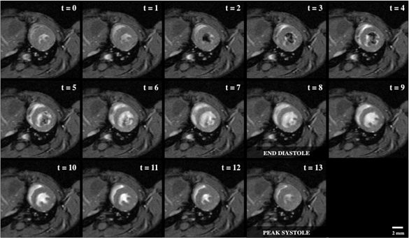
Figure 1: Bright blood cine imaging of a mouse heart with 14 short-axis view snaps
Application and Summary
Here, cardiac MRI is used in conjunction with cardiac- and respiration-gating to acquire cine loop data of the murine heart. While the heart was the focus of demonstration, additional regions of the cardiovascular system can be imaged following the same methodology. Even though MRI does not suffer from the same artifacts commonly seen with other imaging modalities, there is a noticeable trade-off with spatial resolution achieved per acquisition duration. This trade-off is of concern when the mouse cannot withstand longer
Tags
Skip to...
Videos from this collection:

Now Playing
Cardiac Magnetic Resonance Imaging
Biomedical Engineering
14.8K Views

Imaging Biological Samples with Optical and Confocal Microscopy
Biomedical Engineering
35.8K Views

SEM Imaging of Biological Samples
Biomedical Engineering
23.8K Views

Biodistribution of Nano-drug Carriers: Applications of SEM
Biomedical Engineering
9.3K Views

High-frequency Ultrasound Imaging of the Abdominal Aorta
Biomedical Engineering
14.5K Views

Quantitative Strain Mapping of an Abdominal Aortic Aneurysm
Biomedical Engineering
4.6K Views

Photoacoustic Tomography to Image Blood and Lipids in the Infrarenal Aorta
Biomedical Engineering
5.7K Views

Computational Fluid Dynamics Simulations of Blood Flow in a Cerebral Aneurysm
Biomedical Engineering
11.8K Views

Near-infrared Fluorescence Imaging of Abdominal Aortic Aneurysms
Biomedical Engineering
8.3K Views

Noninvasive Blood Pressure Measurement Techniques
Biomedical Engineering
11.9K Views

Acquisition and Analysis of an ECG (electrocardiography) Signal
Biomedical Engineering
105.7K Views

Tensile Strength of Resorbable Biomaterials
Biomedical Engineering
7.5K Views

Micro-CT Imaging of a Mouse Spinal Cord
Biomedical Engineering
8.0K Views

Visualization of Knee Joint Degeneration after Non-invasive ACL Injury in Rats
Biomedical Engineering
8.2K Views

Combined SPECT and CT Imaging to Visualize Cardiac Functionality
Biomedical Engineering
11.0K Views
Copyright © 2025 MyJoVE Corporation. All rights reserved