
Dissecting, Fixing, and Visualizing the Drosophila Pupal Notum
April 6th, 2022
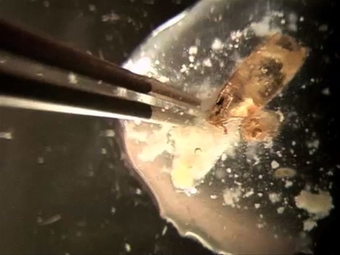
•The present protocol details the preparation and visualization of fixed tissue of the Drosophila pupal notum. It can be used for either intact or wounded tissue, and the original architecture of the tissue is preserved. The procedures for dissecting, fixing, and staining are all described in this article.
Related Videos

In situ Protocol for Butterfly Pupal Wings Using Riboprobes

Visualizing the Live Drosophila Glial-neuromuscular Junction with Fluorescent Dyes

Dissecting and Recording from The C. Elegans Neuromuscular Junction

Dissecting the Non-human Primate Brain in Stereotaxic Space

The Gateway to the Brain: Dissecting the Primate Eye

Visualizing the Beating Heart in Drosophila

Drosophila Pupal Abdomen Immunohistochemistry

Imaging Through the Pupal Case of Drosophila melanogaster

Ex vivo Culture of Drosophila Pupal Testis and Single Male Germ-line Cysts: Dissection, Imaging, and Pharmacological Treatment
Dissection and Mounting of Drosophila Pupal Eye Discs
ABOUT JoVE
Copyright © 2024 MyJoVE Corporation. All rights reserved