
Expanding the Comprehension of the Tumor Microenvironment using Mass Spectrometry Imaging of Formalin-Fixed and Paraffin-Embedded Tissue Samples
June 29th, 2022
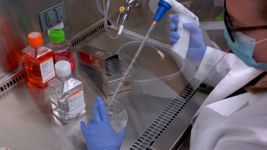
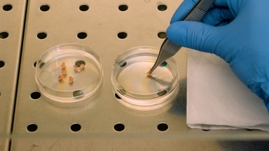
•In the era of cancer immunotherapy, interest in elucidating tumor microenvironment dynamics has increased strikingly. This protocol details a mass spectrometry imaging technique with respect to its staining and imaging steps, which allow for highly multiplexed spatial analysis.
Related Videos

Four-color Fluorescence Immunohistochemistry of T-cell Subpopulations in Archival Formalin-fixed, Paraffin-embedded Human Oropharyngeal Squamous Cell Carcinoma Samples

Two-dimensional Gel Electrophoresis Coupled with Mass Spectrometry Methods for an Analysis of Human Pituitary Adenoma Tissue Proteome

Phosphopeptide Enrichment Coupled with Label-free Quantitative Mass Spectrometry to Investigate the Phosphoproteome in Prostate Cancer

Elastic Staining on Paraffin-embedded Slides of pT3N0M0 Gastric Cancer Tissue

Automated Multiplex Immunofluorescence Panel for Immuno-oncology Studies on Formalin-fixed Carcinoma Tissue Specimens

Measuring Bone Remodeling and Recreating the Tumor-Bone Microenvironment Using Calvaria Co-culture and Histomorphometry

Capturing Small Molecule Communication Between Tissues and Cells Using Imaging Mass Spectrometry

Assessing Tumor Microenvironment of Metastasis Doorway-Mediated Vascular Permeability Associated with Cancer Cell Dissemination using Intravital Imaging and Fixed Tissue Analysis
Pathological Analysis of Lung Metastasis Following Lateral Tail-Vein Injection of Tumor Cells
Isolation of Proximal Fluids to Investigate the Tumor Microenvironment of Pancreatic Adenocarcinoma
ABOUT JoVE
Copyright © 2024 MyJoVE Corporation. All rights reserved