
Method Article
在肠神经系统的原位 钙成像
摘要
The enteric nervous system (ENS) is a network of neurons and glia located in the gut wall that controls intestinal reflexes. This protocol describes methods for recording the activity of enteric neurons and glia in live preparations of ENS using Ca2+ imaging.
摘要
Reflex behaviors of the intestine are controlled by the enteric nervous system (ENS). The ENS is an integrative network of neurons and glia in two ganglionated plexuses housed in the gut wall. Enteric neurons and enteric glia are the only cell types within the enteric ganglia. The activity of enteric neurons and glia is responsible for coordinating intestinal functions. This protocol describes methods for observing the activity of neurons and glia within the intact ENS by imaging intracellular calcium (Ca2+) transients with fluorescent indicator dyes. Our technical discussion focuses on methods for Ca2+ imaging in whole-mount preparations of the myenteric plexus from the rodent bowel. Bulk loading of ENS whole-mounts with a high-affinity Ca2+ indicator such as Fluo-4 permits measurements of Ca2+ responses in individual neurons or glial cells. These responses can be evoked repeatedly and reliably, which permits quantitative studies using pharmacological tools. Ca2+ responses in cells of the ENS are recorded using a fluorescence microscope equipped with a cooled charge-coupled device (CCD) camera. Fluorescence measurements obtained using Ca2+ imaging in whole-mount preparations offer a straightforward means of characterizing the mechanisms and potential functional consequences of Ca2+ responses in enteric neurons and glial cells.
引言
肠神经系统(ENS)被组织成嵌入消化道1的壁内的两个神经节丛。这些肌肉的神经回路中,肠肌丛(MP)和粘膜下丛(SMP),是由神经元和神经胶质肠溶( 图1)2。在MP和SMP调节胃肠(GI)功能,例如肠能动性和上皮的吸收和分泌,分别为3。肠溶胶质位于靠近内神经节的神经元,但肠溶胶质细胞种群内互连纤维束和肠壁3,4的外神经节部也存在。肠神经胶质细胞原本被认为只提供给神经元营养支持。然而,最近的研究有力地表明,神经元的神经胶质细胞的相互作用是必不可少的ENS功能5,6。例如,数据显示,肠溶胶质"收听"神经元活动7和调节神经元电路6,8,保护肠神经元免受氧化应激9,并且能够响应于损伤10,11产生新的肠神经元的。在此技术审查提出的协议提供了一种简单而可靠的方法来检查使用原位细胞内Ca 2+成像的神经元和神经胶质肠溶之间复杂的相互作用。
钙离子是在可兴奋细胞中普遍存在的信号分子,并起着突触信号事件在神经系统12中起重要作用。神经元或神经胶质细胞肠道激发无论是通过大量涌入的Ca 2+ -permeable通道或钙诱发细胞质中钙离子浓度的升高,从细胞内钙库离子释放 。成像钙瞬变的神经元和神经胶质细胞与荧光染料是既定的和广泛使用的技术,研究功能组织和动态在ENS 13-17。的Ca 2+成像已经被证明是在研究完整的GI组织片段通过ICC起搏器网络18和肠平滑肌19,20阐明兴奋性传播的重要工具。它使研究人员能够探测生理参数的广谱,并提供有关他们俩的空间分布和时空动态的信息。细胞可以通过使用膜可渗透荧光指标和优化的染色方案21被有效地染色以微创方式。这提供了机会,以监视大量的神经元和神经胶质肠溶在功能上保留制剂14-16,22,以及在体内 23。整个安装组织标本是散装装载有高亲和力的Ca 2+指示剂染料如荧光4当结合的Ca 2+,增加其荧光。变化的荧光用CCD照相机和ANA记录裂解数字6。钙离子的出现提供了机会以监测神经元和神经胶质细胞的相互作用,响应于各种刺激,而这些细胞类型的胃肠道的过程中实时的参与。
在原位 钙成像已经取得了巨大的洞察肠道神经元和神经胶质细胞的信号传导机制和过细胞培养模型6,24具有几个明显的优势。首先, 在原地准备维持神经元和神经胶质细胞的原生基质环境,让他们的大部分连接到靶组织完好无损。二,遗传学和培养肠道神经胶质细胞的形态是显著已发生变化相比, 在体内 6,24。第三,许多异型相互作用丢失在原代细胞培养,这限制评估细胞 - 细胞相互作用。虽然培养细胞非常适合于调查基本性质,它们的usefulneSS学习肠神经胶质细胞和神经元之间复杂的相互作用是有限的。使用原位方法研究神经元的神经胶质细胞相互作用更生理有关的突触途径保持不变25。相比于细胞培养方法, 原位方法提供了改进的条件,系统地了解神经元和神经胶质细胞肠道之间错综复杂的相互作用。此外,在整个安装制剂的神经节丛的平面组织是理想的细胞内Ca 2+瞬变的荧光成像和该技术提供了用于评估在ENS神经元-神经胶质细胞活性的简单的方法。
研究方案
注:涉及实验动物组织中的下列程序与动物2013年安乐死的AVMA准则一致,是由美国密歇根州立大学IACUC批准。
1.组织准备
- 麻醉动物研究中含有氧或通过将3-5毫升液体异氟烷到吸收材料上的腔室的地板上,以确保物理屏障防止动物从与异氟烷直接接触2.5%异氟烷的腔室。测试麻醉按捏足垫的深度。
注:麻醉深度被认为是合适的时候有后肢没有撤回反射。一旦适当麻醉,安乐死鼠标颈椎脱位 - 将动物仰卧位置和清洁腹部皮肤,用70%的乙醇。使用镊子夹住腹部皮肤在中线,用手术剪刀做出6厘米媒体升切口,沿白线,露出内部的消化器官。
- 使用钝钳找到并揭露腹膜内回肠。切回肠/结肠肠系膜用剪刀,开始解开肠道。
- 一旦肠的长度被充分地解开,切开肠远侧到胃和近端至盲肠为回肠制备。对于大肠的准备,切开结肠远端盲肠和近端到直肠。
- 迅速取出肠段,并将其放置在与补充有3微米尼卡地平盐酸化物和1μM的东莨菪碱盐酸盐的DMEM / F12介质的烧杯中(以下简称为"介质")上的冰。添加这些抑制剂被麻痹肠道平滑肌有利于显微切割和随后的成像。
- 感兴趣的基础上建立的解剖标记切段( 例如 ,空肠,回肠,远端或近端结肠)。通常情况下市利用Ë组织从远端回肠和结肠。然而,使用相同的基本程序来分离,负载和图像肌间神经元和神经胶质细胞中的所有肠道地区。
- 在一个的Sylgard涂覆培养皿填充有冷却介质除去所需肠段和地点的一小部分(4-6厘米)。
- 固定的近端和肠段与昆虫销的远端并且通过使直打开肠管,沿肠系膜边界纵向切割。
- 针组织在轻张力粘膜侧扁了,仔细用细镊子(#5,#四十五分之五工作最好的)和很细的弹簧剪刀解剖走粘膜层。
注:粘膜的除去可以相当创伤为ENS如果没有正确完成。对于质量的准备,照顾通过剥离或刮限制突然去除粘膜。最好的做法是解除粘膜切下的细剪刀。 - 切割组织成更小的准备S(约为0.5 平方厘米)和引脚到成像菜(4个角有圆形肌肉层朝上)置于冰上新鲜的媒体。
- 仔细剖析走环形肌与精细镊子戏弄除了揭露肌间神经丛。避免过度伸展。
- 将成像菜重回冰变化的解决方案,新的设备。
- 准备每盘2毫升酶混合物[分散酶1单位/毫升(4.48毫克/ 8毫升)中,胶原酶II 150 U / ml的(5.45毫克/ 8毫升)中的介质。
- 从冰中取出的菜肴和步骤1.13添加酶混合物。
- 孵育菜在RT 15分钟,用5%的CO 2/95%空气。
- 洗涤组织标本与媒体的3倍,并重新销角落。
2.装载荧光4染料
注:避免用有限的光,同时处理装有指示剂染料荧光染料和组织工作的漂白。
- 准备4μM荧光4加载的解决方案。
- 添加1.5毫升媒体和1.2微升250mM的丙磺舒股票4毫的Fluo-4的库存1.5微升等分试样。到50的Fluo-4,AM微克; 4毫的Fluo-4的库存通过加入11.4微升尼克F-127的(补充有0.25%cremaphor-EL在DMSO中的20%)来制备。
- 在37℃下在黑暗培养箱孵育的Preps中的Fluo-4加载溶液45分钟。
- 从孵化器中取出并清洗与媒体3倍的准备。
- 交换介质为含有介质200μM的丙磺舒孵育15分钟,在37℃前成像。
注:丙磺舒是一种药物,抑制多药耐药转运体中的神经元。此外,利用这种药物的抑制神经元的挤出染料的能力并增强神经元标记。在没有丙磺舒批量装载的钙指标染料生产的主要胶质细胞负荷。加入丙磺舒允许对神经元和神经胶质反应可视化。 - 制备改性贵安BS缓冲区。
- 使修饰的Krebs缓冲液,使得所述组分的终浓度(以mM计)是如下:121氯化钠,氯化钾5.9,2.5 氯化钙 ,1.2的MgCl 2,1.2的NaH 2 PO 4,10 HEPES,21.2 碳酸氢钠 ,1丙酮酸酸和8个葡萄糖(pH值调节至7.4,用NaOH)。加入3μM尼卡和1μM东莨菪碱钙在抑制肌肉收缩2+成像和整个安装解剖。
3.成像和分析
注:使用至少一个基本的成像钻机用荧光光源,显微镜,品质CCD摄像头和适当的采集软件。改变加法取决于光源和特定应用的其他组件。滤光轮和快门必须使用传统的氙弧光源。然而,LED光源和照明系统不需要那些组件。
- 定位REC奥尔丁显微镜下腔室,并用重力流灌注系统具有多个加热注射器储层建立的在37℃的Krebs缓冲液的2-3毫升/分钟的连续灌注速率。要确保防止在输入和吸入管线连接到真空捕集空气气泡的形成。
- 将所需丛成在明亮的视场照明的重点。避免过度曝光的组织,这可能会导致光漂白性。
- 检查神经节内的荧光4加载和选择健康的神经节成像。不健康的/损坏的神经节将呈现自发荧光或点状的形态,而不应被用于成像。
- 一旦神经节被选中,疏导光路摄像头,并获得与图像采集软件的实时图像。确保节是重点,设定图像采集速度和曝光时间。
注:图像采集速度和时间将取决于调查人员希望记录的事件各不相同。对于大多数实验中,图像传统上,在收购0.5-1赫兹胶质细胞和高达2-10赫兹的神经元,因为胶质的Ca 2+的反应并不像快速的钙瞬变的神经元。 - 开始试验并建立基线活动,持续30秒。
- 应用预热药物的兴趣,例如使用重力流灌注系统以2-3毫升/分钟的速率受体激动剂和拮抗剂。按照应用的激动剂/拮抗剂通过返回到正常缓冲的灌注,并允许至少10分钟的洗涤/恢复期。
注:该药物的施用时间将取决于个体化合物和试验设计而变化。一般来说,激动剂20-30秒的应用是足以激活G-蛋白偶联受体的神经元和神经胶质细胞。然而,配体门控离子通道(如烟碱乙酰胆碱受体)要求不超过5-10秒施用时间。而且持续时间曝光会导致配体门控离子通道迅速DESEnsitize。拮抗剂应适用于约3-15分钟,以确保受体途径的完整封锁。然而,这是一个总的概括和研究者应该总是优化任何实验性药物在其特定的模式。 - 停止实验记录和查看时间推移电影。感兴趣的精心选择的区域(ROI的)使用适当的图像分析软件。
- 使用软件来规范和比较投资回报率的荧光强度对初始基线荧光值。变化标准化荧光成正比变化的[Ca 2+]。
- 使用的方法的使用ΔF/ F =((F 1 - f 0的)/ F 0)的变形例前面所述的ROI 26 - ((F 1 - f 0的)/ F 0)的背景下,其中,F1是所述荧光在任何给定点和F0是基线荧光,以改善评估的准确性。此修改辅助从组织筹备荧光变化表现出的肌间神经丛背后的肌肉层的运动降低了噪声。
结果
正确使用这种技术可以让研究人员准确地测量细胞内钙的[Ca 2+] i的瞬态肠神经元和神经胶质细胞在整个安装组织准备。激动剂诱发的Ca 2+在神经胶质细胞内从小鼠结肠肌间神经节反应的代表性实例示于视频1。下面的结果是为了说明,我们使用这种方法获得的一些有代表性的结果。首先, 图2显示了一个实验测量肠神经胶质结果的[Ca 2+] i变化的范围内豚鼠结肠纵肌间神经丛的肌肉(LMMP)准备应对刺激ATP。具体地,该图显示了该方法用于上述包括所分析的肌间神经节和星号表示肠神经元的位置的轮廓中列出的实验方案的适当分析。这些结果还我llustrate的百微摩尔ATP最佳剂量的动员[ 钙 ]在我豚鼠肌间神经胶质细胞。此响应可以被用于校准肠溶胶质刺激和标准化的响应测试刺激。接下来, 图3阐明了如何正确选择感兴趣(投资回报)周围的神经胶质细胞的区域,显示与周围的黄色圆圈。这些结果还表明在基础条件下,并且响应于药理刺激所需的荧光变化。最后, 图4展示的空间考虑选择肠神经胶质细胞和神经元的[Ca 2+] i的整个安装准备响应。

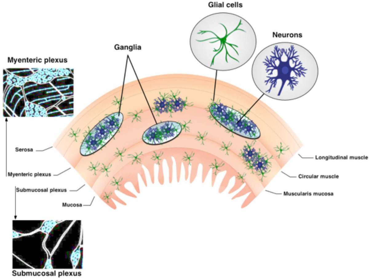
图1.组织的ENS的 ENS包含两个主要的神经节神经丛。肌间神经丛位于升之间ongitudinal和圆形肌肉层。黏膜下神经丛位于粘膜和圆形肌肉层之间。该ENS仅仅由神经元和神经胶质细胞肠道的。神经纤维束连接神经节。 请点击此处查看该图的放大版本。

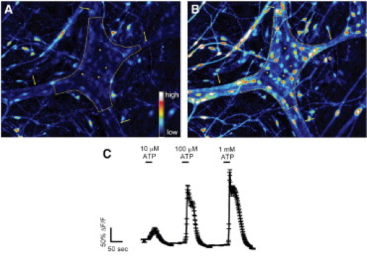
图2.肠道神经胶质细胞在豚鼠结肠肌间神经丛响应到ATP 原位 。 (A)的肌间神经节荧光4荧光基础条件下(虚线所述)。箭头指向厚interganglionic纤维束(B)的刺激后用100微摩尔/ L的ATP,神经胶质细胞,但不是神经元,迅速 增加的Fluo-4的荧光指示增加的[Ca 2+]>我。需要注意的是响应细胞体积小,周围的大得多的神经元(标记星号黑暗的空间)。(C)肠神经胶质细胞具有剂量依赖的方式与1 mmol / L的引发最大反应24回应ATP。 请点击这里查看更大的版本这个数字。

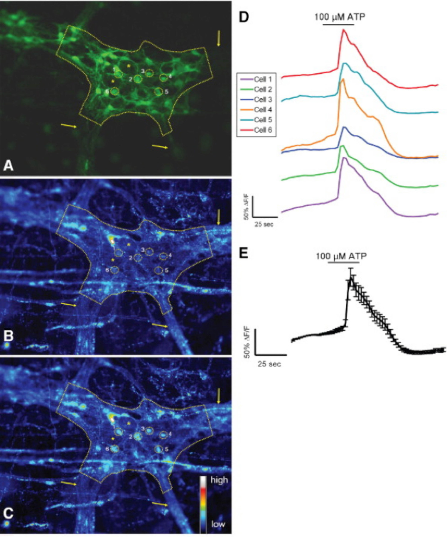
图3.小鼠的S-100-GFP +细胞在结肠肠肌丛响应原位到ATP。(A)的 S-100-GFP +的小鼠结肠肌间神经节的神经胶质细胞(绿色)(由虚线画出的)。六个地区的利益(投资回报)周围神经节内的神经胶质细胞显示为黄色圆圈。箭头表示厚的纤维束领先进入神经节。星号ð2肠神经元的刺激后用100微摩尔/ L的ATP enote位置。(B)中相同的神经节表示基础条件下RHOD-2荧光。(℃),神经胶质细胞具有增加的[Ca 2+] i的如通过增加Rhod-所示响应2荧光。(D)的痕迹对应于A-C中所示的每个投资回报率。(E)的平均值24 D所示的6 ROI的反应(平均值±SEM)。 请点击此处查看该图的放大版本。

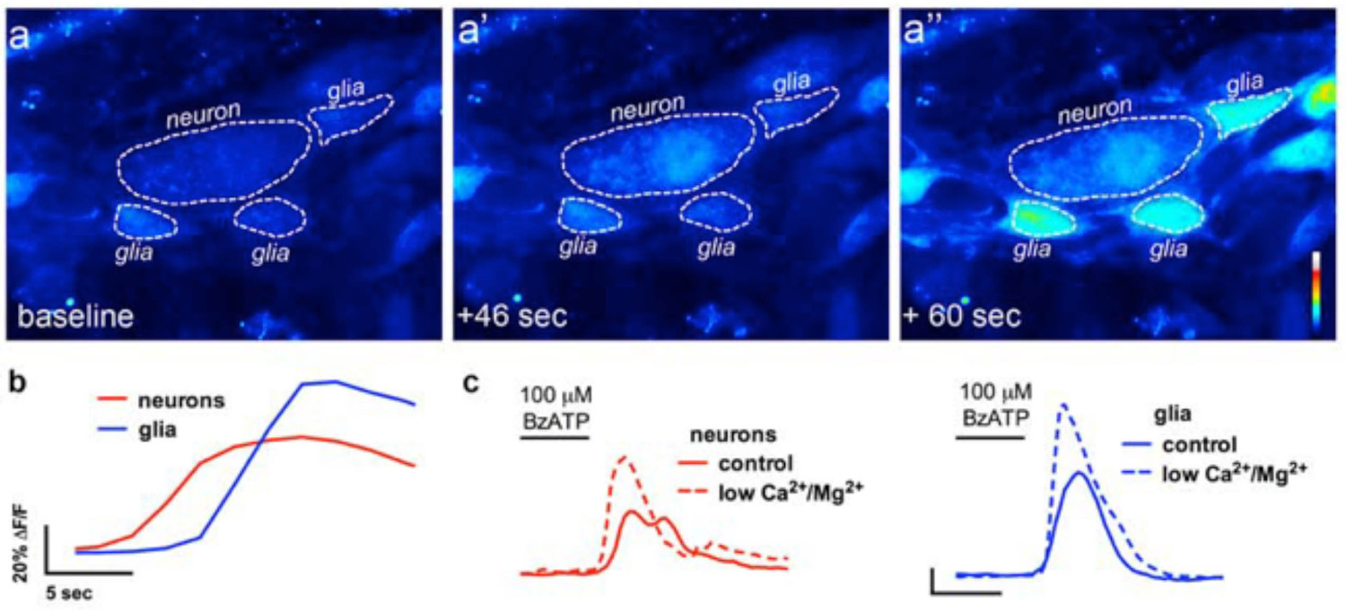
图4. 在肠神经元对胶质细胞通信的现场影像。从钙成像实验(A)代表的图像(伪彩色),其中整体式安装准备肌间神经丛的神经元P2X7受体激动剂BzATP(100微米,30秒)的挑战。需要注意的是神经激动剂导致前周边肠溶胶质细胞(A") 的增加Fluo4荧光的神经元(A')。(B)的荧光随时间的变化中的神经胶质细胞(蓝色)和神经元分析(红)以下应用神经激动剂BzATP(℃)的神经元和神经胶质反应BzATP在正常缓冲液(实线),并在缓冲液含低的Ca 2+和Mg 2+(虚线),以使可能的神经性P2X 7受体13。 请点击此处查看该图的放大版本。
在肠神经胶质视频1激动剂诱发的Ca 2+响应现场,该视频显示,从装有钙指示剂染料,荧光4鼠标远端结肠肌间神经节。胶质细胞激动剂,ADP添加到浴中时表示。 ADP引发2+在荧光4荧光性升高观察到增加细胞内钙在肠道神经胶质细胞。 请点击此处观看该视频。
讨论
The methodologies described in this manuscript provide a consistent approach to effectively study neurons and enteric glia in the ENS. Although imaging neurons and enteric glia in culture has yielded a wealth of insight into the function of individual cells, studying these cells in their native, multi-cellular environment is crucial for our understanding their physiology and pathophysiology. Fluorescence microscopy is a crucial technique for assessing multidirectional interactions of cells in the ENS. Loading cells of the ENS with selective fluorescent markers and image acquisition with high-resolution microscopy permits quantitative observations of cellular activity in the ENS. Imaging live tissue samples of the ENS is performed relatively quickly and generates large amounts of in-depth functional and spatial data. Mouse myenteric and submucosal plexus preparations used in these experiments allow for molecular and genetic manipulation approaches. Ca2+ imaging in whole-mount preparations provides a useful tool for the assessment of neuron-glia interactions.
In advanced experimental paradigms, several probes can be combined to obtain information about different events within the cells. Fluorescence microscopy can record images with enhanced contrast of specific molecules, if an appropriate fluorescent label is used. Fluo-4 was chosen because it possesses a large dynamic range. Sufficient incubation time is vital when using the AM dyes in ENS. Dye concentration and loading method may need to be adjusted to achieve best results. Ideal preparations should be loaded with sufficient dye to visualize changes in fluorescence but not so much so that the dye chelates the target ions and interferes with intracellular signaling. Exposure to fluorescent light should be limited to prevent phototoxicity in cells and photobleaching of dyes.
Investigators must be careful with several steps of this experiment, especially solution and tissue preparation. Particular care has to be taken during processing and dissection of ENS tissue in order to maintain cellular functions. The GI tract contains several layers and tissue varieties, which pose challenges for dissection and imaging quality in these whole-mount preparations 27. Furthermore, the interconnecting fiber tracts of the MP are wider and ganglia are larger than those of the SMP 2. The neuronal density of the myenteric plexus is higher compared to that of the submucosal plexus 28. Slow and imprecise dissections will have detrimental effects on the quality of the plexus preparations and thus the overall success of the experiments. Therefore, clean/undamaged tools, practice and manual dexterity are critical to this procedure.
In whole-mount tissue preparations, careful consideration should be taken when drawing the regions of interest (ROI) to correctly assess the kinetics and degree of observed change in fluorescence intensity of the desired cell type. As the ganglia are located on a contractile muscle layer, motion artifacts caused by gut motility are a primary concern during in situ imaging. Thus, suppressing these motion disturbances through re-pinning tissue preparations after incubation with enzymes and the addition of pharmacological inhibition (nicardipine/scopolamine) to buffers permits clear and reliable image acquisition. Aside from pharmacology and mechanical approaches to prevent tissue movement, recent studies illustrate the application of advanced software methodologies and cell type response characteristics to correct for residual tissue movement in the recordings and improve the accuracy of analysis 29. Barring these technical hurdles, this method provides physiologically relevant conditions to assess morphologic and quantitative characteristics of neurons and enteric glia in the ENS.
披露声明
The authors have nothing to disclose.
致谢
This work was supported by grants from the Pharmaceutical Research and Manufacturers Association of America (PhRMA) Foundation (to B. Gulbransen), National Institutes of Health (Building Interdisciplinary Research Careers in Women’s Health) grant K12 HD065879 (B. Gulbransen) and start-up funds from Michigan State University (B. Gulbransen).
材料
| Name | Company | Catalog Number | Comments |
| BubbleStop Syringe Heater | AutoMate Scientific | 10-4-35-G | |
| CaCl2 | Sigma | C3306 | |
| Collagenase, Type II, powder | Gibco | 17101-015 | |
| Dispase | Sigma-Aldrich | 42613-33-2 | |
| Dissection tools | Roboz | ||
| DMSO | Sigma-Aldrich | D5879 | |
| Fixed-stage microscope | Olympus | BX51WI | |
| Fluo-4 AM dye | Invitrogen | F-14201 | |
| Glucose | Sigma | G8270 | |
| Insect pins | Fine Science Tools | Minutien Pins | |
| iQ Live Cell Imaging Software | Andor | Andor iQ3 | |
| KCl | Sigma | P3911 | |
| MgCl2 | Sigma | M9272 | |
| NaCl | Sigma | S9888 | |
| NaH2PO4 | Sigma | S8282 | |
| NaHCO3 | Sigma | S6014 | |
| Neo sCMOS camera | Andor | Neo 5.5 sCMOS | |
| Nicardipine | Sigma | N7510 | |
| Perfusion chamber | Custom | ||
| Peristaltic pump | Harvard Apparatus | Model 720 | |
| Pluronic F-127 | Invitrogen | P3000MP | |
| Probenecid | Molecular Probes | P36400 | |
| Scopolamine | Sigma | S1013 | |
| Sutter Lambda DG-4 | Sutter | DG-4 | |
| Sylgard | Dow Corning | 184 | |
| Temperature Controller | Warner Instruments | TC-344C |
参考文献
- Furness, J. B. Types of neurons in the enteric nervous system. Journal of the Autonomic Nervous System. 81 (1-3), 87-96 (2000).
- Furness, J. B. The organization of the autonomic nervous system: peripheral connections. Neuroscience. 130 (1-2), 1-5 (2006).
- Pham, T. D., Gershon, M. D., Rothman, T. P. Time of origin of neurons in the murine enteric nervous system: sequence in relation to phenotype. Journal of Comparative Neurology. 314 (4), 789-798 (1991).
- Nasser, Y., et al. Role of enteric glia in intestinal physiology: effects of the gliotoxin fluorocitrate on motor and secretory function. Am J Physiol Gastrointest Liver Physiol. 291, G912-927 (2006).
- Glial cells in the gut. Neurogastroenterology & Motility. 17 (6), 777-790 (2005).
- Broadhead, M. J., Bayguinov, P. O., Okamoto, T., Heredia, D. J., Smith, T. K. Ca2+ transients in myenteric glial cells during the colonic migrating motor complex in the isolated murine large intestine. J. Physiol. 590, 335-350 (2012).
- Gulbransen, B. D., Bains, J. S., Sharkey, K. A. Enteric glia are targets of the sympathetic innervation of the myenteric plexus in the guinea pig distal colon. J. Neurosci. 30, 6801-6809 (2010).
- McClain, J. L., et al. Ca2+ Responses in Enteric Glia Are Mediated by Connexin-43 Hemichannels and Modulate Colonic Transit in Mice. Gastroenterology. 146 (2), 497-507 (2014).
- Chandrasekharan, B., et al. Colonic motor dysfunction in human diabetes is associated with enteric neuronal loss and increased oxidative stress. Neurogastroenterology & Motility. 23 (2), e131-e126 (2011).
- Abdo, H., et al. Enteric glial cells protect neurons from oxidative stress in part via reduced glutathione. FASEB J. 24, 1082-1092 (2010).
- Aube, A. C., et al. Changes in enteric neurone phenotype and intestinal functions in a transgenic mouse model of enteric glia disruption. Gut. 55, 630-637 (2006).
- Berridge, M. J., Lipp, P., Bootman, M. D. The versatility and universality of calcium signalling. Nature Reviews Molecular cell biology. 1 (1), 11-21 (2000).
- Gulbransen, B. D., et al. Activation of neuronal P2X7 receptor-pannexin-1 mediates death of enteric neurons during colitis. Nat Med. 18, 600-604 (2012).
- Bayguinov, P. O., Hennig, G. W., Smith, T. K. Calcium activity in different classes of myenteric neurons underlying the migrating motor complex in the murine colon. J Physiol. 588, 399-421 (2010).
- Okamoto, T., Bayguinov, P. O., Broadhead, M. J., Smith, T. K. Ca(2+) transients in submucous neurons during the colonic migrating motor complex in the isolated murine large intestine. Neurogastroenterol Motil. 24 (8), 769-778 (2012).
- Kunze, W. A., Clerc, N., Furness, J. B., Gola, M. The soma and neurites of primary afferent neurons in the guinea-pig intestine respond differentially to deformation. J Physiol. 526, 375-385 (2000).
- Schemann, M., Michel, K., Peters, S., Bischoff, S. C., Neunlist, M. Imaging and the gastrointestinal tract: mapping the human enteric nervous system. Am J Physiol. 282, G919-G925 (2002).
- Hennig, G. W., et al. Visualization of spread of pacemaker activity in through ICC in guinea-pig antrum. Neurogastro Motil. 14, 575(2001).
- Stevens, R. J., Publicover, N. G., Smith, T. K. Induction and organization of Ca2+ waves by enteric neural reflexes. Nature. 399, 62-66 (1999).
- Stevens, R. J., Publicover, N. G., Smith, T. K. Propagation and neural regulation of calcium waves in longitudinal and circular muscle layers of guinea-pig small intestine. Gastroenterology. 118, 982-984 (2000).
- Jessen, K. R., et al. Astrocyte-like glia in the peripheral nervous system: an immunohistochemical study of enteric glia. Journal of Neuroscience. 3 (11), 2206-2218 (1983).
- Gulbransen, B. D., Sharkey, K. A. Novel functional roles for enteric glia in the gastrointestinal tract. Nat Rev Gastroenterol Hepatol. 9, 625-632 (2012).
- Gomes, P., et al. ATP-dependent paracrine communication between enteric neurons and glia in a primary cell culture derived from embryonic mice. Neurogastroenterology & Motility. 21 (8), e870-e862 (2009).
- Gulbransen, B. D., Sharkey, K. A. Purinergic neuron-to-glia signaling in the enteric nervous system. Gastroenterology. 136, 1349-1358 (2009).
- Ren, J., Bertrand, P. P. Purinergic receptors and synaptic transmission in enteric neurons. Purinergic Signal. 4, 255-266 (2008).
- Takahashi, A., Camacho, P., Lechleiter, J. D., Herman, B. Measurement of intracellular calcium. Physiol Rev. 79, 1089-1125 (1999).
- Dongcheng, Z., et al. Neural crest regionalisation for enteric nervous system formation: implications for Hirschsprung's disease and stem cell therapy. Developmental Biology. 339 (2), 280-294 (2010).
- Gershon, M. D. Behind an enteric neuron there may lie a glial cell. J Clin Invest. 121, 3386-3389 (2011).
- Boesmans, W., et al. Imaging neuron-glia interactions in the enteric nervous system. Frontiers in Cellular Neuroscience. 7, (2013).
转载和许可
请求许可使用此 JoVE 文章的文本或图形
请求许可探索更多文章
This article has been published
Video Coming Soon
版权所属 © 2025 MyJoVE 公司版权所有,本公司不涉及任何医疗业务和医疗服务。