Method Article
从皮肤活检中生成患者来源的足细胞
摘要
本手稿描述了一个两步方案,通过游离体重编程为人诱导的多能干细胞 (hiPSC) 并随后分化为足细胞 ,从 真皮成纤维细胞生成患者特异性足细胞。
摘要
足细胞是位于肾小球滤过屏障泌尿部位的上皮细胞,有助于肾小球的选择性过滤功能。足细胞特异性基因突变可引起局灶节段性肾小球硬化症 (FSGS),足细胞也受许多其他原发性和继发性肾病的影响。由于其差异化性质,原代细胞培养模型对足细胞的限制。因此,通常使用有条件的永生化细胞。然而,这些有条件永生化的足细胞(ciPodocytes)有几个局限性:细胞可以在培养中去分化,特别是当它们达到汇合时,并且几种足细胞特异性标志物要么只是轻微表达,要么根本不表达。这使得ciPodocytes的使用及其在生理,病理生理和临床范围内的适用性受到质疑。在这里,我们描述了一种通过皮肤穿刺活检产生人足细胞(包括患者特异性足细胞)的方案,方法是将真皮成纤维细胞游离体重编程为 hiPSC,随后分化为足细胞。这些足细胞在形态特征方面与 体内 足细胞相似得多,例如足突的发育和足细胞特异性标志物的表达。最后,但重要的是,这些细胞维持患者的突变,从而改进了 离体 模型,以个体化方法研究足细胞疾病和潜在的治疗物质。
引言
足细胞是专门的有丝分裂后肾上皮细胞,与肾小球基底膜 (GBM)、肾小球内皮细胞和糖萼一起形成肾脏的肾小球滤过屏障。表型上,足细胞由细胞体和原代微管驱动的膜延伸以及称为足突的次级延伸组成1,2。从血液中过滤尿液的肾小球滤过屏障由开窗内皮、GBM 和连接邻近足细胞足突的特殊类型的细胞间连接构成,称为足细胞裂缝隔膜3。在健康条件下,比白蛋白大的蛋白质由于其大小和电荷而从过滤屏障中保留下来4。
已知细胞骨架或足细胞特异性基因突变以及影响足细胞信号通路的循环因子可诱导足细胞消失、脱离或凋亡,导致蛋白尿和肾小球硬化。特别是,细胞骨架重排、足细胞极性变化或足突损伤以及相关的狭缝连接缺失是关键因素5.由于其终末分化状态,足细胞在GBM脱离后几乎无法更换。然而,如果足细胞附着在GBM上,它们仍然可以从消失中恢复并改革叉指间足突6,7,8。进一步了解导致各种肾小球疾病中足细胞损伤的事件可能提供新的治疗靶点,有助于开发这些疾病的治疗方法。足细胞损伤是不同肾小球疾病的标志,包括局灶节段性肾小球硬化症(FSGS)、糖尿病肾病、微小病变疾病和膜性肾小球肾病,需要可靠的足细胞离体模型来研究这些疾病的病理机制和潜在的治疗方法9,10。足细胞可以通过基于通过差异筛分离肾小球的经典原代细胞培养离体研究11。然而,由于终末分化状态和有限的增殖能力,大多数研究人员使用表达SV40大T抗原的温度敏感变体的小鼠或人类ciPodocyte细胞系。或者,从携带SV40标签永生化基因1,12的转基因小鼠中分离出ciPodocys。
CiPodocys在33°C增殖,但在37°C进入生长停滞并开始分化13,14。必须记住,用这些细胞获得的实验数据应该谨慎地解释,因为这些细胞是使用非自然基因插入产生的15。由于这些细胞含有永生基因,细胞生理学由于持续的增殖而改变12。这种方法产生的足细胞系最近受到质疑,因为与肾小球表达相比,小鼠、人和大鼠的细胞在蛋白质水平上表达不到5%的突触足素和肾单位,在mRNA水平上表达NPHS1和NPHS216。此外,大多数足细胞系不表达肾单位17,18。Chittiprol等人还描述了ciPodocytes16中细胞运动和对嘌呤霉素和多柔比星的反应的显着差异。在不同的肾小球疾病中,从GBM分离后可以在尿液中发现足细胞19,20,21,22。活的尿足细胞可以在体外培养长达2-3周,但大多数细胞经历细胞凋亡23,24。有趣的是,足细胞不仅存在于肾小球疾病患者的尿液中,还存在于健康受试者的尿液中,最有可能的是当它们再次衰老时,在培养物中复制的潜力有限24。此外,泌尿源足细胞数量有限,细胞在培养中去分化,足突减少,形态改变,最重要的是增殖能力有限。足细胞特异性基因的表达不存在,在几周内消失,或者在这些细胞克隆之间发生变化。一些足细胞特异性标志物阳性的细胞共表达肾小管上皮细胞或肌成纤维细胞和系膜细胞的标志物,这表明培养的尿足细胞去分化和/或转分化24,25。
最近,通过用热敏SV40大T抗原和hTERT转导从患者和健康志愿者的尿液中衍生的ciPodocyte细胞系的产生已被报道26。检测到突触足素、巢蛋白和CD2相关蛋白的mRNA表达,但所有克隆中均不存在podocin mRNA。除了尿足细胞的问题外,这些细胞还含有插入的永生化基因,导致上述缺点。
相比之下,人类诱导多能干细胞(hiPSC)具有巨大的自我更新能力,并在适当的条件下分化成多种细胞类型。之前已经证明,hiPSCs可以作为足细胞的几乎无限的来源27,28。
这里描述了一个两步方案,用于从皮肤穿刺活检的真皮成纤维细胞中生成患者特异性足细胞,随后将游离体重编程为 hiPSC,并最终分化为 hiPSC 来源的足细胞(图 1)。
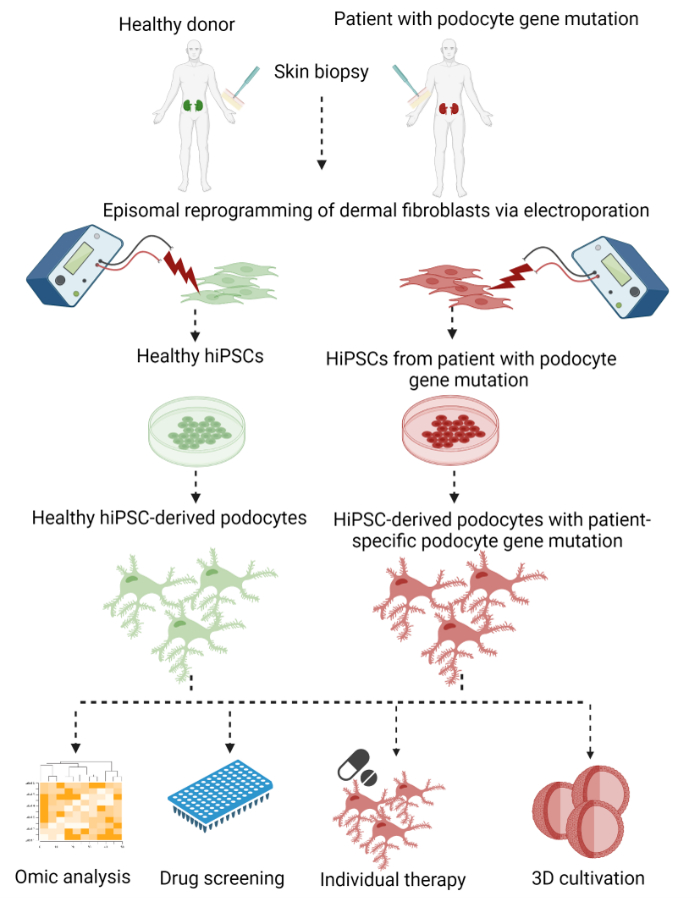
图 1:生成患者特异性 hiPSC 来源足细胞的方案。 通过重编程为 hiPSC 并分化为足细胞,从皮肤活检的真皮成纤维细胞生成患者特异性足细胞的方案的图形概述。 请点击此处查看此图的大图。
作为第一步,体细胞真皮成纤维细胞从皮肤穿刺活检中脱落,并通过电穿孔与表达转录因子OCT3/4、KLF4、SOX2和c-MYC29,30,31的质粒电穿孔,使用无整合方法重编程为hiPSC。随后选择并扩增了产生的hiPSC菌落。分化始于通过激活WNT信号通路诱导中胚层谱系,然后产生仍然能够增殖的肾单位祖细胞。最后,将细胞分化为足细胞。在这个过程中,我们修改并结合了以前发表的由Bang等人32和Okita等人33发表的用于产生hiPSCs的游离体重编程方案,以及Musah等人将hiPSC分化为足细胞的方案28,34,35。
事实上,我们的方案产生的足细胞具有更接近 体内足细胞的表型,关于初级和次级足突的独特网络的发展以及足细胞特异性标志物(如突触、足素和肾素)的表达。通过使用hiPSC来源的足细胞,患者的遗传背景在重编程和分化过程中得以维持。这使得患者特异性足细胞疾病建模和体 外 发现几乎无限细胞数量的潜在治疗物质成为可能。此外,该协议是微创的,具有成本效益的,在道德上可以接受的,并且可以促进药物开发的新途径。
研究方案
该方案得到了埃尔朗根-纽伦堡弗里德里希-亚历山大大学伦理委员会(251_18B)的批准,所有患者和先证者都给予了书面同意。所有实验均按照相关指南和规定进行。有关此处使用的所有介质和溶液的组成,请参见 表1。
1. 皮肤穿刺活检中真皮成纤维细胞的生长
- 将皮肤穿刺活检转移到带有 10 mL 预热成纤维细胞培养基的无菌 15 mL 锥形管中。
- 取出培养基,用 5 mL 无菌、预热的 1x 磷酸盐缓冲盐水 (PBS) 清洗皮肤穿刺活检 3 次。用无菌镊子将皮肤穿刺活检转移到10cm细胞培养板上,并用无菌手术刀将其横向切成三到四块,留下表皮和真皮。
- 将每块转移到无菌的35 mm塑料细胞培养皿中,并将其轻轻按压到培养皿上。去除活检片周围的多余培养基。干燥5-10分钟,直到液体蒸发并将活检附着在细胞培养塑料上。
- 在活检周围用 1,000 μL 移液管滴加 1 mL 成纤维细胞培养基,并小心填充至最终体积为 3 mL。在37°C,5%CO2 下培养7天,不喂食和移动培养皿。小心地将培养基更换为新鲜的预热成纤维细胞培养基。7天后,可以使用相差显微镜36监测皮肤活检周围生长的纺锤形成纤维细胞。
注意:当生长的成纤维细胞到达培养皿边缘时,再次从步骤1.3继续生成另一批成纤维细胞。 - 为了扩增生长的成纤维细胞,用2mL预热的1x PBS洗涤,用1mL的1x胰蛋白酶 - 乙二胺四乙酸(EDTA)解离成纤维细胞,并在37°C,5%CO2下孵育5分钟。将分离的细胞转移到 15 mL 锥形管中,并用 2 mL 新鲜的预热成纤维细胞培养基洗涤细胞培养皿。
- 将洗涤介质聚集在管中以中和解离剂,并在20°C下以200×g离心5分钟。 吸出上清液,并用 1 mL 新鲜的预热成纤维细胞培养基重悬细胞沉淀。使用台盼蓝和新鲜细胞培养瓶中每cm 2的种子2.5 x 10³至5 x 10³个成纤维细胞,用Neubauer室或自动细胞计数器计数细胞。
- 将烧瓶放回培养箱中,并通过在每个方向上移动烧瓶三次来均匀分布细胞。第二天,用新鲜的、预热的成纤维细胞培养基替换培养基。 每周更换培养基两次,并在成纤维细胞达到约80%汇合时分开。
2. 冷冻真皮成纤维细胞
- 要冷冻成纤维细胞,请用 5 mL 预热的 1x PBS 洗涤它们。吸出 PBS 并用 4 mL 的 1x 胰蛋白酶-EDTA 解离成纤维细胞。将烧瓶放回培养箱中,并在37°C下以5%CO2孵育5-7分钟。使用相衬显微镜监测分离,并敲击烧瓶的侧面以分离细胞。
- 将分离的细胞转移到 15 mL 锥形管中。用 5 mL 新鲜的预热成纤维细胞培养基洗涤细胞培养瓶。将洗涤介质沉淀在锥形管中,并在20°C下以200× g 离心5分钟。
- 吸出上清液,并用 2 mL 新鲜的预热成纤维细胞培养基重悬细胞沉淀。计数细胞并将1 x 10⁶ 细胞转移到新的锥形管中。在20°C下以200× g 离心5分钟。
- 吸出上清液并用 1 mL 由 90% 胎牛血清和 10% 二甲基亚砜 (DMSO) 组成的冷成纤维细胞冷冻培养基重悬细胞沉淀。转移到冷冻管中,放入冷冻容器中,在-80°C下冷冻过夜。 为了长期储存,第二天将冷冻管放入液氮罐中。
3. 成纤维细胞的游离体重编程以产生hiPSC
- 对于游离体重编程,从第 4 代到 8 使用 1.5 x 10⁶ 个成纤维细胞。要达到此细胞数,请使用两到三个浓度约为 70% 的 250 mL 烧瓶。
- 在重编程的前一天,以1:2的比例分裂成纤维细胞以确保其增殖状态。因此,吸出培养基并用每个烧瓶5mL预热的1x PBS洗涤烧瓶。吸出 PBS 并用 4 mL 的 1x 胰蛋白酶-EDTA 解离成纤维细胞。将烧瓶放回培养箱中,在37°C和5%CO2下孵育5-7分钟。使用相衬显微镜监测分离,并敲击烧瓶的侧面以将细胞从塑料表面上分离。
- 将分离的细胞转移到 50 mL 锥形管中,并用 5 mL 新鲜的预热成纤维细胞培养基洗涤细胞培养瓶。将洗涤介质沉淀在锥形管中,并在20°C下以200× g 离心5分钟。 吸出上清液并将细胞沉淀重悬于 6 mL 新鲜、预热的成纤维细胞培养基中。
- 通过在每个烧瓶中加入 5 mL 新鲜、预热的成纤维细胞培养基来制备六个新的细胞培养瓶。向每个烧瓶中加入 1 mL 成纤维细胞悬液。将烧瓶放回培养箱中,并通过在每个方向上移动烧瓶三次来均匀分布细胞。
- 对于游离体重编程,用每块板 4 mL 的冷包被溶液包被两个适用于 hiPSC 培养的 10 cm 细胞培养板。将板在37°C孵育1小时。
注意:要培养hiPSC,需要不同的细胞培养塑料和额外的细胞外基质包被(参见 材料表)。 - 从成纤维细胞中取出培养基,并用每个烧瓶中预热的1x PBS洗涤。吸出PBS并通过在37°C孵育5-7分钟,每个烧瓶中用4mL的1x胰蛋白酶-EDTA解离成纤维细胞。 使用相衬显微镜监测分离,如有必要,轻敲烧瓶的侧面以将细胞从塑料表面上分离。
- 将分离的细胞转移到 50 mL 锥形管中。用6mL预热的成纤维细胞培养基洗涤空烧瓶,以收集剩余的细胞并汇集在锥形管中。在20°C下以200× g 离心5分钟。 吸出上清液并将细胞沉淀重悬于 3 mL 新鲜、预热的 1x PBS 中。
- 计数细胞并在新的锥形管中转移1.5 x 10⁶ 个成纤维细胞。在20°C下以200× g 离心5分钟。 弃去上清液并将细胞沉淀重悬于5 mL电穿孔培养基中,然后再次离心。同时,从包被的10cm细胞培养板中吸出包被溶液,并加入7mL预热的成纤维细胞培养基。
- 离心后,弃去上清液并将细胞沉淀重悬于电穿孔培养基中,浓度为1.5 x 10 6个细胞在250μL中。 将250μL细胞悬 液转移到间隙距离为4mm的电穿孔比色皿中(参见 材料表)。
- 通过将 4 μg 每种质粒(pCXLE-hOCT3/4、pCXLE-hSK、pCXLE-hMLN)添加到总体积为 50 μL 的电穿孔培养基中来制备质粒转染混合物。转移到比色皿中,轻轻轻弹混合。在 280 V 下用一个脉冲电穿孔。
- 使用无菌剪刀切割移液器吸头,并将 125 μL 电穿孔成纤维细胞转移到每个制备的 10 cm 细胞培养板上。通过向各个方向搅拌板三次来分配细胞,然后将它们放回培养箱中。在37°C下用5%CO2 孵育过夜,不受干扰。
- 要去除死细胞,第二天用 7 mL 新鲜的预热成纤维细胞培养基替换培养基。 电穿孔后 2 天将培养基更换为 hiPSC 培养基,并在接下来的 20 天内每隔一天更换一次。
4. 生成的hiPSC的选择、扩增和质量控制
- 使用具有10倍或20倍物镜的相差显微镜每天监测细胞至少20天,以观察电穿孔后hiPSC集落的形成。如果hiPSC集落的直径约为300μm,边界明显,并且hiPSC显示出高核体比,则hiPSC菌落可以通过挑选进行选择(图2B,C 和 图3)。
- 在挑选之前,用每孔100μL包被溶液包被适合hiPSC培养的96孔板,并在37°C下孵育1小时。 在孵育过程中,用笔在细胞培养皿底部标记感兴趣的菌落。对于 96 孔板的最终制备,除去包衣溶液并向每个孔中加入 100 μL 含有 10 μM ROCK 抑制剂 Y27632 的预热 hiPSC 培养基。
- 用预热的 1x PBS 洗涤细胞,并加入含有 10 μM ROCK 抑制剂 Y27632 的新鲜预热 hiPSC 培养基,然后挑选去除死细胞。要挑选hiPSC菌落,请使用量规针,并通过在每个菌落中绘制网格将hiPSC菌落分成小块。
- 使用相衬显微镜检查菌落是否成功分成碎片。用 100 μL 移液器将它们转移到准备好的 96 孔板中。将移液器直立在菌落上,不要接触细胞,以避免划伤和菌落丢失。
- 将96孔板置于37°C的培养箱中,5%CO2 ,让细胞附着过夜而不会受到干扰。将培养皿与成纤维细胞和hiPSC混合物一起保存,以防采摘不成功。因此,通过将培养基更换为 hiPSC 培养基来去除 ROCK 抑制剂 Y27632,并将板放回培养箱中。
- 第二天,将 96 孔板的培养基更换为 200 μL 新鲜的 hiPSC 培养基,并每隔一天更换一次。第二天监测96孔板,并用成功挑选的克隆标记孔。
注意:如果在第一次尝试后未完全选择拾取的hiPSC克隆,并且可以在孔中找到成纤维细胞,请从96孔中重复拾取或从板上刮下成纤维细胞。
5. 扩增选定的 hiPSC 克隆
- 使用相衬显微镜监测hiPSC克隆。如果选定的 hiPSC 达到约 70% 的汇合度,则 hiPSC 克隆已准备好进行扩增。
- 在37°C下用每孔250μL包衣溶液涂覆适用于hiPSC培养的48孔板1小时。 用含有 10 μM ROCK 抑制剂 Y27632 的 200 μL 新鲜预热 hiPSC 培养基替换包衣溶液。
- 要将 hiPSC 从 96 孔板上分离,请用 1,000 μL 移液器吸头刮擦塑料表面。将分离的hiPSC从96孔板转移到48孔板。第二天将培养基更换为新鲜的预热hiPSC培养基,以去除死细胞和ROCK抑制剂Y27632。
- 如果 hiPSC 克隆达到约 70% 汇合度,则将 hiPSC 转移到 24 孔板中。因此,在37°C下用每孔400μL包衣溶液涂覆适用于hiPSC培养的24孔板1小时。 用含有 10 μM ROCK 抑制剂 Y27632 的 400 μL 新鲜预热的 hiPSC 培养基替换包衣溶液。 从 48 个孔中吸出培养基,并用 500 μL 预热的 1x PBS 洗涤。
- 用含有10μM ROCK抑制剂Y27632的100μL酶细胞分离溶液替换PBS,并在37°C下孵育4分钟。使用 1,000 μL 移液器冲洗板上的 hiPSC。将解离的细胞转移到 15 mL 锥形管中。用含有10μM ROCK抑制剂Y27632的hiPSC培养基洗涤空的48孔板,并汇集在锥形管中。
- 在20°C下以200× g 离心5分钟。 吸出上清液并将 hiPSC 重悬于 1 mL 含有 10 μM ROCK 抑制剂 Y27632 的 hiPSC 培养基中,并转移到 24 孔板中。将板置于37°C的培养箱中,5%CO2 ,并通过向各个方向搅拌板三次来分配细胞。让细胞附着过夜而不会受到干扰。
- 第二天,将培养基更换为不含ROCK抑制剂Y27632的hiPSC培养基。
注意:如果hiPSC克隆达到约70%汇合度,则通过重复步骤5.4至5.7将hiPSC转移到12孔板中,并增加适合12孔格式的体积。如果来自 12 孔板的 hiPSC 达到约 70% 汇合度,则将 hiPSC 克隆转移到 6 孔板中,增加 6 孔格式的体积。
6. 冷冻选定的hiPSC克隆
- 要冷冻选定的hiPSC克隆,请从6孔板中吸出培养基。用 2 mL 的 1x PBS 洗涤孔。用含有 10 μM Y27632 的 1 mL 酶细胞分离溶液吸出并解离 hiPSC。将板放回培养箱中,在37°C和5%CO2下孵育4分钟。
- 使用 1,000 μL 移液器冲洗板上的 hiPSC,并将分离的细胞转移到 15 mL 锥形管中。用含有 10 μM ROCK 抑制剂 Y27632 的 2 mL 新鲜预热 hiPSC 培养基洗涤板。在锥形管中沉淀,并在20°C下以200× g 离心5分钟。 吸出上清液,并用含有 10 μM ROCK 抑制剂 Y27632 的 2 mL 新鲜预热 hiPSC 培养基重悬细胞沉淀。
- 计数细胞并将1 x 10⁶ 细胞转移到新的锥形管中。在20°C下以200× g 离心5分钟。 吸出上清液,将细胞沉淀重悬于1mL冷的无血清hiPSC冷冻培养基中,并使用冷冻容器在-80°C冷冻过夜。 对于长期储存,第二天将冷冻管放入液氮罐中。
7. HiPSC质量控制
- hiPSC形态学的表征
- 使用具有20倍物镜的相衬显微镜检查特征hiPSC形态。重编程后,细胞形态从长而纺锤状成纤维细胞变为小的圆形hiPSC,呈现在具有不同边界的集落中生长的高细胞核体比(图2C)。
- 如果hiPSC菌落变得太致密并发生自发分化,请使用移液器吸头从板上刮下分化的部件(图3)。由于hiPSC具有高增殖率,因此每天用新鲜的预热hiPSC补料培养基喂养hiPSC培养物。
- 通过免疫荧光染色表征多能性标志物
- 为了通过免疫荧光染色 检查 生成的hiPSC的多能性标志物,将塑料盖玻片置于24孔板中,并在37°C和5%CO2下涂覆250μL包衣溶液1小时。用含有 10 μM ROCK 抑制剂 Y27632 的预热 hiPSC 培养基替换包衣溶液。
- 每 24 孔接种 1.9 x 10⁴ 个解离的 hiPSC。让hiPSC在37°C和5%CO2 下附着过夜而不会受到干扰。请参阅步骤 5.4 至 5.6 了解 hiPSC 的解离。第二天用不含ROCK抑制剂Y27632的hiPSC培养基替换培养基。
- 为了染色,用 1 mL 预热的 1x PBS 洗涤细胞,随后在室温下用 4% 多聚甲醛固定 hiPSC 10 分钟。用 1x PBS 洗涤固定的 hiPSC,并在室温下每孔用 200 μL 预孵育溶液封闭非特异性结合位点 1 小时。在抗体稀释剂中稀释一抗Ki67(1:300),OCT3/4(1:200),SSEA4(1:100)。
- 用70%乙醇清洁塑料箔,并将其放入装有水箱的染色室中。将 30 μL 一抗稀释液的液滴放在箔纸上。将固定样品倒置在稀释液中,并在4°C下孵育过夜。
- 第二天,用 1x PBS 洗涤样品 3 次 5 分钟,并将 30 μL 二抗稀释液液滴 (1:1,000) 放在箔纸上的干净点上。将盖玻片倒置放入稀释液中。在室温下在黑暗中孵育1小时。
- 孵育后,用1x PBS洗涤3次5分钟。将样品安装在含有DAPI的安装介质中的载玻片上。在室温和黑暗中干燥过夜,并使用共聚焦显微镜对样品进行成像(图4)。
- 排除细菌和支原体污染
- 为了排除细菌污染,将 500 μL 解离的 hiPSC 转移到 5 mL 预热的 Luria-Bertani (LB) 培养基中。请参阅步骤 5.4 至 5.6 了解 hiPSC 的解离。在37°C孵育过夜。 如果LB培养基看起来浑浊,则可能发生了细菌污染。丢弃单元格。
- 为了排除支原体污染,从具有约90%汇合hiPSC的6个孔中收集2天未更换的培养基。使用定量聚合酶链反应 (qPCR) 检测支原体污染。许多商用支原体检测试剂盒可用于此目的。
8. hiPSCs分化为足细胞
- 生长因子的制备和储存
- 为了制备10mM Y27632的储备浓度,在3.12mL无菌水中复溶10mgY27632(分子量为320.26g / mol)。将100μL等分试样储存在-20°C,并在细胞培养基中以1:1,000稀释至终浓度10μM。
- 为了制备30mM CHIR99021的储备浓度,在358.2μL无菌DMSO中复溶5mg的CHIR99021(分子量为465.34g / mol)。将50μL等分试样储存在-20°C,并在细胞培养基中以1:10,000稀释至终浓度3μM。
- 为了制备浓度为 100 μg/mL 的活化素 A,在含有 0.1% 牛血清白蛋白 (BSA) 的 1 mL 1x PBS 中复溶 100 μg 活化素 A。将 100 μL 等分试样储存在 -20 °C 下,并在细胞培养基中以 1:2,000 稀释,以达到 50 ng/mL 的终浓度。
- 为了制备 100 μg/mL 骨形态发生蛋白 7 (BMP7) 的储备浓度,请将 100 μg BMP7 复溶在含有 0.1% BSA 的 1 mL 无菌水中。将 100 μL 等分试样储存在 -20 °C 下,并在细胞培养基中以 1:2,000 稀释,以达到 50 ng/mL 的终浓度。
- 为了制备浓度为 100 μg/mL 的血管内皮生长因子 (VEGF),请在 1 mL 无菌水中复溶 100 μg VEGF。将 100 μL 等分试样储存在 -20 °C 下,并在细胞培养基中以 1:4,000 稀释,以达到 25 ng/mL 的终浓度。
- 要制备 10 mM 全反式维甲酸储备浓度,请在 3.33 mL 无菌 DMSO 中复溶 10 mg 全反式维甲酸。将100μL等分试样储存在-20°C,并在细胞培养基中以1:100稀释以达到0.5μM的终浓度。
- 激活WNT信号通路诱导中胚层谱系
- 为了将hiPSC分化为hiPSC来源的足细胞,在37°C和5%CO2下用包衣溶液涂覆适用于hiPSC培养的细胞培养板或烧瓶1小时。包衣溶液的总体积为每6孔培养板1 mL或每10 cm细胞培养板4 mL。
- 吸出培养基并用预热的 1x PBS 洗涤 hiPSC。吸出PBS并加入含有10μM Y27632的酶细胞分离溶液,总体积为每6孔培养板1mL或每10cm细胞培养板4mL。
- 将板放回培养箱中,在37°C和5%CO2下孵育4分钟。使用 1,000 μL 移液器冲洗板上的 hiPSC。将分离的细胞转移到锥形管中。
- 用含有 10 μM ROCK 抑制剂 Y27632 的新鲜预热 hiPSC 培养基清洗板。将洗涤介质汇集在锥形管中以中和解离试剂。在20°C下以200× g 离心5分钟。 吸出上清液,并用含有 10 μM ROCK 抑制剂 Y27632 的 2 mL 新鲜预热 hiPSC 培养基重悬细胞沉淀。
- 计数细胞并接种 1 x 10⁴ hiPSC/cm2。通过向各个方向搅拌三次来分布细胞。让hiPSC在37°C下用5%CO2 附着过夜而不会受到干扰。
- 第二天,用含有 1x B27 补充剂、1% 青霉素-链霉素、3 μM CHIR99021、50 ng/mL 活化素 A 和 10 μM ROCK 抑制剂 Y27632 的预热中胚层分化培养基替换培养基。
- 分化为肾单位祖细胞
- 2 天后,将中胚层培养基更换为 2 mL 预热肾单位祖细胞分化培养基,每 6 孔培养板含有 1x B27 补充剂、1% 青霉素-链霉素、3 μM CHIR99021、50 ng/mL 活化素 A 和 50 ng/mL BMP7 或每 10 cm 细胞培养板 6 mL。在接下来的 14 天内每隔一天更换培养基。
注意:肾单位祖细胞增殖,可以在肾单位祖细胞扩增培养基中分化7天后传代和冷冻。 - 为了分裂肾单位祖细胞,在37°C下用包衣溶液涂覆适用于hiPSC培养的细胞培养塑料1小时。 用肾单位祖细胞膨胀培养基替换包衣溶液。吸出培养基并用预热的 1x PBS 洗涤细胞。取出PBS并用1x胰蛋白酶-EDTA解离细胞。
- 在37°C和5%CO2下孵育5分钟。使用相衬显微镜监测细胞的分离。如有必要,轻拍塑料的侧面以将细胞从表面上分离。冲洗板上的细胞并转移到锥形管中。用相同体积的预热肾单位祖细胞扩增培养基作为解离试剂洗涤空烧瓶或板。
- 将洗涤介质池化在锥形管中。在20°C下以200×g离心5分钟。 吸出上清液,并用 2 mL 新鲜的预热肾单位祖细胞扩增培养基重悬细胞沉淀。计数细胞并接种每cm2的1.5 x 10⁴个肾单位祖细胞以进行进一步分化。
注意:要冷冻肾单位祖细胞,请将 1 x 10⁶ 个细胞重悬于 1 mL 冷无血清冷冻保存培养基中并转移到冷冻管中。在-80°C的冷冻容器中冷冻过夜,然后转移到液氮罐中长期储存。 - 将板放回培养箱中,并通过向各个方向搅拌三次均匀分布细胞。第二天更换培养基,更换为新鲜的预热肾单位祖细胞分化培养基。每隔一天更换培养基,直到细胞在肾单位祖细胞分化培养基中分化14天。
- 2 天后,将中胚层培养基更换为 2 mL 预热肾单位祖细胞分化培养基,每 6 孔培养板含有 1x B27 补充剂、1% 青霉素-链霉素、3 μM CHIR99021、50 ng/mL 活化素 A 和 50 ng/mL BMP7 或每 10 cm 细胞培养板 6 mL。在接下来的 14 天内每隔一天更换培养基。
- 终末分化为hiPSC来源的足细胞
- 吸出培养基,每 6 孔培养板加入 2 mL 预热足细胞分化培养基,其中含有 1x B27 补充剂、1% 青霉素-链霉素、3 μM CHIR99021、50 ng/mL 活化素 A、50 ng/mL BMP7、25 ng/μL VEGF 和 0.5 μM 全反式维甲酸,每 6 cm 细胞培养板。将板放回37°C和5%CO2的培养箱中。每隔一天用新鲜的预热足细胞分化培养基更换培养基4天。
- 为了在分化后保持hiPSC-足细胞在培养物中,每周两次用含有10%胎牛血清、1%青霉素-链霉素和0.1%胰岛素-转铁蛋白-硒的足细胞维持培养基喂养细胞。
注意:终末分化的hiPSC-足细胞不再增殖。但是,可以在细胞培养中将其保存长达4周。
- 冷冻肾单位祖细胞的解冻和扩增
- 在37°C和5%CO2下用包衣溶液涂覆适用于hiPSC培养的新细胞培养塑料烧瓶1小时。用 5 mL 肾单位祖细胞扩增培养基替换包衣溶液。
- 准备一个 15 mL 锥形管,其中包含 7 mL 预热的肾单位祖细胞扩增培养基。将冷冻细胞在37°C的水浴中解冻而不移动约20秒。当一半的细胞解冻时,将冷冻管从水浴中取出,并残留一些冰。
- 用70%乙醇喷洒冷冻管,并放入生物安全柜中。用预热的肾单位祖细胞扩增培养基将解冻的细胞转移到锥形管中。使用 1,000 μL 移液器,让细胞非常缓慢地沿着管壁流下。
- 用 1 mL 肾单位祖细胞扩增培养基清洗空的冷冻管,以收集剩余的细胞并汇集在锥形管中。在20°C下以200× g 离心5分钟,并将细胞沉淀重悬于新鲜的预热肾单位祖细胞扩增培养基中。
- 将解冻的细胞转移到包被的烧瓶中,并在第二天将培养基更换为新鲜的预热肾单位祖细胞扩增培养基。在进一步分化之前,通过每隔一天更换培养基,让细胞在肾单位祖细胞扩增培养基中增殖,并从步骤8.3.5进行。
9. 通过免疫荧光染色表征hiPSC来源的足细胞
- 为了通过免疫荧光染色检查分化的足细胞的足细胞特异性标志物,将塑料盖玻片置于24孔板中,并在37°C和5%CO 2下涂覆250μL包被溶液1小时。 用 1 mL 预热的肾单位祖细胞扩增培养基替换包衣溶液。每 24 孔板接种 2.5 x 10⁴ 个解离的肾单位祖细胞。参见步骤8.3.2至8.3.4了解肾单位祖细胞的解离。
- 让细胞在37°C和5%CO 2下附着过夜而不会受到干扰。第二天用预热的肾单位祖细胞分化培养基替换培养基。
- 通过每隔一天更换培养基来完成分化,直到细胞在肾单位祖细胞分化培养基中分化总共 14 天,在足细胞分化培养基中再分化 5 天。
- 最终分化后,用 1 mL 预热的 1x PBS 洗涤细胞。在室温下用4%多聚甲醛固定hiPSC - 足细胞10分钟。用 1x PBS 洗涤固定细胞,并在室温下每孔用 200 μL 预孵育溶液封闭非特异性结合位点 1 小时。
- 在抗体稀释剂中稀释一抗突触素 (1:200)、鬼臼素 (1:100) 和肾单位 (1:25),并将 30 μL 一抗稀释液滴放在含有储水器的染色室中的干净塑料箔纸上。
- 将带有固定的hiPSC-足细胞的盖玻片倒置在稀释液中。在4°C孵育过夜。 第二天,用 1x PBS 洗涤 3 次,持续 5 分钟。在 1x PBS 中稀释二抗 (1:1,000),并将 30 μL 二抗稀释液的液滴放在箔纸上的干净点上。将盖玻片倒置放入稀释液中。
- 在室温下在黑暗中孵育1小时。孵育后,用1x PBS洗涤3次5分钟。将样品安装在含有DAPI的封片介质中的载玻片上。在室温和黑暗中干燥过夜。使用共聚焦显微镜对样品进行成像。
结果
通过这种结合游离体重编程和分化的分步方案,可以产生携带患者足细胞相关基因突变的足细胞。这使得能够 离体分析疾病特异性足细胞改变。在所有不同细胞阶段的方案中,患者的遗传背景都得以保留。此外,通过使用hiPSC来源的足细胞,可以克服终末分化非增殖足细胞细胞数量不足的限制。虽然通过重编程为hiPSC并随后分化为足细胞,从生长的真皮成纤维细胞产生hiPSC-足细胞需要几个月的时间 , 但可以在方案的三个不同步骤中冷冻细胞(图2)。成纤维细胞、hiPSC 和肾单位祖细胞在肾单位祖细胞分化培养基中分化 7 天后,可以冷冻。因此,工作细胞库的产生和大规模实验是可能的。
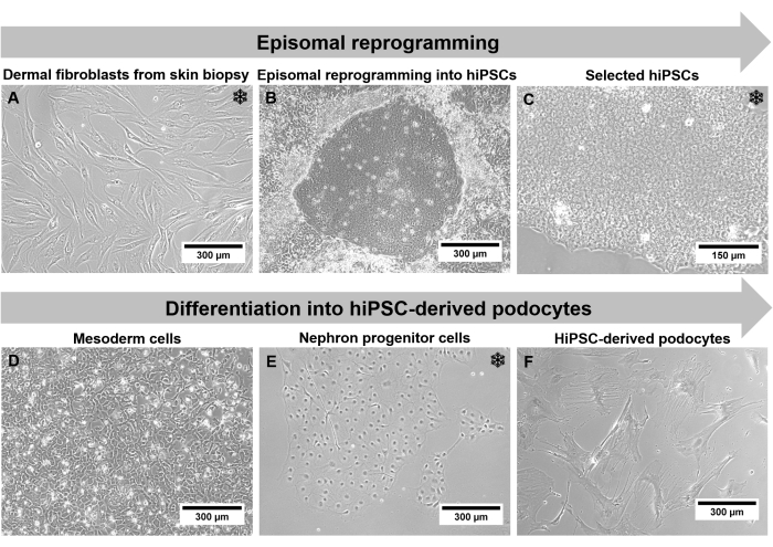
图 2:通过相衬显微镜的协议的各个步骤。 (A)生长的真皮成纤维细胞。(B)重编程后hiPSC集落形成。(C)选定的hiPSC培养物。(D)分化2天后的中胚层细胞。(E)肾单位祖细胞在肾单位祖细胞分化培养基中分化14天后。(F)终末分化的hiPSC衍生足细胞。比例尺代表 300 μm (A,B,D-F) 和 150 μm (C)。雪花突出了可能的冷冻步骤。 请点击此处查看此图的大图。
由于生成的hiPSC衍生足细胞经过一个很长的方案,细胞类型和形态发生了剧烈变化,因此细胞表征是强制性的。细胞形态,如大小和细胞形状,以及生长行为,可以使用相差显微镜进行监测。成纤维细胞呈长纺锤状表型,大小为150μm至300μm(图2A)。重编程后,发生具有50μm小hiPSC的菌落。这些菌落显示出明显的边界和hiPSC,具有高核体比和增加的增殖速率(图2C)。由于足细胞是终末分化的细胞,增殖速率随着进行分化而降低,细胞形态变为具有突出足突的300μm大星形足细胞(图2F)。
融合度是hiPSC培养的关键点,当菌落生长过于密集时,可能会出现自发分化(图3)。在传代之前,必须通过刮擦培养皿的受影响部分来去除这些菌落。
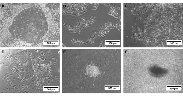
图 3:不同质量的 hiPSC 示例。 成功 (A) 重编程的 hiPSC 集落和 (B) 选定的 hiPSC 的相差图像,以及 (C) 自发分化、(D,E) 非 hiPSC 集落和 (F) 过于密集的 hiPSC 培养物的相差图像。比例尺代表 300 μm。 请点击此处查看此图的大图。
必须根据特定标志物的表达验证该协议的不同细胞类型。重编程后,hiPSC 恢复多能能力并表达增殖和多能性标志物,如 SSEA4、OCT3/4 和 Ki67(图 4)38,39,40。众所周知,重编程以及hiPSCs和分化的漫长培养可导致异常核型,或者更确切地说是诱导突变41。因此,应通过g条带和全外显子组测序来监测培养细胞的遗传背景。
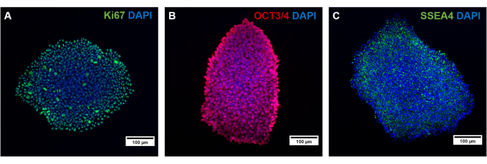
图 4:通过免疫荧光染色表征生成的 hiPSC。 通过染色 (A) 增殖标记物 Ki67 以及多能性标记物 (B) OCT3/4 和 (C) SSEA4 来表征生成的 hiPSC。比例尺代表 100 μm。 请点击此处查看此图的大图。
在形态学上,与ciPodocys相比,hiPSC衍生的足细胞呈星形,并表达长原代和次级足突的独特网络(图5)。HiPSC 来源的足细胞表达足细胞特异性标记蛋白,如突触足蛋白、肾素和足细胞蛋白(图 6A-C)42。
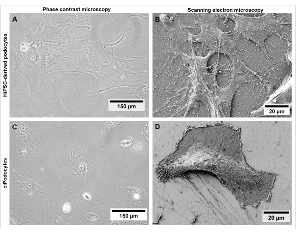
图 5:hiPSC 来源足细胞与 ciPodocys 的形态比较。 使用相衬显微镜(A,C)和扫描电子显微镜(B,D)比较来自健康供体的(A,B)hiPSC来源的足细胞与(C,D)ciPodocys的细胞形态和丝状伪足。比例尺代表 150 μm (A,C) 和 20 μm (B,D)。请点击此处查看此图的大图。
因此,比较hiPSC来源的足细胞不仅来自健康供体,而且来自足细胞特异性基因突变的患者,允许对患者的离 体突变进行个体化表征。
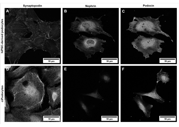
图 6:hiPSC 来源的足细胞和外翻细胞中足细胞特异性标记物的表征。来自健康供体的 (A-C) hiPSC 衍生足细胞与 (D-F) ciPodocys 关于足细胞特异性标记蛋白突触蛋白 (A,D)、肾单位 (B,E) 和鬼臼素 (C,F) 的比较。比例尺代表 50 μm。请点击此处查看此图的大图。
表 1.研究中使用的所有细胞培养基和溶液的组成。请按此下载此表格。
讨论
这种基于细胞培养的方案将人真皮成纤维细胞的游离体重编程结合成患者特异性的hiPSC,随后分化为hiPSC衍生的足细胞。这使我们能够研究遗传性肾小球疾病患者关于足细胞损伤的足细胞突变相关改变。通过电穿孔用无整合方法重新编程真皮成纤维细胞的方案改编自Bang等人32和Okita等人33的已发表工作。区分足细胞和hiPSC的方案改编自Musah等人发表的方案28,34,35。已经有出版物描述了从hiPSCs产生足细胞27,34,35。然而,这里提供的方案在将hiPSC分化为足细胞方面是优化的,并且成本更低。与Musah等人发表的方案相比,我们在不同的包衣试剂上测试了该方案,如玻连蛋白,层粘连蛋白丝-511和可溶的基底膜基质。玻连蛋白和层粘连蛋白丝-511的浓度可以降低到2.5μg/mL,而不是5μg/mL28,34,35。此外,可以将BMP7和激活素A-两种非常昂贵的生长因子的浓度降低50%,从100 ng / mL降低到50 ng / mL。
这使得差异化成本更低。从第7天开始,肾单位祖细胞仍在增殖,并且之前显示了冷冻的可能性。我们在解冻后和最终分化之前在含有Dulbecco修饰的Eagle培养基(DMEM)和B27的碱性培养基中扩增了这些细胞数天,进一步降低了成本。除了分化步骤外,该协议还描述了皮肤活检中成纤维细胞的生长,随后通过游离体重编程 产生 患者特异性hiPSC。这两种方法的结合能够产生患者特异性足细胞。因此,这里提供了一个完整的分步方案来生成患者特异性hiPSC来源的足细胞,这是以前没有详细描述过的。
由于整个实验方案包括几种不同的细胞类型,因此在不同步骤中表征生成的细胞类型至关重要。细胞培养时间较长,因此应在不同的传代中进行质量控制。在使用hiPSC时,日常喂养以及细胞行为和形态监测是必要的。分化培养基的无菌性必须通过0.2μm过滤器进行过滤灭菌来确保。从皮肤活检到hiPSC衍生的足细胞的整个方案需要几个月的时间,但可以在该过程的不同阶段冷冻细胞。成纤维细胞、选定的hiPSC克隆和增殖性肾单位祖细胞在肾单位祖细胞分化培养基中7天后可以冷冻,并且可以生成工作细胞库。
尽管hiPSC衍生的健康足细胞发展出独特的原发性和继发性足突网络(图5A,B)并表达典型的足细胞特异性标志物(图6A-C),但在经典的二维细胞培养模型中很难模仿体内可见的特征狭缝隔膜。此外,在这种单一培养环境中,不可能与其他肾小球细胞类型进行细胞间交流。
由于足细胞的终末分化状态和缺乏增殖能力,很难 体外研究足细胞。在有条件地永生化原代足细胞的帮助下,可以通过插入热敏开关来克服这一限制,从而产生细胞培养模型,其中细胞在33°C增殖并在37°C分化13,14。尽管这些外翻细胞在足细胞研究方面具有很高的潜力,但也存在局限性,例如缺乏标志物表达、去分化形态和无法形成足突15,16。
足细胞与患者来源的体细胞的分化能够产生患病足细胞并将其与健康对照细胞离 体进行比较。这使我们能够研究由于足细胞特异性基因突变引起的足细胞损伤。此外,使用hiPSCs有可能创建三维细胞培养疾病模型,或者更确切地说是类器官43,44。hiPSC来源的足细胞与其他肾小球细胞(如肾小球内皮细胞或系膜细胞)共培养可能会为健康和肾小球疾病中的细胞间通讯带来新的见解。
此外,患者特异性足细胞的表征和治疗可以在高通量分析中进行 离体 。个体化方法为研究特定突变的新治疗靶点和在未来进行个体化医疗提供了机会。
披露声明
作者没有什么可透露的。
致谢
这项工作由埃尔朗根-纽伦堡弗里德里希-亚历山大大学跨学科临床研究中心(IZKF)资助,授权号为M4-IZKF-F009,授予Janina Müller-Deile,由联邦电影和研究部(BMBF)资助,项目名称为STOP-FSGS-Speed Translation-Oriented Progress to Treat FSGS,授权号01GM2202D授予Janina Müller-Deile。我们感谢Annalena Kraus在拍摄SEM图像方面的支持。
材料
| Name | Company | Catalog Number | Comments |
| 0.2 µm sterile filter | Rotilab | P668.1 | for sterilization of differentiation medium |
| all-trans retinoic acid | Stem Cell Technologies | 72262 | supplement for differentiation |
| B27 supplement (50 x), serum free | Gibco | 17504044 | supplement for serum-free differentiation medium |
| BG iMatrix-511 Silk | biogems | RL511S | additional option of extracellular matrix reagent used in coating solution to coat cell culture plastics suitable for hiPSC culture |
| Bovine serum albumin (BSA) | Roth | 8076.4 | |
| CELLSTAR Filter Cap Cell Culture Flasks, T75, 250 mL | Greiner bio-one | 82050-856 | cell culture plastics suitable for fibroblast culture |
| CHIR99021 (5 mg) | Sigma-Aldrich | 252917-06-9 | supplement for differentiation |
| Corning Matrigel hESC qualified matrix | Corning | 354277 | additional option of extracellular matrix reagent used in coating solution to coat cell culture plastics suitable for hiPSC culture (solubilized basement membrane matrix) |
| countess Cell Counting Chamber Slides | Invitrogen | C10283 | to count cells |
| countess II FL Automated Cell Counter | Invitrogen | to count cells | |
| cryoPure tubes, 2 ml, QuickSeal screw cap, white | Sarstedt | 72380 | cryovials for freezing of cells |
| dimethyl sulfoxide (DMSO) | Roth | A994.1 | for fibroblast freezing medium |
| DMEM/F12 (1:1) (1 x) | Gibco | 11320074 | basic medium for differentiation |
| DMEM/F12 + Glutamax | Gibco | 10565018 | basic medium for fibroblast medium |
| EVOS M5000 Imaging System | Thermo Fisher Scientific | AMF5000 | phase contrast microscope |
| fetal bovine serum premium, inactivated (FCS) | PAN Biotech | P301902 | serum for fibroblast medium, fibroblast freezing medium and podocyte maintenance medium |
| fisherbrand Electroporation Cuvettes Plus, 4 mm gap, 800 µL capacity, sterile | Fisherbrand | FB104 | cuvette used for electroporation/episomal reprogramming of fibroblasts (4mm gap) |
| fluoromount-G Mounting Medium, with DAPI | Invitrogen | 00-4959-52 | mounting medium containing dapi |
| gauge needle (0.6 x 30 mm) | BD Microlance3 | 300700 | for separation of hiPSC colonies into small pieces |
| human Recombinant Activin A Protein | 78001.1 | Stem cell technologies | supplement for differentiation |
| human recombinant bone morphogenetic protein 7 (BMP7) | Peprotech | 120-03P | supplement for differentiation |
| human VEGF-165 Recombinant Protein | Thermo Scientific | PHC9394 | supplement for differentiation |
| insulin-transferrin-selenium (ITS -G) (100 x) | Gibco | 41400045 | supplement for podocyte maintenance medium |
| LB medium | Roth | X964.1 | for sterility test of hiPSC culture |
| lookOut Mycoplasma PCR Detection Kit | Sigma Aldrich | MP0035-1KT | commercial mycoplasma detection kit |
| microscope slides | Diagonal GmbH & Co.KG | 21,102 | |
| microtube 1.5 mL | Sarstedt | 72706400 | |
| mTeSR1 Complete Kit | Stem Cell Technologies | 85850 | basic medium for serum-free hiPSC culture medium |
| nalgene freezing container Mr.Frosty | Roth | AC96.1 | to ensure optimal freezing conditions |
| normal goat serum | abcam | ab 7481 | for preincubation solution and antibody diluent |
| nunc 24 well plates | Thermo Scientific | 142485 | cell culture plastics suitable for hiPSC culture |
| nunc 48 well plates | Thermo Scientific | 152640 | cell culture plastics suitable for hiPSC culture |
| nunc 6 well plates | Thermo Scientific | 140685 | cell culture plastics suitable for hiPSC culture |
| nunc EasYDish Dishes 100 mm | Thermo Scientific | 150466 | cell culture plastics suitable for hiPSC culture |
| nunc MicroWell 96-Well, Nunclon Delta-Treated, Flat-Bottom Microplate | Thermo Scientific | 167008 | cell culture plastics suitable for hiPSC culture |
| nutriFreez D10 Cryopreservation Medium | Sartorius | 05-713-1E | serum-free cryopreservation medium for cryopreservation of hiPSC and nephron progenitor cells |
| Opti-MEM | Gibco | 11058021 | electroporation medium |
| pCXLE-hMLN | Addgene | #27079 | plasmid for episomal reprogramming |
| pCXLE-hOCT3/4 plasmid | Addgene | #27077 | plasmid for episomal reprogramming |
| pCXLE-hSK plasmid | Addgene | #27078 | plasmid for episomal reprogramming |
| penicillin-streptomycin | Sigma-Aldrich | P4333-100ML | to avoid bacterial contamination |
| plastic coverslips | Sarstedt | 83.1840.002 | for immunofluorescent stainings of hiPSCs and hiPSC-derived podocytes |
| ROTI Histofix | Roth | P087.3 | commercial paraformaldehyde (4 %) for fixation of cells |
| RPMI 1640 + L-Glutamine | Gibco | 21875034 | basic medium for podocyte maintenance medium |
| staining chamber StainTray Black lid | Roth | HA51.1 | |
| stemPro Accutase Cell Dissociation Reagent | Gibco | A1110501 | enzymatic cell detachment solution used for dissociation of hiPSCs |
| sterile phosphate buffered saline (PBS) (1 x) | Gibco | 14190094 | used for washing and coating |
| sterile water | Roth | T1432 | |
| syringe without needle 20 mL | BD Plastipak | 300629 | to filter sterilize differentiation medium |
| TC dish 100 mm | Sarstedt | 8,33,902 | sterile cell culture plastics used for cutting the skin biopsy and fibroblast culture |
| TC dish 35 mm | Sarstedt | 8,33,900 | sterile cell culture plastics used for outgrowing fibroblasts from skin biopsy |
| triton X 100 | Roth | 3051.3 | for preincubation solution |
| trypan Blue Stain (0.4 %) for use with the Countess Automated Cell Counter | Invitrogen | T10282 | to count cells |
| trypsin-EDTA (10 x) | Biowest | X0930-100 | dissociation reagent used for fibroblasts and nephron progenitor cells |
| tube 15 mL | Greiner bio-one | 188271-N | |
| tube 50 mL | Greiner bio-one | 227261 | |
| vitronectin ACF | Sartorius | 05-754-0002 | extracellular matrix reagent used in coating solution to coat cell culture plastics suitable for hiPSC culture |
| Y-27632 dihydrochloride (10 mg) | Tocris | 1254 | to avoid apoptosis of hiPSCs during splitting |
| Primary antibodies | |||
| OCT4 | Stem Cell Technologies | 60093.1 | pluripotency marker, dilution 1:200 |
| SSEA-4 | Stem Cell Technologies | 60062FI.1 | pluripotency marker, dilution 1:100 |
| Ki67 | Abcam | ab15580 | proliferation marker, dilution 1:300 |
| synaptopodin | Proteintech | 21064-1-AP | podocyte-specific marker, dilution 1:200 |
| nephrin | Progen | GP-N2 | podocyte-specific marker, dilution 1:25 |
| podocin | proteintech | 20384-1-AP | podocyte-specific marker, dilution 1:100 |
| Secondary antibodies | |||
| goat anti-Guinea Pig IgG (H+L) Highly Cross-Adsorbed Secondary Antibody, Alexa Fluor 555 | Invitrogen | A21435 | secondary anditbody, dilution 1:1000 |
| alexa Fluor 647 Goat Anti-Rabbit SFX Kit, highly cross-adsorbed | Invitrogen | A31634 | secondary anditbody, dilution 1:1000 |
| donkey anti-Rabbit IgG (H+L) Highly Cross-Adsorbed Secondary Antibody, Alexa Fluor 488 | Invitrogen | A21206 | secondary anditbody, dilution 1:1000 |
| goat anti-Mouse IgG (H+L) Cross-Adsorbed Secondary Antibody, Alexa Fluor 555 | Invitrogen | A21422 | secondary anditbody, dilution 1:1000 |
| goat anti-Mouse IgG (H+L) Cross-Adsorbed Secondary Antibody, Alexa Fluor 488 | Invitrogen | A11001 | secondary anditbody, dilution 1:1000 |
参考文献
- Mundel, P., et al. Rearrangements of the cytoskeleton and cell contacts induce process formation during differentiation of conditionally immortalized mouse podocyte cell lines. Experimental Cell Research. 236 (1), 248-258 (1997).
- Grgic, I., et al. Imaging of podocyte foot processes by fluorescence microscopy. Journal of the American Society of Nephrology. 23 (5), 785-791 (2012).
- Grahammer, F., Schell, C., Huber, T. B. The podocyte slit diaphragm-from a thin grey line to a complex signalling hub. Nature Reviews Nephrology. 9 (10), 587-598 (2013).
- Deen, W. M., Lazzara, M. J., Myers, B. D. Structural determinants of glomerular permeability. American Journal of Physiology. Renal Physiology. 281 (4), F579-F596 (2001).
- Schell, C., Huber, T. B. The evolving complexity of the podocyte cytoskeleton. Journal of the American Society of Nephrology. 28 (11), 3166-3174 (2017).
- Muller-Deile, J., Schiffer, M. Podocyte directed therapy of nephrotic syndrome-can we bring the inside out. Pediatric Nephrology. 31 (3), 393-405 (2016).
- Boehlke, C., et al. Hantavirus infection with severe proteinuria and podocyte foot-process effacement. American Journal of Kidney Diseases. 64 (3), 452-456 (2014).
- Schiffer, M., et al. Pharmacological targeting of actin-dependent dynamin oligomerization ameliorates chronic kidney disease in diverse animal models. Nature Medicine. 21 (6), 601-609 (2015).
- Kopp, J. B., et al. Podocytopathies. Nature Reviews. Disease Primers. 6 (1), 68 (2020).
- Wiggins, R. C. The spectrum of podocytopathies: a unifying view of glomerular diseases. Kidney International. 71 (12), 1205-1214 (2007).
- Mundel, P., Reiser, J., Kriz, W. Induction of differentiation in cultured rat and human podocytes. Journal of the American Society of Nephrology. 8 (5), 697-705 (1997).
- Jat, P. S., et al. Direct derivation of conditionally immortal cell lines from an H-2Kb-tsA58 transgenic mouse. Proceedings of the National Academy of Sciences. 88 (12), 5096-5100 (1991).
- Saleem, M. A., et al. A conditionally immortalized human podocyte cell line demonstrating nephrin and podocin expression. Journal of the American Society of Nephrology. 13 (3), 630-638 (2002).
- Eto, N., et al. Podocyte protection by darbepoetin: preservation of the cytoskeleton and nephrin expression. Kidney International. 72 (4), 455-463 (2007).
- Krtil, J., Platenik, J., Kazderova, M., Tesar, V., Zima, T. Culture methods of glomerular podocytes. Kidney & Blood Pressure Research. 30 (3), 162-174 (2007).
- Chittiprol, S., Chen, P., Petrovic-Djergovic, D., Eichler, T., Ransom, R. F. Marker expression, behaviors, and responses vary in different lines of conditionally immortalized cultured podocytes. American Journal of Physiology. Renal Physiology. 301 (3), F660-F671 (2011).
- Shih, N. Y., et al. CD2AP localizes to the slit diaphragm and binds to nephrin via a novel C-terminal domain. The American Journal of Pathology. 159 (6), 2303-2308 (2001).
- Yan, K., Khoshnoodi, J., Ruotsalainen, V., Tryggvason, K. N-linked glycosylation is critical for the plasma membrane localization of nephrin. Journal of the American Society of Nephrology. 13 (5), 1385-1389 (2002).
- Sir Elkhatim, R., Li, J. Y., Yong, T. Y., Gleadle, J. M. Dipping your feet in the water: podocytes in urine. Expert Review of Molecular Diagnostics. 14 (4), 423-437 (2014).
- Camici, M. Urinary detection of podocyte injury. Biomedicine & Pharmacotherapy. 61 (5), 245-249 (2007).
- Muller-Deile, J., et al. Overexpression of preeclampsia induced microRNA-26a-5p leads to proteinuria in zebrafish. Scientific Reports. 8 (1), 3621 (2018).
- Schenk, H., et al. Removal of focal segmental glomerulosclerosis (FSGS) factor suPAR using CytoSorb. Journal of Clinical Apheresis. 32 (6), 444-452 (2017).
- Petermann, A., Floege, J. Podocyte damage resulting in podocyturia: a potential diagnostic marker to assess glomerular disease activity. Nephron. Clinical Practice. 106 (2), c61-c66 (2007).
- Vogelmann, S. U., Nelson, W. J., Myers, B. D., Lemley, K. V. Urinary excretion of viable podocytes in health and renal disease. American Journal of Physiology. Renal Physiology. 285 (1), F40-F48 (2003).
- Petermann, A. T., et al. Podocytes that detach in experimental membranous nephropathy are viable. Kidney International. 64 (4), 1222-1231 (2003).
- Sakairi, T., et al. Conditionally immortalized human podocyte cell lines established from urine. American Journal of Physiology. Renal Physiology. 298 (3), F557-F567 (2010).
- Rauch, C., et al. Differentiation of human iPSCs into functional podocytes. PLoS One. 13 (9), e0203869 (2018).
- Musah, S., et al. Mature induced-pluripotent-stem-cell-derived human podocytes reconstitute kidney glomerular-capillary-wall function on a chip. Nature Biomedical Engineering. 1, 0069 (2017).
- Takahashi, K., Okita, K., Nakagawa, M., Yamanaka, S. Induction of pluripotent stem cells from fibroblast cultures. Nature Protocols. 2 (12), 3081-3089 (2007).
- Takahashi, K., Yamanaka, S. Induction of pluripotent stem cells from mouse embryonic and adult fibroblast cultures by defined factors. Cell. 126 (4), 663-676 (2006).
- Teshigawara, R., Cho, J., Kameda, M., Tada, T. Mechanism of human somatic reprogramming to iPS cell. Laboratory Investigation. 97 (10), 1152-1157 (2017).
- Bang, J. S., et al. Optimization of episomal reprogramming for generation of human induced pluripotent stem cells from fibroblasts. Animal Cells and Systems. 22 (2), 132-139 (2018).
- Okita, K., et al. A more efficient method to generate integration-free human iPS cells. Nature Methods. 8 (5), 409-412 (2011).
- Musah, S., Dimitrakakis, N., Camacho, D. M., Church, G. M., Ingber, D. E. Directed differentiation of human induced pluripotent stem cells into mature kidney podocytes and establishment of a Glomerulus Chip. Nature Protocols. 13 (7), 1662-1685 (2018).
- Burt, M., Bhattachaya, R., Okafor, A. E., Musah, S. Guided differentiation of mature kidney podocytes from human induced pluripotent stem cells under chemically defined conditions. Journal of Visualized Experiments. (161), e61299 (2020).
- Vangipuram, M., Ting, D., Kim, S., Diaz, R., Schule, B. Skin punch biopsy explant culture for derivation of primary human fibroblasts. Journal of Visualized Experiments. (77), e3779 (2013).
- Hoffding, M. K., Hyttel, P. Ultrastructural visualization of the Mesenchymal-to-Epithelial Transition during reprogramming of human fibroblasts to induced pluripotent stem cells. Stem Cell Research. 14 (1), 39-53 (2015).
- Bharathan, S. P., et al. Systematic evaluation of markers used for the identification of human induced pluripotent stem cells. Biology Open. 6 (1), 100-108 (2017).
- Scholzen, T., Gerdes, J. The Ki-67 protein: from the known and the unknown. Journal of Cellular Physiology. 182 (3), 311-322 (2000).
- Sun, X., Kaufman, P. D. Ki-67: more than a proliferation marker. Chromosoma. 127 (2), 175-186 (2018).
- Vaz, I. M., et al. Chromosomal aberrations after induced pluripotent stem cells reprogramming. Genetics and Molecular Biology. 44 (3), 20200147 (2021).
- Reiser, J., Altintas, M. M. Podocytes. F1000Research. 5, 114 (2016).
- Ohmori, T., et al. Impaired NEPHRIN localization in kidney organoids derived from nephrotic patient iPS cells. Scientific Reports. 11 (1), 3982 (2021).
- Morizane, R., Bonventre, J. V. Generation of nephron progenitor cells and kidney organoids from human pluripotent stem cells. Nature Protocols. 12 (1), 195-207 (2017).
转载和许可
请求许可使用此 JoVE 文章的文本或图形
请求许可探索更多文章
This article has been published
Video Coming Soon
版权所属 © 2025 MyJoVE 公司版权所有,本公司不涉及任何医疗业务和医疗服务。