用连续波多普勒的外围血管考试
资料来源: 约瑟夫 Donroe,MD、 内科、 儿科,耶鲁大学医学院,纽黑文,康涅狄格
周围血管病 (PVD) 是一种常见的疾病,影响老年人,包括外周动脉和静脉疾病。虽然历史和物理考试提供其诊断的线索,多普勒超声已成为常规的床头血管检查。题为"外围血管考试"的视频给了外围的动脉和静脉系统体检详细的审查。这个视频专门评论床边评估慢性静脉功能不全使用手持的连续波多普勒与外周动脉疾病 (PAD)。
手持多普勒 (HHD) 是超声波的一种简单的仪器,它利用连续的传输和接收 (也称为连续波多普勒) 来检测中血流速度的变化,当它流经一艘船。多普勒探测器包含发出超声发射元素和一个接收元素,检测超声波 (图 1)。发出的超声波被反映从血液回频率直接关系到血流速度的探测器。被反射的信号检测到并可听到的声音频率直接关系到它接收到的多普勒信号转导 (所以,血流速度产生更高频率的声音)。

图 1。一代的多普勒信号。手持多普勒发出超声波信号,然后反射回血液,并最后收到的多普勒探针。
HHD 容易在办公室或医院设置用来探测脉冲,屏幕为垫使用踝臂压力指数 (肱) 和本地化静脉功能不全。这个视频回顾这些程序;然而,它不是要全面检讨的无创性血管检查。
1.编制
- 获得血压袖带、 HHD 机、 多普勒凝胶和皮肤标记。
- 在检查病人之前要洗手。
- 病患者的一件袍子,舒舒服服地仰卧在考试桌上开始。
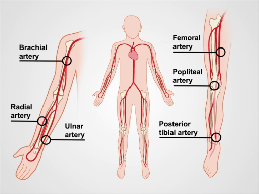
图 2。上肢和下肢主要动脉。
2.降低下肢动脉评估
- 对于弱或无脉冲通过触诊或外周动脉疾病 (PAD) 的历史风险因素的患者,使用 HHD 评估血流量。开始采用凝胶在预期领域的动脉被调查 (图 2)。
- 置于 45 度角到皮肤,头侧指动脉的多普勒频率。如果未检测到的多普勒信号,慢慢地向内侧移动多普勒探头和侧向,并且偶尔地,远端动脉的路径可以会发生变化。请记住,一小部分人可能会有天生缺乏足踏板 (DP) 动脉。
- 如果遇到一个

跳至...
此集合中的视频:

Now Playing
用连续波多普勒的外围血管考试
Physical Examinations I
38.3K Views

物理考试的一般方法
Physical Examinations I
115.1K Views

观察和检查
Physical Examinations I
92.8K Views

触诊
Physical Examinations I
82.4K Views

打击乐
Physical Examinations I
99.2K Views

听诊
Physical Examinations I
60.0K Views

病人的服装物理考试期间的适当调整
Physical Examinations I
82.8K Views

血压测量
Physical Examinations I
106.7K Views

测量生命体征
Physical Examinations I
113.5K Views

呼吸道考试 i: 检查和触诊
Physical Examinations I
155.5K Views

呼吸道考试 II: 打击乐和听诊
Physical Examinations I
211.1K Views

心脏考试 i: 检查和触诊
Physical Examinations I
174.9K Views

心脏考试 II: 听诊
Physical Examinations I
138.9K Views

心脏的考试 III: 异常心音
Physical Examinations I
91.0K Views

外围的血管考试
Physical Examinations I
67.6K Views
版权所属 © 2025 MyJoVE 公司版权所有,本公司不涉及任何医疗业务和医疗服务。