Exame Vascular Periférico Usando Doppler de Onda Contínua
Visão Geral
Fonte: Joseph Donroe, MD, Medicina Interna e Pediatria, Yale School of Medicine, New Haven, CT
A doença vascular periférica (PVD) é uma condição comum que afeta idosos e inclui doença das artérias e veias periféricas. Enquanto a história e o exame físico oferecem pistas para seu diagnóstico, o ultrassom Doppler tornou-se uma parte rotineira do exame vascular de cabeceira. O vídeo intitulado "O Exame Vascular Periférico" fez uma revisão detalhada do exame físico dos sistemas arterial e venoso periféricos. Este vídeo revisa especificamente a avaliação de cabeceira da doença arterial periférica (PAD) e da insuficiência venosa crônica usando uma onda contínua portátil Doppler.
O Doppler portátil (HHD) é um instrumento simples que utiliza transmissão contínua e recepção de ultrassom (também chamado de Doppler de onda contínua) para detectar alterações na velocidade sanguínea à medida que percorre um vaso. A sonda Doppler contém um elemento transmissor que emite ultrassom e um elemento receptor que detecta ondas de ultrassom(Figura 1). O ultrassom emitido é refletido a partir do sangue em movimento e de volta à sonda em uma frequência diretamente relacionada com a velocidade do fluxo sanguíneo. O sinal refletido é detectado e transduzido para um som audível com uma frequência diretamente relacionada com a do sinal Doppler recebido (assim, o fluxo sanguíneo mais rápido produz um som de maior frequência).

Figura 1. Geração de um sinal Doppler. O Doppler portátil emite um sinal de ultrassom, que é então refletido de volta por sangue em movimento, e finalmente recebido pela sonda Doppler.
O HHD é facilmente usado no consultório ou no ambiente hospitalar para detectar pulsos, tela para PAD usando o índice de pressão braquial do tornozelo (ABPI) e localização da insuficiência venosa. Este vídeo revisa esses procedimentos; no entanto, não se destina a ser uma revisão abrangente dos testes vasculares não invasivos.
Procedimento
1. Preparação
- Obtenha um manguito de pressão arterial, uma máquina de HHD, gel Doppler, e marcador de pele.
- Lave as mãos antes de examinar o paciente.
- Comece com o paciente em um vestido, deitado confortavelmente supino na mesa de exame.

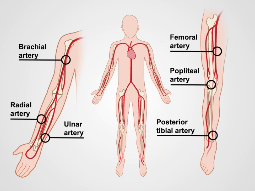
Figura 2. As principais artérias das extremidades superior e inferior.
2. Avalia
Aplicação e Resumo
Um histórico cuidadoso e exame físico são importantes para qualquer pessoa com suspeita de doença vascular periférica com base em sintomas ou fatores de risco. O HHD passou a fazer parte do exame vascular de rotina e deve ser usado para complementar o exame físico, caso haja suspeita de PVD. Não é uma ferramenta tecnicamente difícil de usar, e as manobras descritas no vídeo podem ser realizadas por médicos em geral. Assim como para o exame físico, o conhecimento da anatomia vascular é fundamental para o suce...
Pular para...
Vídeos desta coleção:

Now Playing
Exame Vascular Periférico Usando Doppler de Onda Contínua
Physical Examinations I
38.6K Visualizações

Abordagem Geral para o Exame Físico
Physical Examinations I
117.6K Visualizações

Observação e Inspeção
Physical Examinations I
95.1K Visualizações

Palpação
Physical Examinations I
84.5K Visualizações

Percussão
Physical Examinations I
101.8K Visualizações

Auscultação
Physical Examinations I
62.3K Visualizações

Ajuste adequado da vestimenta do paciente durante o exame físico
Physical Examinations I
83.4K Visualizações

Aferição da pressão arterial
Physical Examinations I
108.8K Visualizações

Medição de Sinais Vitais
Physical Examinations I
115.0K Visualizações

Exame Respiratório I: Inspeção e Palpação
Physical Examinations I
157.4K Visualizações

Exame Respiratório II: Percussão e Auscultação
Physical Examinations I
213.0K Visualizações

Exame Cardíaco I: Inspeção e Palpação
Physical Examinations I
176.6K Visualizações

Exame Cardíaco II: Auscultação
Physical Examinations I
140.1K Visualizações

Exame Cardíaco III: Sons cardíacos anormais
Physical Examinations I
91.9K Visualizações

Exame Vascular Periférico
Physical Examinations I
68.8K Visualizações
Copyright © 2025 MyJoVE Corporation. Todos os direitos reservados
