
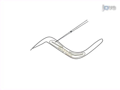
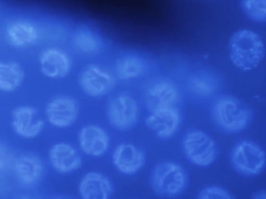
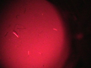
Label-Free Imaging of Lipid Storage Dynamics in Caenorhabditis elegans using Stimulated Raman Scattering Microscopy
May 28th, 2021
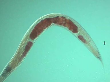
•Stimulated Raman scattering (SRS) microscopy allows selective, label-free imaging of specific chemical moieties and it has been effectively employed to image lipid molecules in vivo. Here, we provide a brief introduction to the principle of SRS microscopy and describe methods for its use in imaging lipid storage in Caenorhabditis elegans.
Related Videos

Label-free in situ Imaging of Lignification in Plant Cell Walls

Differential Imaging of Biological Structures with Doubly-resonant Coherent Anti-stokes Raman Scattering (CARS)

Isolation and In vitro Activation of Caenorhabditis elegans Sperm
Time-lapse Microscopy of Early Embryogenesis in Caenorhabditis elegans
Basic Caenorhabditis elegans Methods: Synchronization and Observation
Prostaglandin Extraction and Analysis in Caenorhabditis elegans
Using Caenorhabditis elegans as a Model System to Study Protein Homeostasis in a Multicellular Organism
Quantification of Lipid Abundance and Evaluation of Lipid Distribution in Caenorhabditis elegans by Nile Red and Oil Red O Staining

Oligomerization Dynamics of Cell Surface Receptors in Living Cells by Total Internal Reflection Fluorescence Microscopy Combined with Number and Brightness Analysis

Using Caenorhabditis elegans to Screen for Tissue-Specific Chaperone Interactions