Method Article
Modelado avanzado de aneurisma de aorta abdominal en ratones mediante combinación de elastasa tópica y ß-aminopropionitrilo oral
En este artículo
Resumen
Este protocolo describe un enfoque quirúrgico metódico para modelar aneurismas de aorta abdominal avanzados en ratones mediante una combinación de aplicación directa de elastasa a la aorta infrarrenal y administración de ß-aminopropionitrilo a través del agua potable.
Resumen
El modelo murino de elastasa tópica de aneurisma de aorta abdominal (AAA) mejora cuando se combina con agua potable suplementada con ß-aminopropionitrilo (BAPN) para producir de manera confiable verdaderos aneurismas infrarrenales con comportamientos que imitan los AAA humanos. La aplicación tópica de elastasa a la adventicia de la aorta infrarrenal provoca daño estructural en las capas elásticas de la pared aórtica e inicia la dilatación del aneurisma. La coadministración de BAPN, un inhibidor de la lisil oxidasa, promueve la degeneración sostenida de la pared al reducir la reticulación de colágeno y elastina. Esta combinación da lugar a grandes AAA que se expanden progresivamente, forman trombos intraluminales y son capaces de romperse. El perfeccionamiento de las técnicas quirúrgicas, como el aislamiento circunferencial de todo el segmento aórtico infrarrenal, puede ayudar a estandarizar el procedimiento para una aplicación consistente y completa de la elastasa pancreática porcina a pesar de los diferentes operadores y las variaciones anatómicas entre ratones. Por lo tanto, el modelo de elastasa/BAPN es un enfoque refinado para la inducción quirúrgica de AAA en ratones, que puede recapitular mejor los aneurismas humanos y proporcionar oportunidades adicionales para estudiar el crecimiento de los aneurismas y el riesgo de ruptura.
Introducción
Un aneurisma se define como una dilatación patológica de un vaso sanguíneo mayor del 50% del diámetro del vaso sano1. A pesar de que los aneurismas de aorta abdominal (AAA) son una afección común en la población envejecida, con una incidencia de aproximadamente el >5% de los hombres > 65 años de edad, no existen estrategias terapéuticas dirigidas para tratar el AAA1. El tratamiento actual del AAA se limita a la reducción de los factores de riesgo y a la reparación quirúrgica con cirugía abierta o endovascular basada en el diámetro aórtico o la tasa de crecimiento2. El mayor peligro del AAA es la rotura del aneurisma, que es mortal si no se trata, y la reparación en este entorno emergente puede conllevar riesgos de mortalidad de hasta el 90%1.
La fisiopatología del AAA es complicada, multifactorial y no se comprende completamente3. Las características del AAA humano incluyen una verdadera dilatación aneurismática de la pared aórtica con infiltración de células inflamatorias, la presencia de trombo intraluminal y una dilatación progresiva que conduce a una eventual ruptura 3,4. Además, los AAA se asocian con la edad avanzada, tienen un predominio sexual masculino:femenino de 9:1 y ocurren con mayor frecuencia en la aorta infrarrenal5. Modelar todas las características y comportamientos de los AAA humanos en animales sigue siendo un desafío continuo6.
El modelo actual de AAA se lleva a cabo principalmente en ratones, y los aneurismas se inducen comúnmente utilizando uno de tres métodos: por infusión de angiotensina II (AngII) a través de una bomba osmótica implantada subcutáneamente, y por aplicación directa de cloruro de calcio (CaCl2) o elastasa a la aorta7. En este último método, la elastasa pancreática porcina (EPP) se aplica a un segmento de la aorta infrarrenal y provoca la degradación enzimática de las fibras de elastina dentro de la lámina elástica de la túnica media. Este daño estructural resulta en el debilitamiento de la pared aórtica y la dilatación externa del aneurisma. Sin embargo, el uso de elastasa tópica sola produce aneurismas infrarrenales relativamente pequeños, que no se agrandan ni se rompen progresivamente con el tiempo. Más recientemente, Lu et al. mejoraron este modelo mediante la administración adicional de β-aminopropionitrilo (BAPN), un inhibidor irreversible de la lisil oxidasa, a sus ratones tratados con elastasa. Al evitar la reticulación de las fibras de elastina y colágeno, la suplementación con BAPN hace que las aortas dañadas por la elastasa se dilaten progresivamente hasta el punto de ruptura. Además, el modelo de elastasa/BAPN tiene una mayor tasa de incidencia de AAA que el modelo de elastasa tópica, y los aneurismas producidos también son más grandes y contienen trombo intraluminal8.
En el modelo de elastasa/BAPN, el grado de disección quirúrgica y la exposición de la aorta a la elastasa pueden afectar el éxito y la replicabilidad de este modelo. En este manuscrito, describimos que la co-administración de agua potable de BAPN y la aplicación tópica de elastasa a la aorta después del aislamiento circunferencial de todo el segmento aórtico infrarrenal mejora la replicabilidad, explica las diferencias anatómicas entre animales y da como resultado una mayor tasa de inducción de AAA, tamaños de aneurismas e incidencia de rupturas. En este artículo, describiremos un enfoque estandarizado para inducir de manera confiable aneurismas de aorta abdominal avanzados en ratones utilizando una combinación de elastasa tópica y agua suplementada con BAPN.
Protocolo
Los protocolos para animales están aprobados por el Comité Institucional de Cuidado y Uso de Animales (M005792) de la Universidad de Wisconsin-Madison.
1. Mantenimiento de animales
- Cría ratones en comida de mantenimiento estándar. Use ratones adultos o ratones adultos jóvenes (8-12 semanas de edad).
NOTA: El uso de adultos asegura que los animales hayan alcanzado la madurez completa y limita cualquier posibilidad de que los cambios en el diámetro aórtico puedan estar relacionados con el crecimiento del animal. Para este estudio, utilizamos ratones machos y hembras C57BL/6J con una edad de 22 a 24 semanas en el momento de la cirugía. Lu y sus colegas no observaron diferencias significativas en la respuesta aneurismática entre ratones más jóvenes ymayores. Además, mientras que la mayoría de los modelos de aneurismas se realizan en ratones machos, este modelo induce con éxito AAA tanto en ratones machos como hembras9. - Determine la duración del estudio y asigne a los animales a grupos de tratamiento o simulacros (control). Administrar agua potable con BAPN al 0,2% a los ratones del grupo de tratamiento y someterlos a cirugía con aplicación tópica de elastasa activa en la aorta infrarrenal. Administrar agua no tratada a los animales de control y someterlos a cirugía con la aplicación de elastasa desnaturalizada en la aorta infrarrenal.
2. Inicio del tratamiento de agua potable suplementada con B-aminopropionitrilo (BAPN)
- Dos días antes de la cirugía, comience el tratamiento con ratones con agua potable BAPN al 0,2%. Prepare el agua BAPN en grandes volúmenes y almacene en la oscuridad a 4 °C durante un máximo de 28 días. Asegúrese de que el agua de BAPN alcance la temperatura ambiente antes de dársela a los ratones.
NOTA: Recomendamos que el agua de BAPN se reemplace en jaulas cada 7 días durante la duración del estudio.
3. Día de preparación del material quirúrgico
- Corte los guantes quirúrgicos en tiras de 5 mm x 10 mm, que se utilizarán posteriormente para ayudar a aislar la aorta antes del tratamiento con elastasa. Prepare un paño quirúrgico cortando un óvalo de ~ 1,5 cm x 3 cm en el centro de un paño quirúrgico. Despliega el 2 en x 2 en gasa y córtalo por la mitad para crear tiras de gasa de aproximadamente 2,5 cm x 10 cm que se utilizarán más tarde para la retracción del contenido abdominal. Autoclave todos los instrumentos quirúrgicos (consulte la Tabla de materiales) y configure un campo quirúrgico estéril como se muestra en el ejemplo de la Figura 1.
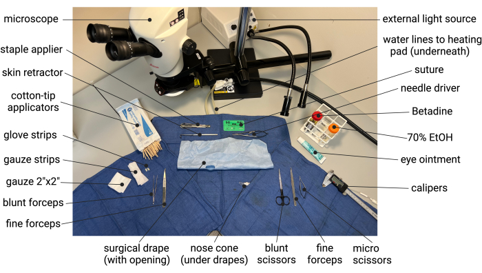
Figura 1: Ejemplo de la configuración de la cirugía estéril en preparación para el modelo murino de elastasa/BAPN de AAA. Abreviaturas: BAPN = ß-aminopropionitrilo; AAA = aneurisma de aorta abdominal. Haga clic aquí para ver una versión más grande de esta figura.
- Prepare una jaula de recuperación postoperatoria colocando una jaula limpia debajo de una lámpara de calor y coloque solución salina cerca de la lámpara para calentar a la temperatura corporal (37 °C). Asegúrese de que la lámpara de calor esté colocada de forma segura de modo que la jaula de recuperación y la solución salina estén calientes pero no superen los 37 °C. Encienda la bomba de agua para comenzar a hacer circular agua tibia a través de la almohadilla térmica.
4. Preparación animal para la cirugía
- Colocar los ratones en una cámara de inducción y anestesiarlos con isoflurano al 5% a 200 mL/min utilizando un vaporizador electrónico de bajo flujo. Mientras están anestesiados, pesar a cada ratón y administrar 0,6 mg/kg de buprenorfina ER y 20 mg/kg de carprofeno por vía subcutánea para la analgesia. Use recortadoras de pelo eléctricas para cortar el pelaje en el abdomen, desde la parte inferior del abdomen hasta la parte inferior de la apófisis xifoidea. Use una gasa o una toallita de laboratorio para cepillar el exceso de vello. Regrese a los ratones a su jaula y espere al menos 20 minutos para que la analgesia haga efecto antes de proceder con la cirugía.
- Después de al menos 20 minutos desde que se administró la analgesia, coloque al ratón en una cámara de inducción de anestesia y vuelva a administrar isoflurano al 5% a 200 mL/min utilizando un vaporizador electrónico de bajo flujo hasta que el ratón esté sedado.
- Retire el ratón sedado de la cámara de inducción y colóquelo en decúbito supino sobre el campo quirúrgico. Aplique gel para los ojos y asegure el cono de la nariz con cinta quirúrgica. Reducir el isoflurano inhalado suministrado a una tasa de mantenimiento del 1-2% a 50 mL/min. Asegure las patas delanteras y traseras del ratón con cinta quirúrgica.
- Examina la parte inferior del abdomen del ratón en busca de vejiga. Aplique suavemente presión externa a la vejiga entre los dedos pulgar, índice y medio para inducir la micción; Mientras tanto, use un trozo de gasa para absorber la orina.
NOTA: Tenga cuidado de no contaminar el campo quirúrgico. - Comience a desinfectar el abdomen aplicando un exfoliante a base de yodo o clorhexidina y alcohol al 70% con hisopos con punta de algodón. Comience en el centro del abdomen y trabaje hacia afuera con un movimiento circular 3x. Deje que el área se seque brevemente entre aplicaciones.
- Verifique la falta de una respuesta de pellizco de los dedos del pie para asegurarse de que la anestesia sea adecuada. Asegúrese de que el cono de la nariz y las extremidades estén asegurados. Coloque un paño quirúrgico sobre el ratón, con la abertura directamente sobre el abdomen preparado quirúrgicamente.
NOTA: No arrastre la cortina por el ratón para evitar una posible contaminación.
5. Inducción quirúrgica del AAA
- Entrada a la cavidad abdominal:
- Lávese las manos y use guantes quirúrgicos limpios de nitrilo o estériles. Antes de entrar en contacto con el campo quirúrgico, rocíe siempre los guantes con un 70% de EtOH y frote las manos enguantadas hasta que se sequen.
- Use pinzas romas para cubrir la piel en la línea media del abdomen. Use tijeras quirúrgicas para hacer una pequeña muesca en la piel, luego extienda la incisión longitudinalmente, de aproximadamente 2-3 cm de longitud.
- Use fórceps para levantar los músculos rectos para identificar la línea alba translúcida. Utilice unas tijeras para entrar en la cavidad abdominal a través de la línea alba, luego extiéndase a lo largo de la línea alba proximal y distalmente.
- Exposición de la aorta abdominal:
- Humedece una tira de gasa y dos bastoncillos con punta de algodón con solución salina tibia. Crea un rollo abdominal enrollando firmemente un extremo de la gasa hasta la mitad, dejando una cola generosa.
- Use un retractor de piel para retraer la pared abdominal derecha.
- Usando hisopos humedecidos con punta de algodón, realice una rotación visceral derecha-medial barriendo suavemente los intestinos delgado y grueso hacia el cuadrante superior izquierdo y visualice la aorta y la vena cava inferior (IVC). Use un rollo abdominal para retraer el intestino fuera de la vista: meta el extremo enrollado de la gasa debajo del intestino y luego lleve el extremo de la cola alrededor y fuera del cuerpo para envolver suavemente el intestino. Aplique una tensión suave a la cola de la gasa para mantener el intestino fuera del campo de visión. Ajuste el balanceo abdominal y el retractor cutáneo para obtener una visión óptima de los órganos retroperitoneales, como se muestra en la Figura 2A.
NOTA: El rollo abdominal ayuda a mantener el intestino húmedo y a protegerlo de ser dañado inadvertidamente por instrumentos quirúrgicos. Asegúrese de que la gasa permanezca húmeda durante el procedimiento para evitar que el intestino se seque. Tenga cuidado de no retraer el intestino con fuerza, ya que esto puede causar retorcimiento de la arteria mesentérica superior y la vasculatura intestinal, lo que puede causar daño isquémico. Además, al barrer inicialmente el intestino delgado, tenga cuidado con una unión delgada y translúcida entre el intestino grueso y el hígado inferior (ligamento hepatocólico), que si no se cuida, puede arrancar fácilmente la cápsula hepática y causar sangrado. Si hay tensión en este ligamento durante la retracción, divídalo bruscamente con unas tijeras.
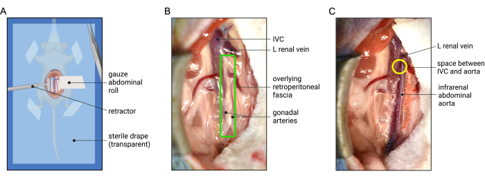
Figura 2: Representación de la retracción abdominal y la vista quirúrgica óptima para la exposición de la aorta infrarrenal del ratón. (A) La colocación de un rollo abdominal de gasa ayuda a retraer los órganos intraabdominales, mientras que un retractor opuesto ayuda a proporcionar visualización del retroperitoneo. Se coloca un paño quirúrgico estéril (transparente para mostrar la orientación del animal) sobre el animal anestesiado para ayudar a mantener la esterilidad. (B) La fascia retroperitoneal (caja verde) se superpone a la aorta anteriormente. (C) Ejemplo de la aorta infrarrenal después de la disección de la fascia retroperitoneal. El aislamiento de la aorta de la VCI se puede lograr comenzando en un espacio potencial entre la aorta y la VCI ubicado justo distal a la vena renal izquierda a medida que cruza anteriormente (círculo amarillo). Abreviatura: IVC = vena cava inferior. Haga clic aquí para ver una versión más grande de esta figura.
- Disección circunferencial y aislamiento de la aorta infrarrenal:
- Confirmar que la VCI y la aorta infrarrenal estén a la vista. Comience a exponer la aorta ingresando y dividiendo primero la fascia retroperitoneal (RP) (Figura 2B). Identificar las arterias gonadales (testiculares u ováricas) que corren paralelas a lo largo de la aorta infrarrenal anterior (Figura 2B y Figura 3). Use fórceps para dividir la fascia sin rodeos entre las arterias gonadales y continúe longitudinalmente para exponer la aorta anteriormente (Figura 2C).
NOTA: La fascia RP es una capa delgada y translúcida de tejido conectivo que contiene linfáticos y el plexo esplácnico. Es necesario diseccionar a través de la fascia RP para exponer la aorta. Sin embargo, no diseccione a través del tejido conectivo de la adventicia aórtica. Un desgarro en la adventicia (tejido conectivo blanco) expondrá el medio (aparece de color rojo brillante) y es probable que la aorta se rompa en este sitio una vez que se aplique elastasa. - A continuación, comience a aislar la aorta abdominal de la VCI. Comience esta disección en un pequeño espacio entre la VCI y la aorta, ubicado justo debajo del borde inferior de la vena renal izquierda a medida que cruza la aorta (Figura 2C). Use las puntas de las pinzas para separar suavemente las fibras del tejido conectivo entre la aorta y la VCI y continúe trabajando circunferencialmente alrededor de la aorta a este nivel.
NOTA: La VCI es de paredes muy delgadas y se adhiere estrechamente a la aorta por una fina capa de tejido conectivo fibroso. Tenga cuidado de evitar manipular la VCI o limpiarla tanto como sea posible. Diseccionar primero el lado derecho de la aorta de la VCI (antes de diseccionar el lado izquierdo de la aorta de la musculatura circundante) ayudará a que la aorta se "desprenda" de la VCI. - Continúe diseccionando sin rodeos el plano entre la aorta y la VCI, trabajando caudalmente hacia la bifurcación aórtica. Detener la disección distal una vez alcanzada la bifurcación aórtica.
NOTA: Tome precauciones adicionales al diseccionar alrededor de la arteria mesentérica inferior (IMA), que generalmente se encuentra cerca de la sección media de la aorta infrarrenal y viaja lateralmente a través de la VCI. - Una vez que el borde derecho de la aorta esté separado de la VCI, regrese proximalmente al nivel de la vena renal izquierda. Diseccionar la fascia RP del borde lateral izquierdo de la aorta, trabajando circunferencialmente hasta que la aorta esté completamente aislada. En la Figura 3 se muestra la anatomía relevante de la disección retroperitoneal.
NOTA: Tenga cuidado al diseccionar detrás de la aorta, ya que existe una gran variabilidad en la ubicación y el número de venas y arterias lumbares. Consulte la Figura 4 para obtener una referencia de las áreas con alto riesgo de sangrado con esta disección. - Inspeccione cuidadosamente que la aorta esté circunferencialmente aislada de la VCI y de la musculatura circundante tanto como sea posible, con una disección cuidadosa alrededor de los segmentos aórticos atados por el IMA y las arterias lumbares.
- Coloque una tira de guante a lo largo de los bordes derecho e izquierdo de la aorta, como se muestra en la Figura 5A. Trate de cubrir la mayor parte posible de la VCI.
- Use calibradores de mano para medir el diámetro aórtico más ancho y registre tres mediciones. Rocíe las puntas de las pinzas con 70% de EtOH antes y después de las mediciones. Evite el contacto directo de la aorta con las puntas de las pinzas para evitar la contaminación.
NOTA: También se pueden utilizar fotos con un microscopio calibrado con capacidad de cámara.
- Confirmar que la VCI y la aorta infrarrenal estén a la vista. Comience a exponer la aorta ingresando y dividiendo primero la fascia retroperitoneal (RP) (Figura 2B). Identificar las arterias gonadales (testiculares u ováricas) que corren paralelas a lo largo de la aorta infrarrenal anterior (Figura 2B y Figura 3). Use fórceps para dividir la fascia sin rodeos entre las arterias gonadales y continúe longitudinalmente para exponer la aorta anteriormente (Figura 2C).
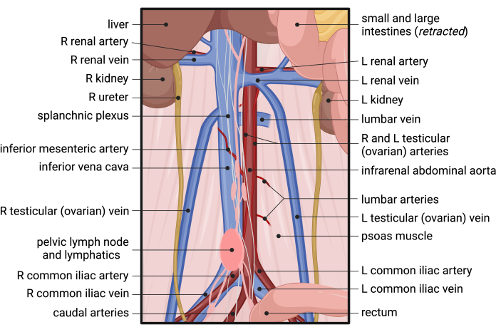
Figura 3: Anatomía del suministro de sangre a la parte inferior del abdomen, la pelvis y el retroperitoneo del ratón. Abreviaturas: R = derecha; L = izquierda. Haga clic aquí para ver una versión más grande de esta figura.
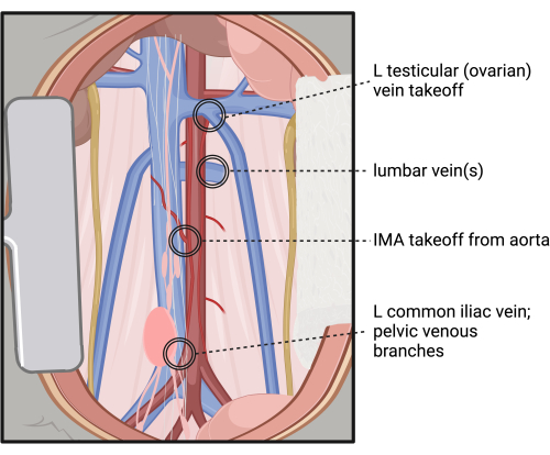
Figura 4: Sitios de alto riesgo de lesión y hemorragia durante la disección retroperitoneal y aislamiento circunferencial de la aorta infrarrenal. Abreviaturas: L = izquierda; IMA = arteria mesentérica inferior. Haga clic aquí para ver una versión más grande de esta figura.
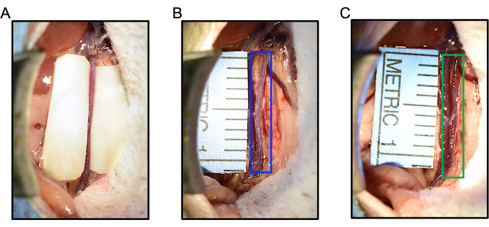
Figura 5: Respuestas intraoperatorias a la aplicación de elastasa o simulacro durante el modelo AAA murino de elastasa/BAPN. (A) Se colocan segmentos de guante a lo largo de la aorta antes de la aplicación de elastasa para ayudar a proteger la VCI y el intestino de la exposición a la elastasa mientras se mantiene la aorta empapada en elastasa (B) La aplicación de elastasa desnaturalizada no causa dilatación de la aorta (caja azul). El diámetro aórtico máximo midió 0,627 mm al inicio del estudio, luego 0,607 mm después de 5 min de elastasa desnaturalizada tópica. (C) La aplicación de elastasa provoca dilatación aórtica después de 5 min de tratamiento. En este ejemplo, la aorta (verde) se dilató de 0,607 mm a 0,953 mm, un aumento del 57% en el diámetro. Abreviaturas: BAPN = ß-aminopropionitrilo; AAA = aneurisma de aorta abdominal. Haga clic aquí para ver una versión más grande de esta figura.
- Aplicación de elastasa:
- Use un hisopo con punta de algodón para eliminar el exceso de sangre o líquido de la aorta.
- A continuación, coloque un trozo de gasa seca de 10 mm x 2 mm encima de la aorta. Utilice una pipeta para dispensar 5 μL de elastasa (o elastasa desnaturalizada de control) para saturar la gasa y la aorta. Dobla suavemente las piezas del guante alrededor de la aorta.
NOTA: Para preparar elastasa desnaturalizada para su uso en grupos simulados o de control, hierva la elastasa a 100 °C durante 30 min. - Espere 5 minutos para que la elastasa actúe sobre la aorta. Durante este período de incubación, si es necesario, libere parte de la tensión ejercida por el rodillo abdominal y el retractor cutáneo.
NOTA: Debido al efecto de lote con elastasa, alentamos a los investigadores a usar la misma botella de elastasa para todos los experimentos dentro de un estudio determinado. Con cada nuevo frasco de elastasa, recomendamos realizar una dosis-respuesta para asegurarse de que no haya un número abrumador de rupturas tempranas (antes de las 4 semanas). La duración de la aplicación de elastasa también se puede ajustar entre 4 y 6 min dependiendo de la respuesta a la elastasa. - Después de 5 minutos, restablezca la retracción intestinal y despliegue las piezas del guante. Irrigar la cavidad abdominal con 1 mL de solución salina normal estéril tibia al 0,9%, mientras se retira cuidadosamente la gasa y los trozos de guante de la aorta. Absorber la solución salina en el abdomen con una gasa de 10 cm x 10 cm. Repita la irrigación del abdomen para un total de 3 x 3 mL.
- Utilice calibradores de mano para volver a medir el diámetro aórtico más amplio después de la aplicación de elastasa y registre 3 veces. Ver Figura 5B,C para ejemplos de la dilatación aórtica al tratamiento con elastasa simulada y activa.
NOTA: Los promedios de las tres mediciones previas y posteriores a la elastasa se pueden utilizar para calcular el cambio porcentual en el diámetro aórtico con el tratamiento. Por lo general, hay una dilatación notable ~ 30-50% inmediatamente después del tratamiento con elastasa, lo que puede ayudar a garantizar que la elastasa sea funcional y que la aorta se haya tratado adecuadamente. El diámetro de la aorta no debe cambiar con la aplicación de elastasa desnaturalizada o puede ser ligeramente más pequeño (probablemente debido a un espasmo).
- Cierre de la cavidad abdominal:
- Retire con cuidado el rollo abdominal de debajo del intestino y fuera del cuerpo. Si es necesario, aplique solución salina adicional al intestino para evitar que se pegue al rollo abdominal durante la extracción. Verifique que el intestino se vea rosado y adecuadamente perfundido.
NOTA: No es necesario intentar reposicionar el intestino de nuevo a su ubicación original; Intentarlo puede correr el riesgo de torcerse el intestino o hernias internas. - Reaproxima la fascia abdominal utilizando una sutura de monofilamento no absorbible 5-0 en funcionamiento. Cierre la piel con 3-4 grapas cutáneas.
- Retire con cuidado el rollo abdominal de debajo del intestino y fuera del cuerpo. Si es necesario, aplique solución salina adicional al intestino para evitar que se pegue al rollo abdominal durante la extracción. Verifique que el intestino se vea rosado y adecuadamente perfundido.
6. Cuidados postoperatorios de los animales
- Coloque el ratón en la jaula de recuperación con una lámpara de calor. Asegúrese de que la temperatura de la jaula sea tibia, no caliente.
- Administrar un bolo de 0,5-1 mL de líquido subcutáneo con solución salina normal al 0,9%.
- Permita que el ratón se recupere por sí solo en la jaula calentada durante ~ 20 minutos hasta que esté activo según el protocolo institucional, luego regrese a una jaula de alojamiento.
- Según el protocolo institucional, administrar 20 mg/kg de carprofeno a las 24 h después de la cirugía en el día 1 del postoperatorio, y continuar diariamente durante 3 días.
7. Medición aórtica y extracción de tejidos
- Tras la eutanasia con isoflurano y la luxación cervical, reabrir la cavidad abdominal. Extienda la incisión a través del esternón para acceder al tórax. Extirpar las aurículas derechas y perfundir el ventrículo izquierdo con 10 ml de solución fría de DPBS al 1% durante 2 min. Reseca los pulmones, el hígado y el bazo.
NOTA: Tenga cuidado de no lesionar el intestino; El derrame de contenido entérico puede afectar el análisis de tejidos. - Exponga la aorta abdominal y mida el diámetro máximo de la aorta infrarrenal, como se describió anteriormente. Continúe diseccionando toda la aorta y el corazón. Una vez que el corazón y la aorta estén aislados, corte todas las ramas arteriales y las arterias ilíacas comunes, dejando intactos los segmentos cortos en la aorta. Coloque el corazón y la aorta sobre un fondo contrastado junto a una regla y una imagen.
8. Análisis de datos e informes
- Para ayudar a tener en cuenta el error humano, mida los diámetros aórticos al menos 3 veces cada uno cuando use calibradores de mano, luego informe el diámetro como el valor promedio.
- Defina el AAA como un aumento del 50% en el diámetro saludable de la aorta. Asegúrese de incluir tanto los diámetros aórticos macroscópicos como el cambio porcentual en el diámetro en los resultados del estudio.
Resultados
En este estudio se utilizaron ratones machos y hembras C57BL/6J de 22 a 24 semanas de edad. Las aortas infrarrenales se trataron con 5 μL de enzima elastasa (6,9 mg de proteína/mL, 6 unidades/mg de proteína) o elastasa desnaturalizada durante 5 min. Los ratones machos tratados con elastaso mostraron un aumento del 43,4% en el diámetro aórtico después de 5 minutos de exposición a la elastasa en comparación con los diámetros aórticos basales no tratados, mientras que las aortas femeninas tratadas aumentaron un 33,6% (P = 0,0342). Los diámetros aórticos de las mascarillas no mostraron cambios durante 5 minutos de exposición a la elastasa desnaturalizada (hombres 0,5%; mujeres -2,8%). No hubo muertes relacionadas con la cirugía entre los 12 ratones tratados y los 6 ratones simulados. Los datos del estudio de 28 días se muestran en la Tabla 1. De los ratones hembra tratados, 3 de 6 murieron por rotura de AAA; uno el día 20 del postoperatorio y dos el día 25 (Figura 6). No hubo rupturas de AAA entre los hombres tratados. Los AAA (definidos como un aumento >50% del diámetro basal de la aorta o la muerte por ruptura de AAA) se indujeron con éxito en todos los ratones tratados (12 de 12). A los 28 días, el diámetro promedio de AAA de los machos tratados fue de 2,86 ± 0,31 mm, con un cambio porcentual promedio de 257 ± 54%, mientras que los diámetros de AAA de los ratones hembra tratados sobrevivientes fueron de 3,60 ± 1,87 mm, con un cambio porcentual promedio de 417 ± 286% (Figura 7). Los ratones simulados no mostraron relativamente ningún cambio en los diámetros aórticos.
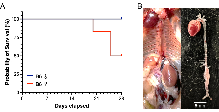
Figura 6: Supervivencia de ratones B6 machos y hembras durante un modelo de elastasa/BAPN de AAA de 28 días. (A) La ruptura de AAA ocurrió en 3 de los 6 ratones hembra tratados (un ratón a los 20 días, luego dos ratones a los 25 días), mientras que no hubo rupturas entre los 6 ratones machos tratados a los 28 días. (B) Imágenes representativas en la necropsia de un ratón hembra que murió por rotura de AAA. La rotura del AAA se manifiesta por un gran hematoma retroperitoneal (izquierda) y la presencia de un AAA infrarrenal con un defecto de la pared (derecha). Abreviaturas: BAPN = ß-aminopropionitrilo; AAA = aneurisma de aorta abdominal. Haga clic aquí para ver una versión más grande de esta figura.
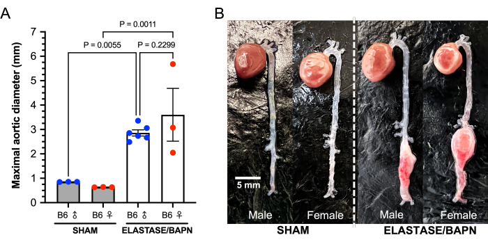
Figura 7: Diámetros aórticos máximos de elastasa/BAPN y ratones B6 machos y hembras simulados a los 28 días. (A) Los ratones tratados exhiben diámetros infrarrenales significativamente mayores a los 28 días en comparación con los simulados. (B) La combinación de elastasa y BAPN produce con éxito grandes AAA infrarrenales en ratones B6 machos y hembras. Abreviaturas: BAPN = ß-aminopropionitrilo; AAA = aneurisma de aorta abdominal. Haga clic aquí para ver una versión más grande de esta figura.
| 86 hombres Sham | 86 Mujer Sham | 86 elastaso macho/8APN | 86 elastaso hembra/8APN | |||
| Número de ratones | 3 | 3 | 6 | 6 | ||
| Edad (semanas) | 22,3 ± 0,0 | 22,7 ± 0,7 | 23,1 ± 0,2 | 23,2 ± 0,2 | ||
| Peso (g; en el momento de la cirugía) | 36,3 ± 2,5 | 23,7 ± 1,2 | 32,8 ± 1,7* | 23,7 ± 0,8 | ||
| Diámetro aórtico previo al tratamiento (mm) | 0,89 ± 0,02 | 0,75 ± 0,04 | 0,81 ± 0,07 | 0,73 ± 0,09 | ||
| Diámetro aórtico post-tratamiento (mm) | 0,90 ± 0,03 | 0,73 ± 0,01 | 1,15 ± 0,03** | 0,98 ± 0,12** | ||
| Cambio porcentual después de 5 min de tratamiento (%) | 0,5 ± 4,4 | -2,8 ± 5,3 | 43,4 ± 10,2*** | 33,6 ± 4,5*** | ||
| Incidencia de AAA (%) | 0 / 3 | 0 / 3 | 6 / 6 | 6 / 6 | ||
| El AAA se rompe a los 28 días | 0 / 3 | 0 / 3 | 0 / 6 | 3 / 6 | ||
| Supervivencia hasta 28 días | 3 / 3 | 3 / 3 | 6 / 6 | 3 / 6 | ||
| Diámetro aórtico máximo a los 28 días (mm) | 0,85 ± 0,01 | 0,64 ± 0,01 | 2,86 ± 0,31* | 3,60 ± 1,87** | ||
| Cambio porcentual del diámetro aórtico a los 28 días (%) | -4 ± 2 | -16 ± 2 | 257 ± 54* | 417 ± 286** | ||
Tabla 1: Resultados de un modelo de 28 días del modelo murino de elastasa/BAPN de AAA. Los datos se ± media de DE. *P<0.05, **P<0.005, ***P<0.0001 en comparación con simulacros del mismo sexo a través de la prueba de Fischer ANOVA de un factor. Abreviaturas: BAPN = ß-aminopropionitrilo; AAA = aneurisma de aorta abdominal.
Discusión
Comprender la compleja fisiopatología del AAA es fundamental para mejorar el tratamiento de la enfermedad por aneurisma aórtico. Si bien se desarrollan activamente nuevas estrategias para mejorar los resultados quirúrgicos, los AAA siguen siendo prevalentes en nuestra sociedad envejecida y la ruptura de un aneurisma sigue siendo una de las principales causas de muerte enlos Estados Unidos. Por lo tanto, las necesidades insatisfechas en las estrategias de detección, prevención y tratamiento de AAA justifican una mayor investigación fundamental sobre el aneurisma11.
Los modelos animales que recapitulan de manera precisa y eficiente las características y comportamientos de los AAA humanos son esenciales para los estudios mecanicistas de la fisiopatología de los aneurismas y la identificación de posibles objetivos terapéuticos. Si bien los modelos animales actuales pueden imitar los aspectos principales de los cambios aneurismales que ocurren en las enfermedades humanas, ningún modelo único representa completamente la verdadera complejidad de los AAA humanos. En la actualidad, los ratones son la especie más ampliamente aceptada para el modelado animal de AAA. Los investigadores deben considerar las diversas fortalezas y debilidades de cada modelo murino para su estudio particular de aneurisma, como los descritos por expertos en las revisiones de Daugherty et al. y Busch et al.12,13.
El uso de elastasa para inducir AAA en roedores fue descrito por primera vez por Anidjar et al. en 1990:14. La perfusión de la aorta con elastasa pancreática porcina mediante una bomba de jeringa crea una dilatación inicial de aproximadamente entre el 50% y el 70%, y los segmentos dilatados muestran favorablemente características patológicas similares a las de los AAA humanos, como la degeneración medial y la inflamación adventicia. Sin embargo, el modelo clásico de perfusión es posiblemente el modelo de aneurisma más desafiante desde el punto de vista técnico, y los aneurismas que generalmente se forman en la segunda semana comienzan a resolverse gradualmente a partir de entonces. Bhamidipati et al. en 2012 demostraron que la aplicación adventicial de elastasa también podría inducir con éxito aneurismas similares que son más reproducibles en tamaño15. Un modelo mucho menos desafiante, el modelo de elastasa tópica se adoptó ampliamente en la investigación de aneurismas. La metodología adicional y las ventajas del modelo de elastasa tópica se discuten en el artículo de métodos de Xue y colegas16.
El modelo elastasa/BAPN del AAA murino fue desarrollado por Lu y sus colegas en 20178. La introducción del agua potable con un 0,2% de BAPN mejoró muchas de las críticas al modelo clásico de elastasa tópica, que ahora produce aneurismas que se expanden continuamente hasta el punto de la ruptura del AAA. En su estudio de 2017, demostraron que los ratones del grupo tratado con elastasa/BAPN tenían tasas de formación de AAA significativamente más altas en comparación con el grupo de elastasa (93% frente a 65%, P < 0,01), que también eran AAA en etapas más avanzadas. Durante un período de estudio de 100 días, los AAA en el grupo de elastasa/BAPN continuaron dilatándose hasta un diámetro basal del >800% y formaron trombo intraluminal (53,8%), y el 46,2% se rompieron espontáneamente antes del final del experimento. Este modelo ha permitido a los investigadores investigar factores que pueden afectar la progresión y la estabilidad del aneurisma a lo largo del tiempo.
Berman et al. exploraron más a fondo el modelo de elastasa/BAPN variando la concentración de elastasa tópica, la duración del estudio, el momento de la administración de BAPN y el impacto del sexo del animal9. El tratamiento con 5 μL de elastasa de mayor concentración (5 mg/mL o 10 mg/mL) produjo aneurismas más grandes que 2,5 mg/mL durante 56 días. La prevalencia de la formación de trombos intraluminales también dependió de la concentración de elastasa, que se produjo en el 28,6% de los ratones tratados con 5 mg/mL y en el 62,5% de los ratones tratados con 10 mg/mL. También demostraron que el modelo de elastasa/BAPN podía inducir aneurismas en ratones hembra. Aunque solo se estudiaron unos pocos ratones hembra (n = 5), encontraron que los aneurismas en las hembras eran más propensos a romperse (2 de 5 ratones) y eran significativamente más grandes que los AAA masculinos a los 56 días.
En este artículo, nuestro objetivo es proporcionar un método para abordar una de las mayores limitaciones del modelado quirúrgico, que es la variación en el procedimiento quirúrgico. Sin un consenso claro sobre el grado de disección y el área de la aorta tratada con elastasa, los resultados de este modelo podrían variar drásticamente entre animales, investigadores e instituciones. Hemos observado numerosas variaciones anatómicas entre ratones, incluyendo el número y tamaño de las arterias y venas lumbares, y la localización del IMA, el despegue de la vena gonadal izquierda, entre otras, que pueden ser limitantes cuando se intenta tratar solo una porción o segmento específico de la aorta infrarrenal. Aquí, demostramos que la disección circunferencial de toda la longitud de la aorta infrarrenal desde la arteria renal izquierda proximalmente a la bifurcación aórtica distalmente ayuda a proporcionar grados reproducibles de exposición aórtica a pesar de las diferencias anatómicas, al tiempo que aumenta el éxito de la inducción del aneurisma y proporciona límites claros para el operador. Además, el tamaño y la posición más anterior de la VCI tienden a cubrir la mayor parte de la aorta, lo que puede afectar la cantidad de aorta tratada si no se aísla de la VCI. Si bien es necesario extirpar la fascia retroperitoneal para exponer la aorta, es importante no diseccionar completamente el tejido conectivo de la adventicia de la aorta y exponer ninguna de las capas medias, ya que esto generalmente resulta en una ruptura durante el período de incubación de elastasa de 5 minutos. Esto podría servir como un control interno adicional al grado de disección con este modelo, pero puede ser una curva de aprendizaje frustrante al adoptar este modelo. Además, los operadores aprenderán las áreas de mayor riesgo (Figura 4) que pueden lesionarse fácilmente durante la cirugía y provocar una hemorragia incontrolable.
Si bien es importante que los pasos del procedimiento de este modelo sean coherentes, la duración del estudio y el momento de la ecografía de intervalo pueden variar según el objetivo de la investigación. La dilatación aórtica comienza inmediatamente con la aplicación de elastasa, sin embargo, los estudios que utilizan este modelo suelen seguir a los ratones durante 28 días después de la cirugía7, como en este experimento de ejemplo. Se debe considerar la extensión de la duración del estudio cuando se estudian AAA avanzados, crecimiento a largo plazo, formación de trombos intraluminales o ruptura.
Las medidas perioperatorias adicionales, como mantener la temperatura corporal y el estado de hidratación de los animales, pueden ayudar a mejorar la supervivencia de los animales a este procedimiento invasivo. El uso de una almohadilla térmica durante la cirugía y la colocación en una jaula de recuperación tibia pueden ayudar a evitar la hipotermia. La solución salina debe calentarse antes de usarla para irrigar la cavidad abdominal. Un bolo de líquido subcutáneo inmediatamente después de la cirugía puede explicar pérdidas insensibles de líquido durante la operación y ayudar al animal a mantener una hidratación adecuada durante la fase de recuperación inmediata. Con un manejo cuidadoso de los tejidos y un enfoque metódico consistente, el modelo de elastasa/BAPN puede ser realizado por un operador experimentado entre 30 min y 45 min por ratón y producir de manera confiable AAA con complicaciones perioperatorias muy bajas.
Nuestros resultados demuestran que la combinación de BAPN además de la disección circunferencial de la aorta infrarrenal antes de la aplicación de elastasa produce AAAs grandes y en continua expansión, con diámetros mayores e incidencia de ruptura en períodos más cortos. En este experimento, los AAA se indujeron con éxito en todos los ratones machos (6 de 6) y hembras (6 de 6) tratados con elastasa activa. La exposición a la elastasa durante 5 minutos resultó en un aumento inmediato del diámetro aórtico en aproximadamente un 30-40%, lo que es útil para confirmar la aplicación exitosa y consistente de elastasa entre los grupos de tratamiento. Al igual que Berman et al., hemos demostrado que este modelo puede inducir AAAs en ratones hembra, que también tienen una mayor respuesta a la ruptura que los machos. La mitad de los ratones hembra (3 de 6) se rompieron dentro de los 28 días, en comparación con 0 de 6 de los machos, sin embargo, los ratones hembra pesan menos que los machos. Los ratones machos demostraron un aumento en el diámetro de AAA en un 257% en comparación con el -4% de los controles masculinos, mientras que las hembras sobrevivientes mostraron un aumento del diámetro del 417%, en comparación con el -16% de las hembras de control. Los diámetros aórticos no fueron significativamente diferentes entre los ratones macho y hembra tratados a los 28 días debido al mayor número de rupturas en el grupo de hembras. Especulamos que los ratones simulados exhiben diámetros aórticos más pequeños al final del estudio, ya que la aorta tiende a dilatarse ligeramente durante la disección inicial y luego forma tejido cicatricial a los 28 días.
El modelo elastasa/BAPN posee ciertas limitaciones. La disección circunferencial de la aorta requiere habilidades quirúrgicas finas, pero ayuda a mejorar la replicabilidad y el grado de inducción del aneurisma. Al igual que el modelo de elastasa tópica, también hay un efecto de lote en la actividad de la enzima elastasa, que, como se mencionó anteriormente, es importante utilizar la misma botella de elastasa para todos los animales en un experimento determinado. Si bien la incidencia de trombo y ruptura intraluminal del AAA aumenta con el tiempo y la gravedad del aneurisma, estos no están garantizados ni son totalmente predecibles en este modelo.
En resumen, el modelo de elastasa/BAPN produce AAAs infrarrenales grandes y verdaderos en ratones machos y hembras, que se expanden progresivamente con el tiempo, forman trombos intraluminales y son capaces de romperse. Estas fortalezas de este modelo murino ayudan a recapitular mejor algunos de los comportamientos y características de los aneurismas en humanos. Aunque técnicamente difícil, la disección cuidadosa y completa de la aorta puede aumentar la respuesta aneurismática. En la actualidad, el método elastasa/BAPN es un modelo avanzado para el estudio de los aneurismas de aorta abdominal infrarrenal.
Divulgaciones
Los autores de este manuscrito no tienen conflictos de intereses que declarar.
Agradecimientos
Esta investigación contó con el apoyo del Instituto Nacional del Corazón, los Pulmones y la Sangre (NHLBI) de los Institutos Nacionales de Salud (NIH) bajo el número 1R01HL149404-01A1 (BL), y el Premio Ruth L. Kirschstein del Servicio Nacional de Investigación T32 HL 007936 al Centro de Investigación Cardiovascular (JB) de la Universidad de Wisconsin-Madison. Las figuras fueron creadas o editadas con Biorender.com. El análisis estadístico se realizó con el software GraphPad Prism 10.
Materiales
| Name | Company | Catalog Number | Comments |
| 0.5 L induction chamber | Kent Scientific Corporation | SOMNO-0530XXS | anesthesia induction chamber |
| 0.9% sodicum chloride injection, USP, 20 mL | Hospira | NDC 0409-4888-03 | normal saline |
| 3 mL syringe Luer-Lok Tip with BD PrecisionGlide Needle 22 G x 3/4 | BD | REF 309569 | syringe, 22 G needle |
| 3-Aminopropionitrile Fumarate | TCI | A0796 | BAPN |
| 3-Aminopropionitrile Fumarate salt | Sigma-Aldrich | A3134-25G | BAPN |
| Avant Delux gauze sponges, 2" x 2" 4-Ply | Medline | NON26224 | gauze sponges |
| Balding clipper | Whal Clipper Corporation | 8110 | hair clippers |
| betadine surgical scrub (povidone-iodine, 7.5%) | Avrio | NCD 67618-154-16 | betadine surgical scrub |
| blunt forceps | ROBOZ | RS-5130 | blunt forceps |
| Buprenorphine ER-lab | ZooPharm | BERLAB0.5 | buprenorphine |
| carprofen | Norbrook | NDC 55529-131-11 | carprofen |
| CASTROVIEJO 5.75" straight with lock | ROBOZ | RS-6412 | Castroviejo needle driver |
| cotton tipped wood applicators, 6" | Dynarex | No. 4302 | cotton tipped wood applicators |
| DESMARRES 5.5' rectractor | ROBOZ | RS-6672 | skin rectractor |
| digital caliper, 0-150 mm | World Precision Instruments | 501601 | digital caliper |
| DPBS (1x) | Gibco | 14190-144 | DPBS |
| Elastase from porcine pancrease Type I | Sigma-Aldrich | E1250-10MG | elastase >4.0 units/mg protein |
| Ethanol 200 proof | Decon Labs, Inc | 2701 | ethanol diluted to 70% |
| eye lube | Optixcare | 14716 | eye lube |
| Germinator 500 dry sterilizer | CellPoint Scientific, Inc | 5-1450 | dry bead sterilizer |
| heat therapy mat | Adroit Medical Systems | V016 | heat therapy mat |
| heat therapy pump | Adroit Medical Systems | HTP-1500 | heat therapy pump |
| isoflurane, USP | Akorn Animal Health | NCD 59399-106-01 | isoflurane |
| L-10 pipette | Rainin | LTS 0.5-10 uL | pipette |
| Low profile anesthesia mask, small | Kent Scientific Corporation | SOMNO-0801 | anesthesia nose cone |
| micro dissector scissors | ROBOZ | RS-5619 | micro dissector scissors |
| microscope | Leica | S9i | microscope |
| Nii-LED high intensity LED illuminatorLED exertnal light | Nikon Instruments, Inc | 83359 NII-LED | external dissection light |
| nylon 5-0 monofilament, black non-absorbable suture | Oasis | MV-661-V | 5-0 nylon suture |
| polyisoprene surgical gloves, GAMMEX Non-Latex PI Micro, size 7.5 | Ansell | 20685975 | non-latex surgical gloves |
| Reflex 7 mm stainless steel wound clips | CellPoint Scientific, Inc | 203-1000 | wound clips |
| scale | Ohaus | Compass CR2200 | scale |
| SomnofFlo Accessory Kit | Kent Scientific Corporation | 10-8000-71 | tubing for electronic vaporizer |
| SomnoFlo electronic vaporizer | Kent Scientific Corporation | SF2992 | low-flow electronic vaporizer |
| SomnoPath Flow Diverter | Kent Scientific Corporation | SP1016 | flow diverter for electronic vaporizer |
| SS/45 sharp forceps | ROBOZ | RS-4941 | sharp forceps |
| surgical scissors | ROBOZ | RS-6010SC | surgical scissors |
| vessel forceps | Dumont | VES 0.35 | vessel forceps |
Referencias
- Kent, K. C. Clinical practice. Abdominal aortic aneurysms. New Engl J Med. 371 (22), 2101-2208 (2014).
- Wanhainen, A., et al. European Society for Vascular Surgery Guidelines on the management of aorto-iliac abdominal aortic aneurysms. Eur J Vasc Endocasc Surg. 57 (1), 8-93 (2019).
- Shimizu, K., Mitchell, R. N., Libby, P. Inflammation and cellular immune responses in abdominal aortic aneurysms. Arterioscler Thromb Vasc Biol. 26 (5), 987-994 (2006).
- Shen, Y. H., et al. Aortic aneurysms and dissections series. ArteriosclerThromb Vasc Biol. 40 (3), e37-e46 (2020).
- Stanley, J. C., Veith, F., Wakefield, T. W. Current Therapy in Vascular and Endovascular Surgery E-Book. Elsevier Health Sciences. , (2014).
- Morgan, S., et al. Identifying novel mechanisms of abdominal aortic aneurysm via unbiased proteomics and systems biology. Front Cardiovasc Med. 9, 889994 (2022).
- Yin, L., Kent, E. W., Wang, B. Progress in murine models of ruptured abdominal aortic aneurysm. Front Cardiovasc Med. 9, 950018 (2022).
- Lu, G., et al. A novel chronic advanced stage abdominal aortic aneurysm murine model. J Vasc Surg. 66 (1), 232-242 (2017).
- Berman, A. G., et al. Experimental aortic aneurysm severity and growth depend on topical elastase concentration and lysyl oxidase inhibition. Sci Rep. 12 (1), 99 (2022).
- Deaths, percent of total deaths, and death rates for the 15 leading causes of death in 5-year age groups, by race, and sex. Centers for Disease Control and Prevention Available from: https://www.cdc.gov/nchs/nvss/mortality/lcwk1.htm (2015)
- Dansey, K. D., et al. Epidemiology of endovascular and open repair for abdominal aortic aneurysms in the United States from 2004 to 2015 and implications for screening. J Vasc Surg. 74 (2), 414-424 (2021).
- Daugherty, A., Cassis, L. A. Mouse models of abdominal aortic aneurysms. Arterioscler Thromb Vasc Biol. 24 (3), 429-434 (2004).
- Busch, A., et al. Translating mouse models of abdominal aortic aneurysm to the translational needs of vascular surgery. JVS Vasc Sci. 2, 219-234 (2021).
- Anidjar, S., et al. Elastase-induced experimental aneurysms in rats. Circulation. 82 (3), 973-981 (1990).
- Bhamidipati, C. M., et al. Development of a novel murine model of aortic aneurysms using peri-adventitial elastase. Surgery. 152 (2), 238-246 (2012).
- Xue, C., Zhao, G., Zhao, Y., Chen, Y. E., Zhang, J. Mouse abdominal aortic aneurysm model induced by perivascular application of elastase. J Vis Exp. (180), e63608 (2022).
Reimpresiones y Permisos
Solicitar permiso para reutilizar el texto o las figuras de este JoVE artículos
Solicitar permisoExplorar más artículos
This article has been published
Video Coming Soon
ACERCA DE JoVE
Copyright © 2025 MyJoVE Corporation. Todos los derechos reservados