Medición de las diferencias de materia gris con Morfometría basada en Voxel: el cerebro Musical
Visión general
Fuente: Laboratorios de Jonas T. Kaplan y Sarah I. Gimbel, University of Southern California
Experiencia moldea el cerebro. Bien se entiende que nuestros cerebros son diferentes como resultado de aprendizaje. Si bien muchos cambios relacionados con la experiencia se manifiestan en el nivel microscópico, por ejemplo por neuroquímicos ajustes en el comportamiento de las neuronas individuales, podemos también examinamos cambios anatómicos de la estructura del cerebro en un nivel macroscópico. Un ejemplo famoso de este tipo de cambio viene desde el caso de los taxistas de Londres, que junto con aprender las complejas rutas de la ciudad muestran mayor volumen en el hipocampo, una estructura cerebral conocida por desempeñar un papel en la memoria de navegación. 1
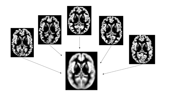
Muchos métodos tradicionales de examinar la anatomía del cerebro requieren meticuloso seguimiento de regiones anatómicas de interés para medir su tamaño. Sin embargo, usando técnicas modernas de neuroimagen, podemos ahora comparar la anatomía de los cerebros a través de grupos de personas que utilizan algoritmos automatizados. Aunque estas técnicas no beneficiarse de los sofisticados conocimientos que http://www.ehu.es/Lance humano puede aportar a la tarea, son rápidas y sensibles a las diferencias muy pequeñas en la anatomía. En una imagen de resonancia magnética estructural del cerebro, la intensidad de cada pixel volumétrico, o voxel, se refiere a la densidad de la materia gris en esa región. Por ejemplo, en una exploración de T1-weighted MRI voxels muy brillantes se encuentran en lugares donde hay haces de fibras de materia blanca, mientras que los vóxeles más oscuros corresponden a materia gris, donde residen los cuerpos celulares de las neuronas. La técnica de cuantificación y comparación de la estructura cerebral en forma de voxel por voxel se denomina Morfometría basada en voxel o VBM. 2 en VBM, primero registramos todos los cerebros a un espacio común, alisar cualquier bruto diferencias en anatomía. Luego comparamos los valores de intensidad de los voxels para identificar escala localizada, pequeñas diferencias en la densidad de materia gris.
En este experimento demostramos la técnica VBM comparando los cerebros de los músicos con los no músicos. Músicos participan en entrenamiento intenso motoric, visual y acústica. Hay evidencia de múltiples fuentes que los cerebros de personas que han pasado a través de la formación musical son funcionalmente y estructurales distintas de las que no. Aquí, seguimos Gaser y Shlaug3 y Bermúdez et al. 4 uso de VBM para identificar estas diferencias estructurales en el cerebro de los músicos.
Procedimiento
1. reclutar 40 músicos y no 40 músicos.
- Músicos deben tener por lo menos 10 años de entrenamiento musical formal. Entrenamiento con cualquier instrumento musical es aceptable. Músicos deben también ser activamente practicar su instrumento al menos una hr/día.
- Sujetos de control deben tener poco entrenamiento formal en tocar un instrumento musical.
- Todos los participantes deben ser diestros.
- Todos los participantes deben no tienen antecedentes de trastornos neurológicos, psiquiátr
Resultados
El análisis VBM reveló aumentos significativos de la localizada en la densidad de materia gris en el cerebro de los músicos en comparación con controles no músico. Estas diferencias en los lóbulos temporales superiores a ambos lados. El racimo más grande, más importante fue en el lado derecho e incluye la parte posterior de la circunvolución de Heschl (figura 2). Circunvolución de Heschl es el lugar de la corteza auditiva primaria y las cortezas circundantes est...
Aplicación y resumen
La técnica VBM tiene el potencial de demostrar diferencias localizadas en la materia gris entre grupos de personas, o en asociación con una medición que varía de un grupo de personas. Además de encontrar las diferencias estructurales que se relacionan con diferentes formas de entrenamiento, esta técnica puede revelar diferencias anatómicas que se asocian a distintas condiciones neuropsicológicas como depresión y esquizofrenia, dislexia5 ,6 . 7
Es impor...
Referencias
- Maguire, E.A., et al. Navigation-related structural change in the hippocampi of taxi drivers. Proc Natl Acad Sci U S A 97, 4398-4403 (2000).
- Ashburner, J. & Friston, K.J. Voxel-based morphometry--the methods. Neuroimage 11, 805-821 (2000).
- Gaser, C. & Schlaug, G. Brain structures differ between musicians and non-musicians. J Neurosci 23, 9240-9245 (2003).
- Bermudez, P., Lerch, J.P., Evans, A.C. & Zatorre, R.J. Neuroanatomical correlates of musicianship as revealed by cortical thickness and voxel-based morphometry. Cereb Cortex 19, 1583-1596 (2009).
- Bora, E., Fornito, A., Pantelis, C. & Yucel, M. Gray matter abnormalities in Major Depressive Disorder: a meta-analysis of voxel based morphometry studies. J Affect Disord 138, 9-18 (2012).
- Richlan, F., Kronbichler, M. & Wimmer, H. Structural abnormalities in the dyslexic brain: a meta-analysis of voxel-based morphometry studies. Hum Brain Mapp 34, 3055-3065 (2013).
- Zhang, T. & Davatzikos, C. Optimally-Discriminative Voxel-Based Morphometry significantly increases the ability to detect group differences in schizophrenia, mild cognitive impairment, and Alzheimer's disease. Neuroimage 79, 94-110 (2013).
Saltar a...
Vídeos de esta colección:

Now Playing
Medición de las diferencias de materia gris con Morfometría basada en Voxel: el cerebro Musical
Neuropsychology
17.4K Vistas

El cerebro dividido
Neuropsychology
68.5K Vistas

Mapas de motor
Neuropsychology
27.7K Vistas

Perspectivas de la neuropsicología
Neuropsychology
12.2K Vistas

Toma de decisiones y la Iowa Gambling Task
Neuropsychology
33.0K Vistas

Función ejecutiva en el trastorno del espectro autista
Neuropsychology
18.0K Vistas

Amnesia Anterógrada
Neuropsychology
30.5K Vistas

Correlatos fisiológicos de reconocimiento de la emoción
Neuropsychology
16.4K Vistas

Potenciales acontecimiento-relacionados y la tarea de Oddball
Neuropsychology
27.6K Vistas

Idioma: La N400 en incongruencia semántica
Neuropsychology
19.7K Vistas

Aprendizaje y la memoria: la tarea de recordar-sabe
Neuropsychology
17.3K Vistas

Descodificación de imágenes auditivas con análisis Multivoxel
Neuropsychology
6.5K Vistas

Atención visual: fMRI Control atencional basado en la investigación del objeto
Neuropsychology
42.2K Vistas

Utilizando imágenes de Tensor de difusión en la lesión cerebral traumática
Neuropsychology
16.9K Vistas

Uso de TMS para medir la excitabilidad motora durante la observación de la acción
Neuropsychology
10.3K Vistas
ACERCA DE JoVE
Copyright © 2025 MyJoVE Corporation. Todos los derechos reservados
