Physical Properties Of Minerals I: Crystals and Cleavage
Source: Laboratory of Alan Lester - University of Colorado Boulder
The physical properties of minerals comprise various measurable and discernible attributes, including color, streak, magnetic properties, hardness, crystal growth form, and crystal cleavage. Each of these properties are mineral-specific, and they are fundamentally related to a particular mineral’s chemical make-up and atomic structure.
This experiment examines two properties that stem primarily from symmetric repetition of fundamental, structural atomic groupings, called unit cells, within a crystal lattice, a crystal growth form, and crystal cleavage.
Crystal growth form is the macroscopic expression of atomic-level symmetry, generated by the natural growth process of adding unit cells (the molecular building blocks of minerals) to a growing crystal lattice. Zones of rapid unit-cell-addition become the edges between the planar surfaces, i.e. faces, of the crystal.
It is important to recognize that rocks are aggregates of mineral grains. Most rocks are polymineralic (multiple kinds of mineral grains) but some are effectively monomineralic (composed of a single mineral). Because rocks are combinations of minerals, rocks are not referred to as having crystal form. In some cases, geologists refer to rocks as having a general cleavage, but here the term is simply used to refer to repetitive breaking surfaces and is not a reflection of atomic crystal structure. So, in general, the terms crystal form and crystal cleavage are used in reference to mineral samples and not rock samples.
All minerals possess physical properties, but specific and easily recognizable features associated with the properties are not always expressed in an individual crystal. For example, quartz crystals have a characteristic hexagonal shape, but if crystal growth occurs in an environment where other minerals block or impinge the natural growth shape (which is commonly the case in most rocks) then the hexagonal shape does not form. So, with this in mind, it’s important to carefully select a suitable group of samples for either crystal growth or crystal cleavage analysis, as not all samples show these key features.
Furthermore, although crystal cleavage is relatively easy to test — by breaking a sample with a hammer — different minerals demonstrate a range of cleavage quality, such that the planar surfaces generated by breaking may be ragged and rough (termed “poor-cleavage”) or extremely smooth (termed “good-” or “excellent- cleavage”). In some cases (e.g. quartz), crystallographic bond strengths are uniform in all directions, and this results in a mineral with a lack of recognizable cleavage planes.
1. Establish a Group of Mineral Samples
- Include as many of the following as possible: quartz, halite, calcite, garnet, biotite, and/or muscovite. Some are chosen for crystal growth features and others for crystal cleavage features.
2. Observe and Analyze Crystal Form
- Place a sample onto the observation surface.
- Rotate in order to observe all sides. Look for crystal faces, crystal edges (lines where faces meet), and crystal vertices (points where edges meet).
- Where possible, measure the interfacial angles using the goniometer. This is done by simply laying one side of the goniometer on a particular crystal face, the other side of the goniometer on an adjoining face, and then reading the angle.
- Compare to the set of characteristic crystalline polyhedra.
- Repeat steps 2.1 – 2.4 for quartz (note hexagonal dipyramidal form (Figure 1)), calcite (note scalenohedron form (Figure 2)), halite (note cubic crystal form (Figure 3)), garnet (note dodecahedron form (Figure 4)), and biotite (note pseudo-hexagonal form (Figure 5)).
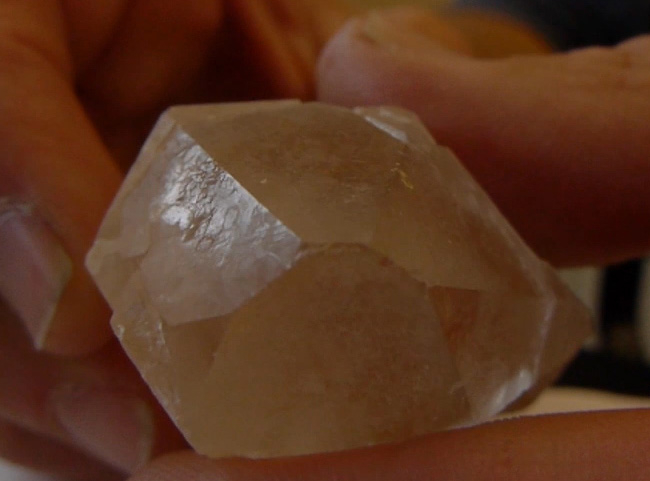
Figure 1. Quartz displaying hexagonal dipyramidal form.
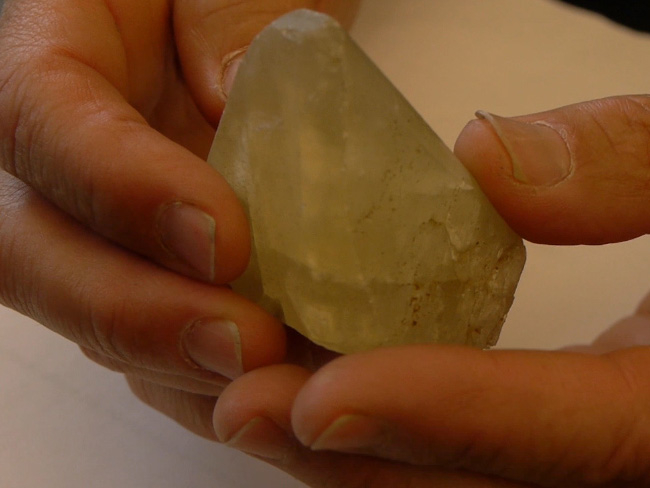
Figure 2. Calcite displaying scalenohedron form. Note how several crystal faces intersect to form crystal edges and the combination of edges forms points known as “vertices.” Symmetric crystal growth forms are generated by repetition of fundamental atomic structures (unit cells) within the crystal lattice. In this case, calcite crystal growth generates the specific polyhedron known as a scalenohedron.
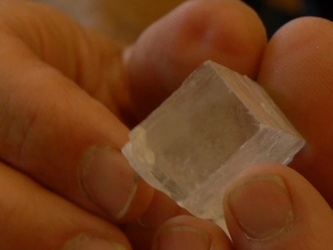
Figure 3. Halite displaying cubic crystal form.
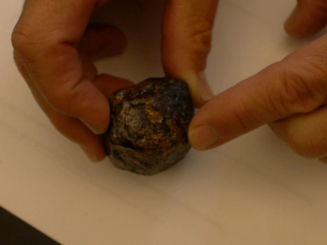
Figure 4. Garnet displaying dodecahedron form.
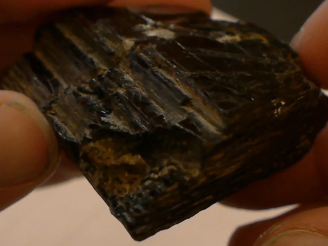
Figure 5. Biotite displaying pseudo-hexagonal form.
3. Observe and Analyze Cleavage
- Put on eye protection.
- Place a piece of quartz on breaking surface.
- Using a hammer, break the piece of quartz in half.
- Using a hand lens, observe broken piece of quartz for cleavage surfaces. Notice that quartz has none. Quartz exhibits conchoidal fracture, but no well-defined cleavage surfaces (Figure 6). This is a consequence of the fact that the unit cells in the quartz crystal lattice (SiO4 groups, called silica tetrahedral) have comparably equal bond strengths in all directions. This uniformity of bond strengths results in a crystal with no preferred breaking planes.
- Repeat steps 3.2 – 3.4 for calcite (should display rhombohedral cleavage (Figure 7)), halite (should display cubic cleavage (Figure 8)), biotite, and/or muscovite (should each display planar cleavage (Figure 9)).
- Use a hand lens to evaluate different cleavage qualities. Cleavage can occur at a variety of levels. When there is a dramatic difference in bond strengths in a particular orientation, such as between sheets of SiO4 groupings in the case of mica, a nearly perfect cleavage is generated between these sheets. As noted above, quartz exhibits a nearly total lack of cleavage. In between these extremes (of perfect cleavage and lack of cleavage), there are minerals that have good cleavage (e.g. feldspar) and poor cleavage (certain faces on amphibole crystals).
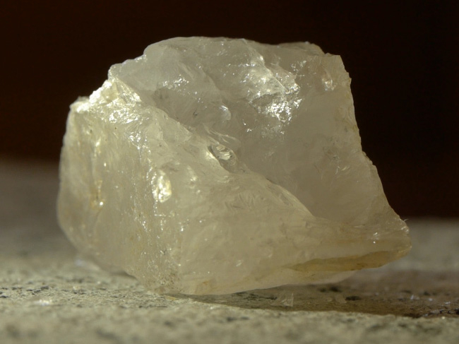
Figure 6. Quartz displaying conchoidal fracture, without cleavage surfaces.
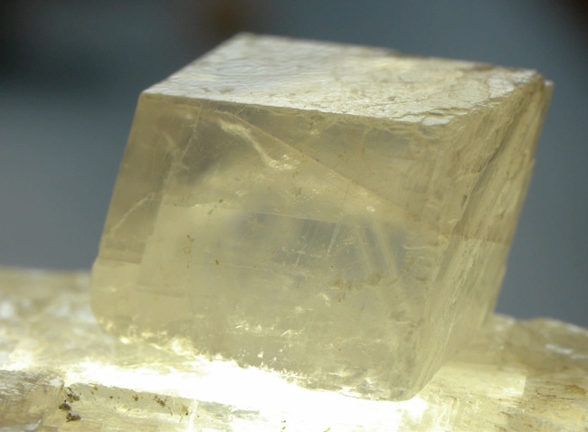
Figure 7. Calcite displaying rhombohedral cleavage. Symmetric breaking and fracture surfaces are generated by zones of relative weakness in atomic bonding within the crystal lattice. Calcite cleavage results in the specific polyhedron known as rhombohedron.
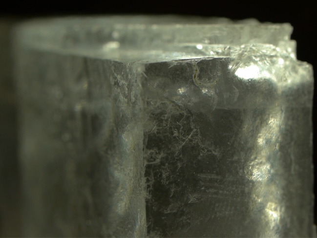
Figure 8. Halite displaying cubic cleavage.
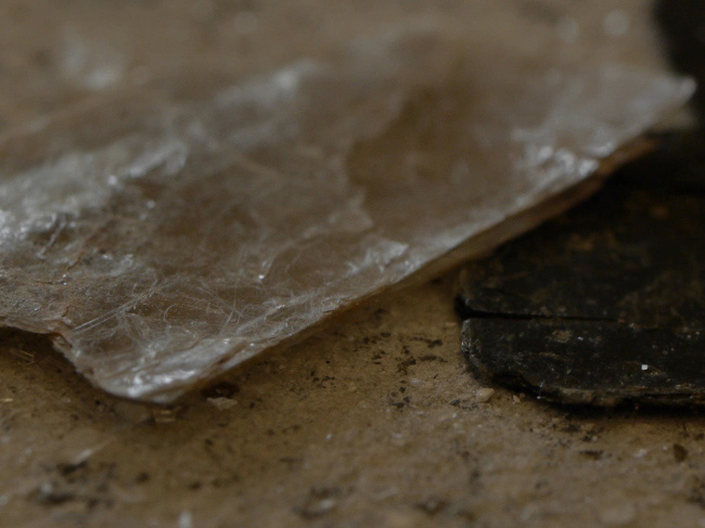
Figure 9. Biotite displaying planar cleavage.
Historically, evaluating the physical properties of minerals has been a key first step in mineral identification. Even today, when lacking microscopic and modern analytical instrumentation (e.g. petrographic microscopy, x-ray diffraction, x-ray fluorescence, and electron microprobe techniques), observed physical properties are still quite useful as diagnostic tools for mineral identification. This is particularly the case in field geologic studies.
Evaluating and observing the physical properties of minerals is an excellent means to demonstrate the critical dependence of macroscopic features on atomic-level structure and arrangement.
The key physical properties of minerals are not always expressed in specific samples. Therefore, actually being able to recognize and use these properties as diagnostic tools requires a combination of science, experience, and craft. Often, the geologist must utilize a hand lens to evaluate relatively small mineral crystals or grains within the matrix of a larger rock. In such cases, it can become a distinct challenge to identify the useful aspects of crystal form and crystal cleavage.
In an academic or teaching setting, the evaluation of minerals via hand sample analysis is an exercise that demonstrates how repetitive patterns and characteristics are imposed by the physical chemistry of natural materials. In other words, for any specific mineral, there are certain crystallographic features (e.g. crystal morphology) and physical properties (e.g. color, hardness, streak) that are imposed by chemical composition and atomic structure.
In the realm of mineral resources and exploration geology, the identification of minerals via hand sample is a key component of fieldwork, aimed at locating potential ores and economically useful deposits. For example, the identification of various metal sulfides (pyrite, sphalerite, galena) in association with hydrothermal iron oxy-hydroxides (hematite, goethite, limonite) can be indicative of potential Au- and Ag-rich veins and regions.
In the context of historical geology (deciphering the deep temporal history of a region), mineral identification can set the stage for interpretations of ancient conditions. For example, certain metamorphic minerals (e.g. the Al2SiO5 polymorphs, kyanite, andalusite, and sillimanite) are markers of particular pressure and temperature conditions in the ancient crust.
Passer à...
Vidéos de cette collection:

Now Playing
Physical Properties Of Minerals I: Crystals and Cleavage
Earth Science
51.6K Vues

Détermination de l'orientation spatiale des couches rocheuses avec la boussole Brunton
Earth Science
25.4K Vues

Utilisation de cartes topographiques pour créer des profils topographiques
Earth Science
32.0K Vues

Faire une coupe géologique
Earth Science
46.9K Vues

Propriétés physiques des minéraux II : analyse polyminéralique
Earth Science
38.0K Vues

Roche ignée volcanique
Earth Science
39.6K Vues

Roche ignée intrusive
Earth Science
32.3K Vues

Un aperçu de l'analyse des biomarqueurs bGDGT pour la paléoclimatologie
Earth Science
5.4K Vues

Un aperçu de l'analyse des biomarqueurs d'alcénones pour la paléothermométrie
Earth Science
7.2K Vues

Extraction par sonication de biomarqueurs lipidiques de sédiments
Earth Science
8.6K Vues

Utilisation de l'extracteur de Soxhlet pour isoler des biomarqueurs lipidiques à partir de sédiments
Earth Science
18.4K Vues

Extraction de biomarqueurs à partir de sédiments - Extraction accélérée par solvant
Earth Science
8.7K Vues

Conversion des esters méthyliques d'acides gras par saponification pour la paléothermométrie Uk'37
Earth Science
10.1K Vues

Purification d'un extrait lipidique par chromatographie sur colonne
Earth Science
12.4K Vues

Élimination des composés ramifiés et cycliques par adduction d'urée pour la paléothermométrie Uk'37
Earth Science
6.4K Vues