Proprietà fisiche dei minerali I: cristalli e clivaggio
Panoramica
Fonte: Laboratorio di Alan Lester - Università del Colorado Boulder
Le proprietà fisiche dei minerali comprendono vari attributi misurabili e distinguibili, tra cui colore, striscia, proprietà magnetiche, durezza, forma di crescita del cristallo e scissione del cristallo. Ognuna di queste proprietà è minerale-specifica e sono fondamentalmente correlate alla composizione chimica e alla struttura atomica di un particolare minerale.
Questo esperimento esamina due proprietà che derivano principalmente dalla ripetizione simmetrica di raggruppamenti atomici strutturali fondamentali, chiamati cellule unitarie, all'interno di un reticolo cristallino, una forma di crescita cristallina e una scissione cristallina.
La forma di crescita dei cristalli è l'espressione macroscopica della simmetria a livello atomico, generata dal processo di crescita naturale dell'aggiunta di cellule unitarie (i mattoni molecolari dei minerali) a un reticolo cristallino in crescita. Le zone di rapida addizione unità-cella diventano i bordi tra le superfici planari, cioè le facce, del cristallo.
È importante riconoscere che le rocce sono aggregati di grani minerali. La maggior parte delle rocce sono polimineraliche (più tipi di grani minerali), ma alcune sono effettivamente monomineraliche (composte da un singolo minerale). Poiché le rocce sono combinazioni di minerali, le rocce non sono indicate come aventi forma cristallina. In alcuni casi, i geologi si riferiscono alle rocce come aventi una scissione generale, ma qui il termine è semplicemente usato per riferirsi a superfici di rottura ripetitive e non è un riflesso della struttura cristallina atomica. Quindi, in generale, i termini forma cristallina e scissione del cristallo sono usati in riferimento a campioni minerali e non a campioni di roccia.
Principi
Tutti i minerali possiedono proprietà fisiche, ma le caratteristiche specifiche e facilmente riconoscibili associate alle proprietà non sono sempre espresse in un singolo cristallo. Ad esempio, i cristalli di quarzo hanno una caratteristica forma esagonale, ma se la crescita dei cristalli avviene in un ambiente in cui altri minerali bloccano o ostacolano la forma di crescita naturale (che è comunemente il caso nella maggior parte delle rocce), allora la forma esagonale non si forma. Quindi, con questo in mente, è importante selezionare attentamente un gruppo adatto di campioni per la crescita dei cristalli o l'analisi della scissione del cristallo, poiché non tutti i campioni mostrano queste caratteristiche chiave.
Inoltre, sebbene la scissione del cristallo sia relativamente facile da testare - rompendo un campione con un martello - diversi minerali dimostrano una gamma di qualità di scissione, in modo tale che le superfici planari generate dalla rottura possono essere lacere e ruvide (definita "scarsa scissione") o estremamente liscia (definita "buona" o "eccellente- scissione"). In alcuni casi(ad esempio il quarzo), le forze di legame cristallografico sono uniformi in tutte le direzioni e questo si traduce in un minerale con una mancanza di piani di scissione riconoscibili.
Procedura
1. Stabilire un gruppo di campioni minerali
- Includere il maggior numero possibile di quanto segue: quarzo, halite, calcite, granato, biotite e / o muscovite. Alcuni sono scelti per le caratteristiche di crescita del cristallo e altri per le caratteristiche di scissione del cristallo.
2. Osserva e analizza la forma cristallina
- Posizionare un campione sulla superficie di osservazione.
- Ruota per osservare tutti i lati. Cerca le facce di cristallo, i bordi di cristallo (linee in cui le facce si incontrano) e i vertici di cristallo (punti in cui i bordi si incontrano).
- Ove possibile, misurare gli angoli interfacciali utilizzando il goniometro. Questo viene fatto semplicemente posando un lato del goniometro su una particolare faccia di cristallo, l'altro lato del goniometro su una faccia adiacente, e quindi leggendo l'angolo.
- Confronta con l'insieme dei poliedri cristallini caratteristici.
- Ripetere i passaggi 2.1 – 2.4 per il quarzo (nota forma dipiramidale esagonale (Figura 1)), calcite (nota forma scalenoedrica (Figura 2)), halite (nota forma cubica di cristallo (Figura 3)), granato (nota forma dodecaedrica (Figura 4)) e biotite (nota forma pseudo-esagonale (Figura 5)).
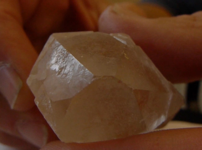
Figura 1. Quarzo che mostra forma dipiramidale esagonale.
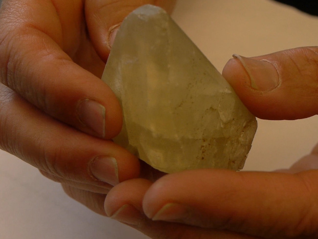
Figura 2. Calcite che mostra la forma di scalenoedro. Si noti come diverse facce di cristallo si intersecano per formare bordi di cristallo e la combinazione di spigoli forma punti noti come "vertici". Le forme di crescita simmetrica dei cristalli sono generate dalla ripetizione di strutture atomiche fondamentali (cellule unitarie) all'interno del reticolo cristallino. In questo caso, la crescita dei cristalli di calcite genera il poliedro specifico noto come scalenoedro.
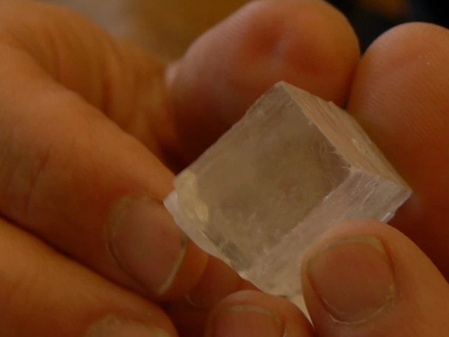
Figura 3. Halite che mostra la forma di cristallo cubico.
Figura 4. Granato che mostra la forma del dodecaedro.
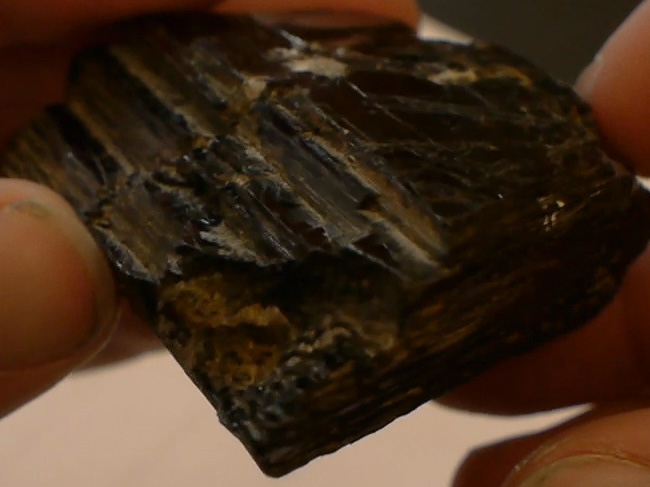
Figura 5. Biotite che mostra forma pseudo-esagonale.
3. Osserva e analizza la scissione
- Indossare la protezione per gli occhi.
- Posizionare un pezzo di quarzo sulla superficie di rottura.
- Usando un martello, rompere il pezzo di quarzo a metà.
- Usando una lente a mano, osserva il pezzo di quarzo rotto per le superfici di scissione. Si noti che il quarzo non ne ha. Il quarzo presenta fratture concoidali, ma nessuna superficie di scissione ben definita (Figura 6). Questa è una conseguenza del fatto che le celle unitarie nel reticolo cristallino di quarzo (gruppi SiO4, chiamati tetraedri di silice) hanno forze di legame comparabilmente uguali in tutte le direzioni. Questa uniformità di forza di legame si traduce in un cristallo senza piani di rottura preferiti.
- Ripetere i passaggi 3.2 – 3.4 per la calcite (dovrebbe mostrare la scissione romboedrica (Figura 7)), l'halite (dovrebbe mostrare la scissione cubica (Figura 8)), la biotite e/o la muscovite (dovrebbero mostrare la scissione planare (Figura 9)).
- Usa una lente a mano per valutare diverse qualità di scissione. La scissione può verificarsi a una varietà di livelli. Quando c'è una differenza drammatica nelle forze di legame in un particolare orientamento, come tra fogli di raggruppamenti SiO4 nel caso della mica, viene generata una scissione quasi perfetta tra questi fogli. Come notato sopra, il quarzo presenta una quasi totale mancanza di scissione. Tra questi estremi (di scissione perfetta e mancanza di scissione), ci sono minerali che hanno una buona scissione(ad esempio feldspato) e una scarsa scissione (alcune facce su cristalli di anfibolo).
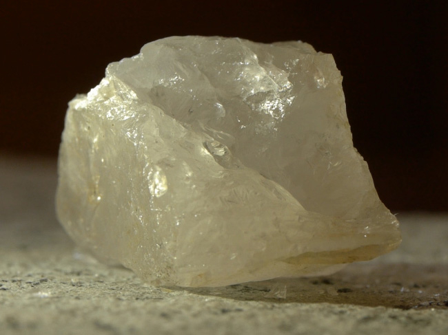
Figura 6. Quarzo che mostra frattura concoidale, senza superfici di scissione.
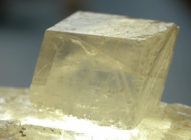
Figura 7. Calcite che mostra scissione romboedrica. Le superfici di rottura e frattura simmetrica sono generate da zone di relativa debolezza nel legame atomico all'interno del reticolo cristallino. La scissione della calcite provoca il poliedro specifico noto come romboedro.
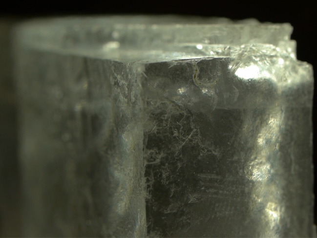
Figura 8. Halite che mostra scissione cubica.
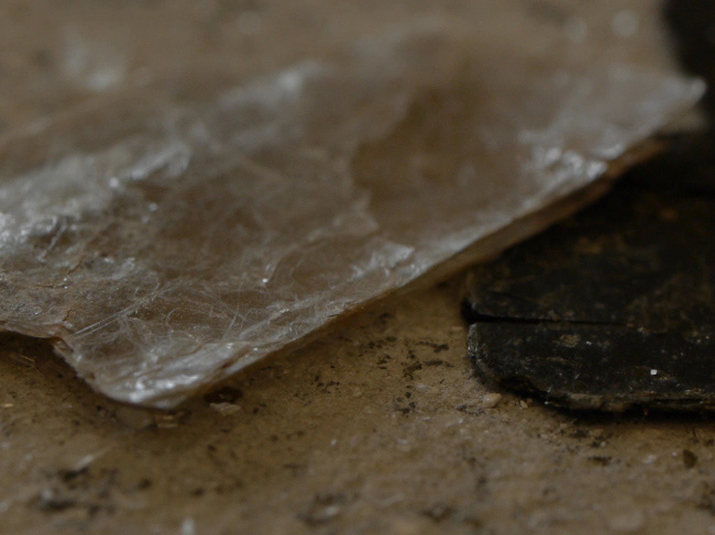
Figura 9. Biotite che mostra scissione planare.
Applicazione e Riepilogo
Storicamente, la valutazione delle proprietà fisiche dei minerali è stato un primo passo fondamentale nell'identificazione dei minerali. Ancora oggi, quando mancano strumentazioni analitiche microscopiche e moderne(ad esempio microscopia petrografica, diffrazione a raggi X, fluorescenza a raggi X e tecniche di microsonde elettroniche), le proprietà fisiche osservate sono ancora molto utili come strumenti diagnostici per l'identificazione dei minerali. Questo è particolarmente vero negli studi geologici sul campo.
Valutare e osservare le proprietà fisiche dei minerali è un mezzo eccellente per dimostrare la dipendenza critica delle caratteristiche macroscopiche dalla struttura e dalla disposizione a livello atomico.
Le principali proprietà fisiche dei minerali non sono sempre espresse in campioni specifici. Pertanto, essere effettivamente in grado di riconoscere e utilizzare queste proprietà come strumenti diagnostici richiede una combinazione di scienza, esperienza e artigianato. Spesso, il geologo deve utilizzare una lente manuale per valutare cristalli minerali relativamente piccoli o grani all'interno della matrice di una roccia più grande. In questi casi, può diventare una sfida distinta identificare gli aspetti utili della forma del cristallo e della scissione del cristallo.
In un contesto accademico o didattico, la valutazione dei minerali tramite l'analisi del campione a mano è un esercizio che dimostra come i modelli e le caratteristiche ripetitive siano imposti dalla chimica fisica dei materiali naturali. In altre parole, per ogni minerale specifico, ci sono alcune caratteristiche cristallografiche(ad esempio la morfologia del cristallo) e proprietà fisiche(ad esempio colore, durezza, striscia) che sono imposte dalla composizione chimica e dalla struttura atomica.
Nel campo delle risorse minerarie e della geologia esplorativa, l'identificazione dei minerali tramite campione manuale è una componente chiave del lavoro sul campo, volto a localizzare potenziali minerali e depositi economicamente utili. Ad esempio, l'identificazione di vari solfuri metallici (pirite, sfalerite, galena) in associazione con ossi-idrossidi di ferro idrotermale (ematite, goetite, limonite) può essere indicativa di potenziali vene e regioni ricche di Au- e Ag.
Nel contesto della geologia storica (decifrando la profonda storia temporale di una regione), l'identificazione minerale può preparare il terreno per interpretazioni di condizioni antiche. Ad esempio, alcuni minerali metamorfici(ad esempio i polimorfi Al2SiO5, la cianite, l'andalusite e la sillimanite) sono marcatori di particolari condizioni di pressione e temperatura nell'antica crosta.
Vai a...
Video da questa raccolta:

Now Playing
Proprietà fisiche dei minerali I: cristalli e clivaggio
Earth Science
51.6K Visualizzazioni

Determinazione dell'orientamento spaziale degli strati rocciosi con la bussola Brunton
Earth Science
25.5K Visualizzazioni

Utilizzo di mappe topografiche per la realizzazione di profili topografici
Earth Science
32.1K Visualizzazioni

Realizzazione di una sezione geologica trasversale
Earth Science
47.1K Visualizzazioni

Proprietà fisiche dei minerali II: analisi poliminerale
Earth Science
38.0K Visualizzazioni

Rocce ignee vulcaniche
Earth Science
39.7K Visualizzazioni

Rocce ignee intrusive
Earth Science
32.4K Visualizzazioni

Una panoramica sull'analisi dei biomarcatori bGDGT per la paleoclimatologia
Earth Science
5.4K Visualizzazioni

Una panoramica sull'analisi dei biomarcatori dell'alchenone per la paleotermometria
Earth Science
7.2K Visualizzazioni

Estrazione tramite sonicazione di biomarcatori lipidici dal sedimento
Earth Science
8.8K Visualizzazioni

Estrazione Soxhlet di biomarcatori lipidici dal sedimento
Earth Science
18.5K Visualizzazioni

Estrazione di biomarcatori dai sedimenti - estrazione accelerata con solvente
Earth Science
9.0K Visualizzazioni

Conversione di esteri metilici di acidi grassi mediante saponificazione per paleotermometria Uk'37
Earth Science
10.1K Visualizzazioni

Purificazione di un estratto lipidico totale con la cromatografia su colonna
Earth Science
12.4K Visualizzazioni

Rimozione di composti ramificati e ciclici mediante adduzione di urea per paleotermometria Uk'37
Earth Science
6.4K Visualizzazioni
Personale delle biblioteche
Copyright © 2025 MyJoVE Corporation. Tutti i diritti riservati