
Accedi
JoVE Science Education
Physical Examinations II
È necessario avere un abbonamento a JoVE per visualizzare questo.
Esame obiettivo dell'occhio
Panoramica
Fonte: Richard Glickman-Simon, MD, Assistant Professor, Dipartimento di Sanità Pubblica e Medicina di Comunità, Tufts University School of Medicine, MA
Una corretta valutazione degli occhi in un ambiente di pratica generale comporta test della vista, ispezione dell'orbita ed esame oftalmoscopico. Prima di iniziare l'esame, è fondamentale avere familiarità con l'anatomia e la fisiologia dell'occhio. La palpebra superiore dovrebbe essere leggermente sopra l'iride, ma non dovrebbe coprire la pupilla quando è aperta; il coperchio inferiore si trova sotto l'iride. La sclera appare normalmente bianca o leggermente di colore buff. L'aspetto della congiuntiva, una membrana trasparente che copre la sclera anteriore e le palpebre interne, è un indicatore sensibile di disturbi oculari, come infezioni e infiammazioni. La ghiandola lacrimale che produce lacrime si trova sopra e lateralmente al bulbo oculare. Le lacrime si diffondono verso il basso e attraverso l'occhio per drenare medialmente in due puncta lacrimali prima di passare nel sacco lacrimale e nel dotto nasolacrimale al naso.
L'iride divide la camera anteriore da quella posteriore. I muscoli dell'iride controllano le dimensioni della pupilla e i muscoli del corpo ciliare dietro di esso controllano la lunghezza focale della lente. Il corpo ciliare produce anche umore acqueo, che determina in gran parte la pressione intraoculare (Figura 1). I nervi cranici II e III controllano la reazione pupillare e l'accomodazione della lente; il nervo cranico III controlla l'elevazione della palpebra superiore; i nervi cranici III, IV e VI controllano il movimento oculare. Le sei direzioni cardinali dello sguardo sono controllate da sei muscoli extraoculari (Figura 2) innervati dai nervi cranici III, IV e VI.
Il test visivo è una parte essenziale dell'esame oftalmologico e viene eseguito anche come parte della valutazione del nervo cranico II durante l'esame neurologico. Un'immagine focalizzata viene proiettata sulla retina dopo che la sua luce passa attraverso la cornea, la pupilla, la lente e il corpo vitreo. La proiezione è capovolta e invertita da destra a sinistra, il che significa che la luce che entra dal campo temporale inferiore della visione colpisce il quadrante nasale superiore della retina. Le cellule fotosensibili della retina rispondono generando impulsi elettrici, che vengono trasmessi al nervo ottico e passati alla corteccia visiva attraverso le vie ottiche. Le cortecce visive destra e sinistra elaborano le immagini che entrano rispettivamente dai campi visivi sinistro e destro.
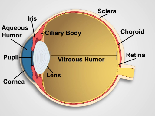
Figura 1. Anatomia dell'occhio. Un diagramma che mostra una vista sagittale dell'occhio umano con le strutture etichettate.
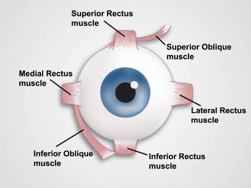
Figura 2. Muscoli dell'occhio. Un cartone animato che mostra una vista frontale dell'occhio umano e dei muscoli extraoculari (etichettati).
Procedura
1. Visione
L'acuità visiva è registrata come due numeri(ad esempio,20/40 corretto). Il numero superiore indica la distanza che il paziente si trovava dal grafico (20 piedi) e il numero inferiore indica la distanza da cui una persona con visione normale (20/20) poteva vedere la più piccola linea di stampa letta con precisione dal paziente (40 piedi con occhiali). Negli Stati Uniti, un paziente con visione di 20/200 o peggio è considerato legalmente cieco.
- Se disponibile, util
Applicazione e Riepilogo
Molte patologie sistemiche e oculari hanno manifestazioni che possono essere identificate durante una visita oculistica. Un semplice test di acuità visiva con un grafico di Snellen, o un sostituto adeguato, consente lo screening per la miopia e la presbiopia - visione lontana e da vicino compromessa, rispettivamente. Le limitazioni nella visione periferica aumentano la possibilità di glaucoma, così come altre condizioni gravi, e dovrebbero sempre richiedere un'ulteriore valutazione oftalmologica. Il gonfiore localizza...
Tags
Vai a...
Video da questa raccolta:

Now Playing
Esame obiettivo dell'occhio
Physical Examinations II
76.5K Visualizzazioni
Esame oftalmoscopico
Physical Examinations II
67.2K Visualizzazioni
Esame obiettivo dell'orecchio
Physical Examinations II
54.4K Visualizzazioni
Esame di naso, seni, cavità orale e faringe
Physical Examinations II
65.1K Visualizzazioni
Esame obiettivo della tiroide
Physical Examinations II
104.2K Visualizzazioni
Esame obiettivo dei linfonodi
Physical Examinations II
384.4K Visualizzazioni
Esame obiettivo dell'addome I: ispezione e auscultazione
Physical Examinations II
201.7K Visualizzazioni
Esame obiettivo dell'addome II: percussione
Physical Examinations II
247.0K Visualizzazioni
Esame obiettivo dell'addome III: palpazione
Physical Examinations II
138.1K Visualizzazioni
Esame obiettivo dell'addome IV: valutazione del dolore addominale acuto
Physical Examinations II
66.9K Visualizzazioni
Esame rettale maschile
Physical Examinations II
113.5K Visualizzazioni
Esame completo del seno
Physical Examinations II
86.5K Visualizzazioni
Esame obiettivo ginecologico I: valutazione dei genitali esterni
Physical Examinations II
303.2K Visualizzazioni
Esame obiettivo ginecologico II: esame con lo speculum
Physical Examinations II
149.2K Visualizzazioni
Esame obiettivo ginecologico III: palpazione rettovaginale e bimanuale
Physical Examinations II
146.6K Visualizzazioni
ISSN 2578-8264
Personale delle biblioteche
Copyright © 2025 MyJoVE Corporation. Tutti i diritti riservati
Utilizziamo i cookies per migliorare la tua esperienza sul nostro sito web.
Continuando a utilizzare il nostro sito web o cliccando “Continua”, accetti l'utilizzo dei cookies.