このコンテンツを視聴するには、JoVE 購読が必要です。 サインイン又は無料トライアルを申し込む。
Method Article
機能イメージングによるヒト聴覚皮質上のシータバースト刺激の後遺症のマッピング
要約
聴覚処理は、音声や音楽関連の処理の基礎となっています。経頭蓋磁気刺激(TMS)を認知、感覚と運動のシステムを研究するために正常に使用されているが、まれにオーディションを受けるために適用されていません。ここでは、聴覚皮質の機能的な組織を理解することは機能的磁気共鳴画像法と組み合わせるTMSを検討した。
要約
Auditory cortex pertains to the processing of sound, which is at the basis of speech or music-related processing1. However, despite considerable recent progress, the functional properties and lateralization of the human auditory cortex are far from being fully understood. Transcranial Magnetic Stimulation (TMS) is a non-invasive technique that can transiently or lastingly modulate cortical excitability via the application of localized magnetic field pulses, and represents a unique method of exploring plasticity and connectivity. It has only recently begun to be applied to understand auditory cortical function 2.
An important issue in using TMS is that the physiological consequences of the stimulation are difficult to establish. Although many TMS studies make the implicit assumption that the area targeted by the coil is the area affected, this need not be the case, particularly for complex cognitive functions which depend on interactions across many brain regions 3. One solution to this problem is to combine TMS with functional Magnetic resonance imaging (fMRI). The idea here is that fMRI will provide an index of changes in brain activity associated with TMS. Thus, fMRI would give an independent means of assessing which areas are affected by TMS and how they are modulated 4. In addition, fMRI allows the assessment of functional connectivity, which represents a measure of the temporal coupling between distant regions. It can thus be useful not only to measure the net activity modulation induced by TMS in given locations, but also the degree to which the network properties are affected by TMS, via any observed changes in functional connectivity.
Different approaches exist to combine TMS and functional imaging according to the temporal order of the methods. Functional MRI can be applied before, during, after, or both before and after TMS. Recently, some studies interleaved TMS and fMRI in order to provide online mapping of the functional changes induced by TMS 5-7. However, this online combination has many technical problems, including the static artifacts resulting from the presence of the TMS coil in the scanner room, or the effects of TMS pulses on the process of MR image formation. But more importantly, the loud acoustic noise induced by TMS (increased compared with standard use because of the resonance of the scanner bore) and the increased TMS coil vibrations (caused by the strong mechanical forces due to the static magnetic field of the MR scanner) constitute a crucial problem when studying auditory processing.
This is one reason why fMRI was carried out before and after TMS in the present study. Similar approaches have been used to target the motor cortex 8,9, premotor cortex 10, primary somatosensory cortex 11,12 and language-related areas 13, but so far no combined TMS-fMRI study has investigated the auditory cortex. The purpose of this article is to provide details concerning the protocol and considerations necessary to successfully combine these two neuroscientific tools to investigate auditory processing.
Previously we showed that repetitive TMS (rTMS) at high and low frequencies (resp. 10 Hz and 1 Hz) applied over the auditory cortex modulated response time (RT) in a melody discrimination task 2. We also showed that RT modulation was correlated with functional connectivity in the auditory network assessed using fMRI: the higher the functional connectivity between left and right auditory cortices during task performance, the higher the facilitatory effect (i.e. decreased RT) observed with rTMS. However those findings were mainly correlational, as fMRI was performed before rTMS. Here, fMRI was carried out before and immediately after TMS to provide direct measures of the functional organization of the auditory cortex, and more specifically of the plastic reorganization of the auditory neural network occurring after the neural intervention provided by TMS.
Combined fMRI and TMS applied over the auditory cortex should enable a better understanding of brain mechanisms of auditory processing, providing physiological information about functional effects of TMS. This knowledge could be useful for many cognitive neuroscience applications, as well as for optimizing therapeutic applications of TMS, particularly in auditory-related disorders.
プロトコル
プロトコルは2日間のセッション(必ずしも連続しない)に分かれています。初日は領域は、TMSと対象とするために、各参加者のために定義するために解剖学的および機能的MRスキャンで構成fMRIのローカライザーで構成されています。二日目は、fMRIのセッションで構成されています事前とTMSは特別なMRコンパチブルTMSコイル(Magstim株式会社、ウェールズ、英国)とフレームレス定位システム(Brainsight)を使用して、スキャナの内部に適用されるポストTMS。後者は、各参加者の解剖学的および機能的なデータに相対皮質領域でリアルタイムTMSコイルの位置に使用されます。
1。ローカライザーセッション
- あなたの参加者の高解像度の解剖学的画像を取得して起動します。
- 次に、任意のBOLD効果やMRIスキャンノイズ14,15に起因する聴覚マスキングを最小限に抑えるために、勾配エコーEPIパルスおよびスパースサンプリングのパラダイムを用いて機能画像を取得する。我々のケースでは、fMRIはdを行っている参加者が2つの連続した5分音符のメロディーは2,16、同一または異なっているかどうかを判断する必要がありますているメロディータスクをuring。被験者は5ノートの等しい2つの長さのパターン、C5の同じピッチですべてを聞いて、第2刺激後の左ボタンをクリックするように指示されている無差別の聴覚制御タスクも、含まれています。沈黙の期間はまた、各実行中のタスクの試験間でランダムに挿入されます。 12分16秒の合計持続時間のために、メロディの弁別、24聴覚制御試験と沈黙の24周期の24試験:合計で72試験は無作為化された順に記載しています。
- 解剖学的および/または機能的なランドマークを用いた刺激部位を定義します。一つは、TMSがあるため深さにおける電界強度の減衰の刺激部位の深さに関する制限されていることを認識する必要があり、3センチメートル6,17よりも深い領域に達することを期待することはできません。重要なステップは、パートごとに類似したランドマークを使用することですなぜなら参加者間の構造と機能に違いがあるため難しいかもしれicipant、。ここでは、解剖学的および機能的なランドマークの両方を使用して配置された各参加者でHeschlの回を、ターゲットにしています。我々は、ハーバード·オックスフォード構造アトラス(が提供するHeschlの回のマスク使用http://www.fmrib.ox.ac.uk/fsl/data/atlas-descriptions.html )とTMSターゲットがピークによって個別に定義されていますHeschlの回2内活性化。加えて、我々はまた、このような音響と体性感覚の成果物としてのTMSの非特異的効果を制御するためのコントロールサイトとして使用される頂点の位置を定義します。頂点はイニオンと鼻筋、左右intertragalノッチから等距離の点の間に中間として解剖学的に定義されています。刺激部位( すなわち Heschlの回または頂点)の順序は全体で相殺される個人。
2。事前·事後のTMSのfMRI実験
プリTMS fMRIのセッション
- スキャナに直接移動する参加者を準備します。これは、金属の除去を含むとTMSとMRスクリーニングフォームを充填。
- 解剖学的および機能的なスキャン(ローカライザーのセッションで行われたものと同じ、第1節を参照)で、MRの取得を開始します。
MRI環境におけるフレームレス定位とTMS
フレームレス定位システムは、赤外線カメラ(ポラリススペクトル)、登録手順とコンピュータのために使用するいくつかのツールとトラッカー(Brainsight)から構成される。コンピュータがスキャナ室の外側に位置するが、スキャナ室の入口に位置し、スキャナのドアは、TMSの適用中に開いたままです。ツールやトラッカーは、MR互換性があるだけでなく、赤外線カメラを支える三脚(自家製)とthアールereforeはスキャナ室の内部で使用。赤外線カメラは、MR-互換性がありませんので、スキャナーベッドから約2メートルで、スキャナのドアの近くスキャナ室、(安全手順についての説明を参照してください)の内側に配置されています。 TMSの刺激システムは、MRIスキャナ室に隣接する部屋に位置しています。我々は、MRI適合性のTMSコイルスキャナ室の内側に位置しており、RFフィルターチューブを通って、7メートルケーブルを介して、TMSシステムに接続を使用しています。
- 定位ソフトウェアパッケージ( 例えば Brainsight)にご参加者の解剖学的および機能画像と刺激ターゲットをロードします。ここでは、右Heschlの回をターゲットにされます。
- プリTMS fMRIの買収後、32チャネル·ヘッド·コイル(シーメンス3Tスキャナと32チャンネルヘッドコイルの構成を使用している場合)の上部MRヘッドのコイル部分を削除します。
- 次に、スキャナのベッドの上で参加者を下にスライドさせます。
- participで設定ヘッドバンドとトラッカーを修正アリの頭部。
- スキャナベッドに多関節アームを取り付け、アームにMRコンパチブルTMSコイルを固定してください。
- すべてのトラッカーとコイルはカメラの視野にあることを確認します。ここでは、カメラが若干右半球をターゲットとした場合、コイルの変位の簡単追跡を有効にする参加者の右側に移動されます。
- 定位ツール( つまり 、ポインタツール)を使用して被検者の頭部をキャリブレーションします。これは、解剖学的データで同じランドマークの参加者の頭(私たちの例では、例えば鼻の先、[後]と両耳の耳珠)でいくつかのランドマークをcoregisteringすることによって行われます。この手順では、2つの実験は、一人の参加者の頭の上にポインタツールを配置するため、参加者の頭の近くにして、コンピュータの登録を行うスキャナ室の入り口に他の実験を必要とされている。
- トンに対する接線のMRコンパチブルのTMSコイルを置き彼頭皮、および赤外線カメラ向けコイルトラッカー。コイルは正中2に後方と平行ポインティングコイルハンドル付き配向されている。多関節アームにネジを使用して、コイルの位置を修正します。
- MRIスキャナに隣接する部屋では、TMSのシステムの電源をオンにし、刺激を開始します。 TMSは、40代のための5Hzので繰り返し50Hzで3パルスで構成されるパターン化されたプロトコル、 すなわち 、連続シータバースト刺激(CTBS)、続いて適用されます。我々は、刺激出力18,19によって定義された固定の刺激強度(41%)を使用します。それは(安全手順については議論のセクションを参照)、健康な集団20の刺激中止後30分間の持続時間のために皮質可塑性を調節することが示されているように我々は、このプロトコルを選択しました。
ポストTMSのfMRIのセッション
- 刺激が完了したら、それはできるだけ早くスキャナに対象を取り戻すことが重要です。 Tを削除するMSがスキャナ室からコイル、及び多関節アームを取り外します。 MRヘッドのコイルに、参加者の頭を後ろにスライドさせます。お使いのスキャナが準備済みで行く準備ができていることを確認します。我々のアドバイスは、全体のTMSのセッション中に発生した身体のプラットフォームを維持し、最小限にローカライザースキャンの回数と期間を削減することです。
- rTMSの効果が一時的なものなので、最終的なスキャンセッションは、機能スキャンで始まらなければなりません。再び、我々はメロディタスクの12分間実行中にfMRIを実施しました。
- 最後のスキャンが完了したら、解剖スキャンで仕上げてください。
3。代表的な結果
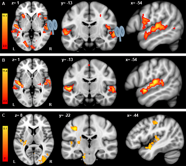
fMRIデータの解析は、事前·事後のTMS fMRIのセッションの両方に対して個別に行われている。各fMRIのセッションのために( すなわち 、事前と事後TMS)は、メロディーと聴覚制御タスクとのコントラストが左右Heschlの脳回、上側頭脳回、下前頭脳回とprecenでタスク関連活性を示す最大スペクトル脳回( 図1のB)。事前·事後のTMS fMRIのセッションとの違いを評価するために、我々はスチューデントのt検定を用いて、ランダム効果分析を行う。意義は、z> 2のしきい値とpの補正されたクラスタのしきい値= 0.05で識別されるクラスタを使用して決定されます。 図1 Cは単一の参加者のためのコントラストポストマイナスプレCTBSを表しています。データは右Heschlの回(黒丸)を標的CTBSは左Heschlの回を含む反対(左)聴覚野におけるfMRIの応答の増加を誘導することを示唆している。 fMRIの応答の変化はまた、二国間で左中心後回、左の島にと横後頭葉皮質で発見されています。しかし、fMRIの応答に有意な変化は、コイルの下に見られません。また、同様の結合されたTMS-fMRIのプロトコルは、頂点(コントロール·サイト)を刺激するために繰り返されます。頂点の上に適用さCTBSと事前·事後fMRIのセッションの比較は、どのsignificaを示さなかったntの効果(データは示さず)。

図1:個々のプレTMS fMRIのデータ()、ポストTMS fMRIデータ(B)とポストマイナスプレTMS fMRIのデータ(C)の分析。プリTMS fMRIのセッション(A)中とポストTMS fMRIのセッション(B)の単一の参加者のためのコントラストメロディ差別マイナス聴覚制御試験のAの結果。左から右へ:軸冠状および矢状ビュー。 (a)も(b)、TMSのコイルは、次の場所に右Heschlの回(黒丸)を標的としたx = 54、Y = -13、Z = 1(MNI152標準スペース)。事前·事後のTMS fMRIのセッションの両方の場合、座標値は、x = -54、Y = -13、Z = 1(MNI152標準スペース)刺激部位( すなわち右Heschlの回で左半球の変化を示すことで表示されます)。スチューデントのt検定を用いたコントラストポストマイナスプレTMS fMRIのセッションの後は、結果。
ディスカッション
我々は、聴覚野の機能的な組織を調査するためにオフラインTMSおよびfMRIを組み合わせるプロトコルを記述します。次のセクションでは、我々はそのようなアプローチを実施する際に考慮すべき方法論的な要因を説明します。
ポストTMS fMRIのセッションの取得とタイミング
事前·事後のTMS fMRIのセッションのスキャンの取得と相殺の順序
開示事項
特別な利害関係は宣言されません。
謝辞
CIBCフェローシップ(ja)およびNSERC助成(RZ)。我々は、赤外線カメラに関する彼の助けのためロッシュM.コモ(Brainsight)、MRコンパチブルトラッカーや他のハードウェアのサポートに感謝しています。我々はまた、コイルホルダ用の多関節アームを設計し、ビデオで表示される数値のいくつかを提供するブライアン·ハインズ(Hybexイノベーション株式会社)に感謝しています。そして、私たちは、実験の設計を最適化する助けモントリオール神経学研究所のマクコーネル脳イメージングセンターからすべてのMR技術とM.フェレイラ氏に感謝します。
資料
| Name | Company | Catalog Number | Comments |
| 材料名 | タイプ | 会社 | |
| 経頭蓋磁気刺激 | MagstimスーパーRapid2刺激すると、Rapid-2プラスワンモジュール | Magstim株式会社、ウェールズ、イギリス | |
| 磁気刺激用コイル | MRI対応70ミリメートル8の字コイル | Magstim株式会社、ウェールズ、イギリス | |
| 磁気共鳴画像 | 3-Tシーメンストリオスキャナ、32チャネル·ヘッドコイル | シーメンス社、ドイツ | |
| フレームレス定位脳手術 | Brainsight | ローグリサーチ社、モントリオール、カナダ | |
| 光学測定システム | ポラリススペクトル | ノーザン·デジタル社は、オンタリオ州、カナダ | |
| コイルホルダ用の多関節アーム | 標準 | Hybex Innovatイオン(株)、アンジュ、カナダ | |
| MRI対応インサートイヤホン | Sensimetrics、モデルS14 | Sensimetricsコーポレーション、米国マサチューセッツ州 |
参考文献
- Winer, J. A., Schreiner, C. E. . The Auditory Cortex. , (2011).
- Andoh, J., Zatorre, R. J. Interhemispheric Connectivity Influences the Degree of Modulation of TMS-Induced Effects during Auditory Processing. Frontiers in psychology. 2, 161 (2011).
- Siebner, H. R., Hartwigsen, G., Kassuba, T., Rothwell, J. C. How does transcranial magnetic stimulation modify neuronal activity in the brain? Implications for studies of cognition. Cortex. 45, 1035-1042 (2009).
- Ruff, C. C., Driver, J., Bestmann, S. Combining TMS and fMRI: from 'virtual lesions' to functional-network accounts of cognition. Cortex; a journal devoted to the study of the nervous system and behavior. 45, 1043-1049 (2009).
- Bestmann, S. Mapping causal interregional influences with concurrent TMS-fMRI. Exp. Brain Res. 191, 383-402 (2008).
- Bohning, D. E. BOLD-fMRI response to single-pulse transcranial magnetic stimulation (TMS. Journal of magnetic resonance imaging : JMRI. 11, 569-574 (2000).
- de Vries, P. M. Changes in cerebral activations during movement execution and imagery after parietal cortex TMS interleaved with 3T MRI. Brain research. 1285, 58-68 (2009).
- Cardenas-Morales, L., Gron, G., Kammer, T. Exploring the after-effects of theta burst magnetic stimulation on the human motor cortex: a functional imaging study. Human brain mapping. 32, 1948-1960 (2011).
- Grefkes, C. Modulating cortical connectivity in stroke patients by rTMS assessed with fMRI and dynamic causal modeling. NeuroImage. 50, 233-242 (2010).
- O'shea, J., Johansen-Berg, H., Trief, D., Gobel, S., Rushworth, M. F. S. Functionally specific in human premotor reorganization cortex. Neuron. 54, 479-490 (2007).
- Pleger, B. Repetitive transcranial magnetic stimulation-induced changes in sensorimotor coupling parallel improvements of somatosensation in humans. The Journal of neuroscience : the official journal of the Society for Neuroscience. 26, 1945-1952 (2006).
- Tegenthoff, M. Improvement of tactile discrimination performance and enlargement of cortical somatosensory maps after 5 Hz rTMS. Plos Biology. 3, 2031-2040 (2005).
- Andoh, J., Paus, T. Combining functional neuroimaging with off-line brain stimulation: modulation of task-related activity in language areas. Journal of cognitive neuroscience. 23, 349-361 (2011).
- Belin, P., Zatorre, R. J., Hoge, R., Evans, A. C., Pike, B. Event-related fMRI of the auditory cortex. Neuroimage. 10, 417-429 (1999).
- Hall, D. A. "Sparse" temporal sampling in auditory fMRI. Human Brain Mapping. 7, 213-223 (1999).
- Foster, N. E., Zatorre, R. J. A role for the intraparietal sulcus in transforming musical pitch information. Cereb Cortex. 20, 1350-1359 (2010).
- Bohning, D. E. Mapping transcranial magnetic stimulation (TMS) fields in vivo with MRI. Neuroreport. 8, 2535-2538 (1997).
- Corthout, E., Uttl, B., Walsh, V., Hallett, M., Cowey, A. Timing of activity in early visual cortex as revealed by transcranial magnetic stimulation. Neuroreport. 10, 2631-2634 (1999).
- Lewald, J., Foltys, H., Topper, R. Role of the posterior parietal cortex in spatial hearing. The Journal of neuroscience : the official journal of the Society for Neuroscience. 22, RC207 (2002).
- Huang, Y. Z., Edwards, M. J., Rounis, E., Bhatia, K. P., Rothwell, J. C. Theta burst stimulation of the human motor cortex. Neuron. 45, 201-206 (2005).
- Loubinoux, I. Within-session and between-session reproducibility of cerebral sensorimotor activation: a test--retest effect evidenced with functional magnetic resonance imaging. Journal of cerebral blood flow and metabolism : official journal of the International Society of Cerebral Blood Flow and Metabolism. 21, 592-607 (2001).
- Lisanby, S. H., Gutman, D., Luber, B., Schroeder, C., Sackeim, H. A. Sham TMS: intracerebral measurement of the induced electrical field and the induction of motor-evoked potentials. Biological psychiatry. 49, 460-463 (2001).
- Loo, C. K. Transcranial magnetic stimulation (TMS) in controlled treatment studies: are some "sham" forms active. Biological psychiatry. 47, 325-331 (2000).
- Robertson, E. M., Theoret, H., Pascual-Leone, A. Studies in cognition: the problems solved and created by transcranial magnetic stimulation. J. Cogn. Neurosci. 15, 948-960 (2003).
- Puschmann, S., Uppenkamp, S., Kollmeier, B., Thiel, C. M. Dichotic pitch activates pitch processing centre in Heschl's gyrus. NeuroImage. 49, 1641-1649 (2010).
- Johnsrude, I. S., Penhune, V. B., Zatorre, R. J. Functional specificity in the right human auditory cortex for perceiving pitch direction. Brain : a journal of neurology. 123, 155-163 (2000).
- Di Lazzaro, V. The physiological basis of the effects of intermittent theta burst stimulation of the human motor cortex. The Journal of physiology. 586, 3871-3879 (2008).
- Stagg, C. J. Neurochemical effects of theta burst stimulation as assessed by magnetic resonance spectroscopy. Journal of neurophysiology. 101, 2872-2877 (2009).
- Todd, G., Flavel, S. C., Ridding, M. C. Priming theta-burst repetitive transcranial magnetic stimulation with low- and high-frequency stimulation. Experimental brain research. Experimentelle Hirnforschung. Experimentation cerebrale. 195, 307-315 (2009).
- Bestmann, S., Baudewig, J., Siebner, H. R., Rothwell, J. C., Frahm, J. Subthreshold high-frequency TMS of human primary motor cortex modulates interconnected frontal motor areas as detected by interleaved fMRI-TMS. Neuroimage. 20, 1685-1696 (2003).
- Bungert, A. TMS combined with fMRI. , (2010).
- Bestmann, S., Baudewig, J., Frahm, J. On the synchronization of transcranial magnetic stimulation and functional echo-planar imaging. Journal of magnetic resonance imaging : JMRI. 17, 309-316 (2003).
- Wassermann, E. M. Use and safety of a new repetitive transcranial magnetic stimulator. Electroencephalogr. Clin. Neurophysiol. 101, 412-417 (1996).
- Oberman, L. M., Pascual-Leone, A. Report of seizure induced by continuous theta burst stimulation. Brain stimulation. 2, 246-247 (2009).
- Rossi, S., Hallett, M., Rossini, P. M., Pascual-Leone, A. Safety, ethical considerations, and application guidelines for the use of transcranial magnetic stimulation in clinical practice and research. Clin. Neurophysiol. 120, 2008-2039 (2009).
- Wassermann, E. M. Risk and safety of repetitive transcranial magnetic stimulation: report and suggested guidelines from the International Workshop on the Safety of Repetitive Transcranial Magnetic Stimulation, June 5-7, 1996. Electroencephalography and clinical neurophysiology. , 1-16 (1998).
- Yamaguchi-Sekino, S., Sekino, M., Ueno, S. Biological effects of electromagnetic fields and recently updated safety guidelines for strong static magnetic fields. Magn. Reson. Med. Sci. 10, 1-10 (2011).
- Bestmann, S. Mapping causal interregional influences with concurrent TMS-fMRI. Experimental brain research. Experimentelle Hirnforschung. Experimentation cerebrale. 191, 383-402 (2008).
- Oberman, L., Edwards, D., Eldaief, M., Pascual-Leone, A. Safety of theta burst transcranial magnetic stimulation: a systematic review of the literature. Journal of clinical neurophysiology: official publication of the American Electroencephalographic Society. 28, 67-74 (2011).
- Kangarlu, A. Cognitive, cardiac, and physiological safety studies in ultra high field magnetic resonance imaging. Magn. Reson. Imaging. 17, 1407-1416 (1999).
- Schenck, J. F. Safety of strong, static magnetic fields. Journal of magnetic resonance imaging : JMRI. 12, 2-19 (2000).
- Lee, V. S. . Cardiovascular MRI: physical principles to practical protocols. , 175 (2006).
- Paus, T. Transcranial magnetic stimulation during positron emission tomography: a new method for studying connectivity of the human cerebral cortex. The Journal of neuroscience: the official journal of the Society for Neuroscience. 17, 3178-3184 (1997).
- Sack, A. T., Linden, D. E. Combining transcranial magnetic stimulation and functional imaging in cognitive brain research: possibilities and limitations. Brain Res. Brain Res. Rev. 43, 41-56 (2003).
- Ilmoniemi, R. J. Neuronal responses to magnetic stimulation reveal cortical reactivity and connectivity. Neuroreport. 8, 3537-3540 (1997).
- Thiel, A. From the left to the right: How the brain compensates progressive loss of language function. Brain Lang. 98, 57-65 (2006).
転載および許可
このJoVE論文のテキスト又は図を再利用するための許可を申請します
許可を申請さらに記事を探す
This article has been published
Video Coming Soon
Copyright © 2023 MyJoVE Corporation. All rights reserved