JoVE 비디오를 활용하시려면 도서관을 통한 기관 구독이 필요합니다. 전체 비디오를 보시려면 로그인하거나 무료 트라이얼을 시작하세요.
A Plate-Based Assay to Measure Monoamine Release from Brain Slices on Test Drug Exposure
Overview
In this video, we demonstrate an assay that investigates monoamine release mechanisms from the neurons of metabolically active ex vivo rat brain slices, when treated with test drugs. The assay demonstrates that neurostimulant drugs induce monoamine release via the cellular monoamine transporters, whereas high KCl concentration induces synaptic vesicle exocytosis-mediated vesicular monoamine release.
프로토콜
All procedures involving animal models have been reviewed by the local institutional animal care committee and the JoVE veterinary review board.
1. Ex-vivo endogenous monoamine release from brain slices or punches
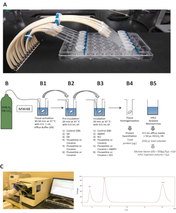
NOTE: The device used for this section consists of a 48-well plate and a tissue holder made of six microcentrifuge filter units without the inset filters connected to a carbogen line (Figure 1). To make the holder, use a sturdy plastic rod (e.g., from a cell scraper) and super glue the microcentrifuge filter units without the inset filters to it. Let it dry for 1-2 days. Time required for the endogenous monoamine release experiment and concentrations of amphetamine, fluoxetine, and cocaine are based on the current literature and previous protocols.
- Tissue activation
- Transfer the brain tissue to each well of the efflux chamber and allow recovery for 30-50 min at 37 °C on a slide warmer in 0.5-1 mL of oxygenated efflux buffer, with constant, gentle bubbling (Figure 1B1).
- During this incubation, dilute the drugs to the desired concentration for the experiment. All the drugs must be dissolved in efflux buffer, and concentrations are based off the current literature.
- First incubation
- Move the tissue holder with brain tissue to wells containing 500 μL of oxygenated efflux buffer and incubate for 20 min at 37 °C. Ensure that minimal to no buffer is transported over by tapping the holder on the edge of the well until no excess buffer is in the holder.
- In experiments with pharmacological agents such as monoamine transporter inhibitors, incubate the tissue samples with the drugs diluted in oxygenated efflux buffer (e.g., 10 μM Fluoxetine, 40 μM cocaine; see Figure 1B2). The final volume in each well will be 500 μL.
- Second incubation
- Move the holder with the tissue to a new set of wells containing 500 μL total efflux buffer with or without the desired concentration of each drug. Ensure there is no excess buffer left over. Each well represents an n= 1 for experimental conditions. Each experimental condition is performed in triplicate.
- One well includes an oxygenated efflux buffer, the next 10-30 μM AMPH, and the final well includes 10-30 μM AMPH plus monoamine transporter inhibitors. Each drug is dissolved in oxygenated efflux buffer.
- Incubate the tissue for 20 min at 37 °C with 500 μL of the drug condition.
NOTE: Additional wells may include an oxygenated high K+ efflux buffer with or without the monoamine transporter inhibitors. Dissolve each drug in the oxygenated efflux buffer (500 μL). - During this second incubation of 20 min, collect the solution from the wells from the first incubation in step 1.2.1 and transfer to microcentrifuge tubes containing 50 μL of 1 N perchloric acid or phosphoric acid (dependent on the type of HPLC, final concentration 0.1 N). The final volume of the sample will be 550 μL. Maintain microcentrifuge tubes on ice and label the tubes #1.
- After the second incubation of 20 min, move the tissue holder with brain sections or punches to an empty well and maintain the plate on ice. Transfer the supernatant to microcentrifuge tubes containing 50 μL of 1 N perchloric acid or phosphoric acid. The final volume of the sample will be 550 μL. Maintain microcentrifuge tubes on ice and label the tubes #2.
- Add 1 mL of ice-cold dissection buffer to each well containing tissue. Collect all of the tissue using small tweezers, and transfer to clean microcentrifuge tubes.
- Maintain tubes with brain tissue at -80 °C. Discard the 1 mL of dissection buffer (Figure 1B4).
- Filter solutions obtained from each incubation using microcentrifuge filter tubes (0.22 μm) at 2,500 x g for 2min. Use the filtrate to determine monoamine content using HPLC with electrochemical detection (Figure 1B5).
결과

Figure 1: Experimental setup for efflux experiment. (A) The efflux chamber consists of a 48-well tissue culture plate and a tissue holder tray connected to the carbogen line. (B) A diagram showing the experimental design for the endogenous monoamine efflux experiment in which the tissue activation (B1), pre-incubation with/without monoamine transporter inhibitors...
공개
자료
| Name | Company | Catalog Number | Comments |
| 48-well plate | NA | NA | Assay |
| Calcium Chloride Anhydrous | Sigma Aldrich | C1016 | Modified Artifical Cerebrospinal Fluid OR Efflux Buffer |
| D-(+)-Glucose | Sigma | 1002608421 | Dissection Buffer |
| L-Ascorbic Acid | Sigma Aldrich | A5960 | Dissection Buffer |
| Magnesium Sulfate | Sigma | 7487-88-9 | KH Buffer |
| Microcentrifuge Filter Units UltraFree | Millipore | C7554 | Assay - 6 to fit in 48-well plate |
| Pargyline Clorohydrate | Sigma Aldrich | P8013 | Modified Artifical Cerebrospinal Fluid OR Efflux Buffer |
| Potassium Chloride | Sigma | 12636 | KH Buffer |
| Potassium Phosphate Monobasic | Sigma | 1001655559 | KH Buffer |
| Sodium Bicarbonate | Sigma | S5761 | Dissection Buffer |
| Sodium Bicarbonate | Sigma Aldrich | S5761 | Dissection Buffer |
| Sodium Chloride | Sigma | S3014 | KH Buffer |
참고문헌
This article has been published
Video Coming Soon
Source: Pino, J. A. et al. A Plate-Based Assay for the Measurement of Endogenous Monoamine Release in Acute Brain Slices. J. Vis. Exp. (2021)
Copyright © 2025 MyJoVE Corporation. 판권 소유