
Method Article
에반스 블루 염료와 Zebrafish의 애벌레 골격 근육 무결성 분석
* 이 저자들은 동등하게 기여했습니다
요약
In this study, we describe a straightforward method to perform Evans Blue Dye (EBD) analysis on zebrafish larvae. This technique is a powerful tool for the characterization of skeletal muscle integrity and delineation of zebrafish models of muscular dystrophy, and is a valuable method for the development of novel therapeutics.
초록
The zebrafish model is an emerging system for the study of neuromuscular disorders. In the study of neuromuscular diseases, the integrity of the muscle membrane is a critical disease determinant. To date, numerous neuromuscular conditions display degenerating muscle fibers with abnormal membrane integrity; this is most commonly observed in muscular dystrophies. Evans Blue Dye (EBD) is a vital, cell permeable dye that is rapidly taken into degenerating, damaged, or apoptotic cells; in contrast, it is not taken up by cells with an intact membrane. EBD injection is commonly employed to ascertain muscle integrity in mouse models of neuromuscular diseases. However, such EBD experiments require muscle dissection and/or sectioning prior to analysis. In contrast, EBD uptake in zebrafish is visualized in live, intact preparations. Here, we demonstrate a simple and straightforward methodology for performing EBD injections and analysis in live zebrafish. In addition, we demonstrate a co-injection strategy to increase efficacy of EBD analysis. Overall, this video article provides an outline to perform EBD injection and characterization in zebrafish models of neuromuscular disease.
서문
Muscular dystrophies constitute a group of prevalent and heterogeneous human muscle diseases with specific histopathological features1,2. Symptoms typically associated with this devastating group of diseases include muscle weakness, muscle degeneration, loss of motility, and early mortality1,3. The primary pathomechanisms of muscular dystrophies are the loss of proteins that stabilize the sarcolemma, anchor transmembrane complexes, and mediate cell signaling across the membrane4-6. For example, complete loss of the protein dystrophin, a primary scaffold protein of the dystrophin-glycoprotein complex, results in destabilization of the muscle membrane in Duchenne muscular dystrophy7. Due to the fact that most muscular dystrophies result from mutations in proteins that participate in the link between the extracellular matrix and the sarcolemmal cytoskeleton, a common observation at the cellular level is the loss of sacrolemmal integrity8,9. This understanding of the primary pathomechanism(s) associated with muscular dystrophies is the product of numerous years of research employing animal model systems2,10-15. However, despite advances in the field, there are still limited therapeutic options for treatment or management of the range of dystrophy subtypes. This limitation of viable therapies is due to several key factors: 1) the difficulty of gene therapy strategies, 2) a high frequency of de-novo disease cases and the corresponding lack of translatable animal models, and 3) the lack of rigorous strategies to test the physiological consequences of putative therapeutic agents with clear and reproducible outcome measures.
To overcome some of these limitations, numerous labs including our own are employing zebrafish as a system to model and study human neuromuscular diseases2. To date, zebrafish have proven a valuable tool in disease research. The zebrafish model has been used to identify and validate novel human disease causing mutations16,17, elucidate uncharacterized disease causing mechanisms17,18, and identify novel therapeutic strategies12,19. These advances were made, in part, by the canonical strengths of the zebrafish system such as their optical clarity, ease of genetic manipulation, and ability to breed in large numbers20. Zebrafish have additionally proven amendable to large-scale drug screens21, a valuable method for the identification of novel therapeutics22-24. Regarding muscle disease research, these strengths are complemented by the ability to isolate single zebrafish skeletal muscle fibers via dissociation25 and by the ability to examine myofiber organization in vivo using the optical phenomenon called birefringence26, which collectively allows for rapid determination of macroscopic muscle integrity. Regardless of these available utilities, further tool development is continuously required to advance investigation.
We, and others, have adapted a protocol for EBD injection and analysis in the zebrafish model. EBD is a vital, cell permeable dye that is taken up by damaged, degenerating, or apoptotic cells and then visualized under fluorescence27. To date, EBD analysis has extensively been used to analyze muscle membrane integrity in mouse models of skeletal muscle and heart diseases8,9,27. However, in mammalian preparations, harvested muscle typically requires laborious sectioning or dissection prior to analysis. In zebrafish, direct analysis is possible in high numbers using live and intact animals. In this video article, we will demonstrate the methodology to perform EBD injection and analysis in live zebrafish larvae, with representative images of EBD uptake in the zebrafish dystrophy mutant line sapje15,28. Furthermore, we present a co-injection strategy that allows for increased quantification of EBD preparations.
프로토콜
한천 사출 플레이트 1. 준비 (시간 : 45 분)
- E3 미디어의 2 % ~ 3 % 아가로 오스를 삶아 및 솔루션은 벤치에 약간 냉각 할 수 있습니다. 참고 제조되는 분사 플레이트의 수는 필요 아가 로스의 양을 지시한다. 각 분사 플레이트 아가 용액 약 35 ㎖에 필요하다.
- 끓는 후, 원하는 온도가 사출 금형 제조 업체의 지침에 따라 (예., 45 ℃로)에 도달 할 때까지 아가로 오스를 냉각 할 수 있습니다.
- 100mm 접시에 아가로 오스 냉각의 약 35 ml에 붓고.
- 솔루션으로 선호하는 사출 금형의 한쪽 끝을 놓고, 다음 (이 기포의 발생을 줄이는 데 도움이됩니다) 아가로 오스 솔루션에 금형의 나머지를하다.
- 아가로 오스 용액을 약 30 분 동안 하나 실온에서 4 ° C를 공고히 할 수 있습니다.
- 고체 아가에서 금형의 일단을 분리 주걱을 사용한다. 천천히 m의 나머지를 제거 늙은.
에반스 블루 염료 (EBD) 사출 믹스 2. 준비 (시간 : 30 분)
- 1X 링거액에 EBD의 1 %의 주식을 확인 (5 mM의 KCl을 155 mM의 NaCl을 2 mM의 염화칼슘 2 1 밀리미터의 MgCl 2, 2 밀리미터 나 2 HPO 4, 10 mM의 HEPES 10 mM의 포도당, 7.2의 pH), 이는 실온에서 저장 될 수있다.
- 25 밀리그램에서 MW 10,000 kDa의 -dextran 형광 염료의 주식 솔루션 (FITC)를 확인 / ㎖ -20 ℃에서 1X 링거액 및 저장소
- (. 즉, 100 μL의 최종 작업 볼륨 : FITC 덱스 트란 주식의 90 μL에 1 % EBD 10 μl를 혼합) FITC-덱스 트란 주식의 원액을 직접 0.1 %에 EBD를 희석하여 주입 믹스를 준비합니다.
- 철저하게 소용돌이 주입 믹스 (이 녹색으로 변합니다) 및 알루미늄 호일에 주입 혼합 튜브를 배치하여 직접 빛이 닿지 않는 곳에 보관.
3. EBD 주입 준비 (시간 : 약 30 분)
ENT "> 참고 : 프로토콜 3-7 일 포스트 수정 (DPF)에서 유충으로 가장 적합합니다.- RT에 대한 사전 따뜻한 주입 판.
- 금속 접시에 미세 조작기를 배치하여 사출 장치를 설정하고 현미경 옆에 사출에 사용되는 서있다. 공기 구동 미세 주입 컨트롤러를 켭니다. 참고 : 선호 분사 시스템은 실험실에 따라 다릅니다 및 분석의 결과를 변경해서는 안됩니다.
- 위로 EBD 믹스의 약 2-4 μL와 주사 바늘을 입력합니다.
- EBD 믹스의 약 5 NL에 주입 볼륨을 보정합니다. 주 : 주입량 교정 교정 방법에 의존 할 것이다. 가스 압력이 인젝터 마이크로 미터를 사용하여 볼륨을 통해 볼 루스 보정 주입 부피를 필요로하는 반면, 피스톤 구동 주입 직접 주어진 주입 부피로 설정 될 수있다.
- 1X 링거액 젖은 주입 판과는 우물에서 초과 제거합니다.
- 0.04 %의 에틸 3- 아미노 벤조 에이트의 메탄 샐 사전 치료 유충분사의 시작에 앞서 유충 고정화 1X 링거액 희석 t (tricaine). 참고 : 적절한 주입 잔여 움직임 어렵 기 때문에 유충을 보장하는 것은 완전히 immotile 있습니다 중요하다.
- 유리 피펫을 사용하여 한천 주입 판 우물에 마취 애벌레를 놓습니다. 유충이 완전히 잘 내에서 자신의 측면에 누워 있는지 확인합니다. 참고 : 물론 당 유충의 수는 실험에 달려있다.
- 유충이 웰에 넣어 후 웰 내에 유충 움직임을 최소화하기 위해 과량의 링거액을 제거한다. 그 애벌레가 탈수되지 않도록 용액의 잔류량을 남겨주세요.
EBD와 Zebrafish의 유충 4. 심막 주입 (시간 : 유충 주입의 수에 따라, 약 1-3 시간)
- 주사가 수행 될 해부 범위에 유충을 포함하는 주입 판을 놓습니다.
- 포함하는 주입 피펫 바늘의 위치를EBD는 제브라 피쉬 애벌레를 통해 섞는다.
- 다시 위치를 회전시킴으로써 분사 플레이트 정도로 주사 바늘은 유충의 중심부 근처이며, 대략 45 ° 복부 전후방 축으로부터.
- 정맥 처음 지느러미 방향으로 돌고 노른자 (그림 1)의 전방 부분에서 정맥의 영역에서 일반 추기경 정맥 (CCV)에 주사 바늘을 삽입합니다. 참고 : 최대 40 배까지의 배율이 명확하게 CCV를 참조하는 것이 유용 할 수 있습니다.
- EBD 믹스 5 NL을 주입하고 EBD 믹스 즉시 누출을 최소화하기 위해 5-8 초 동안 위치에 주사 바늘을 유지. 참고 : 좋은 주입은 심장 챔버에서 볼 수 염료 착색 (그림 1)을해야합니다. EBD 믹스는 심장에서 관찰되지 않으면, EBD 믹스 추가 5 NL 염료 흡수를 유도하기에 충분할 수있다 주입. 또한, 배아는 폐기 될 수있다.
참고 : 어떤 경우에는 심장이 박동을 중지 할 수 있습니다. 이 O를하는 경우ccurs, 20 ~ 40 초 동안 유충을 모니터링하는 것을 계속한다. 염료가 순환 시스템을 통해 이동함에 따라 일반적으로 심장 박동이 다시 시작됩니다. - 다음 유충 반복에 이동합니다.
- 바로 주입 (그림 2) 후 혈관에 FITC-덱스 트란의 존재를 관찰하여 성공적으로 주입 된 배아를 식별합니다.
5. 배양 및 EBD 통풍 관 (시간 : 4 ~ 6 시간)
- 유충의 원하는 번호가 주입 된 후, 반환 100mm 요리에 tricaine없이 1X 링거액에 유충을 주입.
- 알루미늄 호일에 싸서 요리를 유지합니다. 주 : 어두운 주입 유충 크게 늘리지 생존율을 증가시키고, 신호 강도에 큰 일관성을 보장한다. 알루미늄 호일로 포장 유충 인큐베이터 외부의 시간 동안 특히 중요하다.
- 충분한 EBD 흡수를 보장하기 위해 유충은 4 ~ 6 시간 동안 28.5 ℃에서 부화 할 수 있습니다.
근육에 EBD 6. 시각화 (시간 : 유충 주입과 현미경의 종류의 수에 따라, 예상 0.5-3 시간)
- 이전 영상에 움직임을 방지하기 위해 0.04 %의 유충과 tricaine 마취.
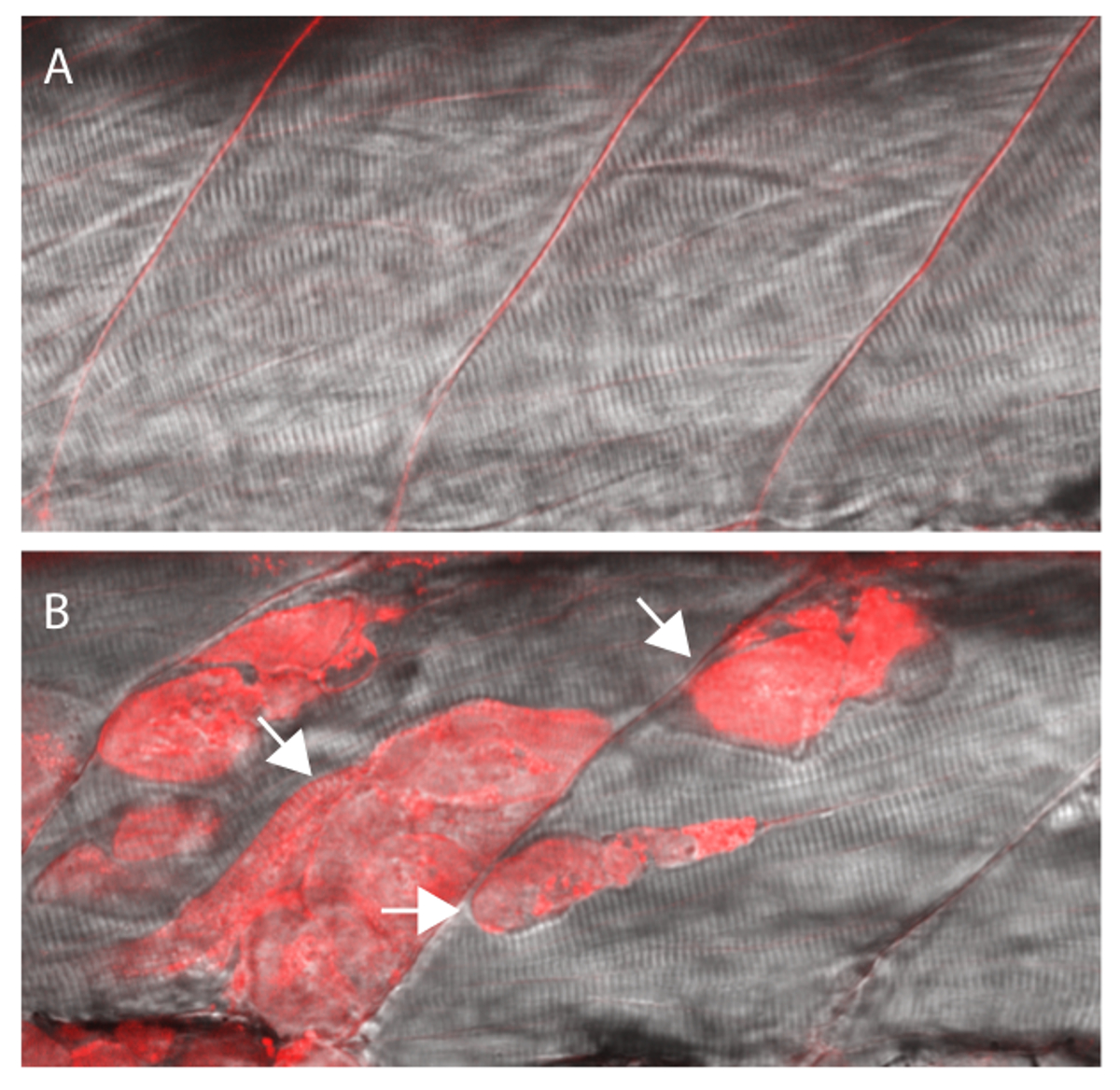
- EBD 흡수가 골격 근육 (그림 3)에서 발생하는 경우 적색 형광에서보기 유충 확인합니다.
결과
EBD 주입 혼합 3 DPF에서 sapje 동형 접합 돌연변이와 야생 형 형제의 CCV에 주입했다. 심장 챔버 (그림 1B)를 가득 주입 후 FITC - 덱스 트란 녹색 형광 아래에있는 혈관에 (그림 2) 시각화하여 성공적으로 주입을 분석 하였다.
4 시간의 잠복기 후, EBD 흡수는 형광 현미경을 사용하여 체절 수준에서 조사 하였다. sapje 동형 접합 돌연변이가 근육 막 (15) (그림 3B)에 손상을 나타내는, EBD 흡수를 보여 주었다 반면 야생 형 형제, 눈에 보이는 근육 섬유 (그림 3A) 내에 EBD 형광을 나타내지 않았다.

제브라 피쉬 엠브리의 일반 추기경 정맥 (CCV)에 그림 1. 주입 EBD 주입 믹스오. (A) Uninjected 배아. 화살표는 CCV 주입을위한 이상적인 위치를 나타낸다. (B) CCV에 성공 주입. 염료는 심장 챔버 (화살표)를 입력하고 혈관을 통해 펌핑하기 시작합니다. (C) 실패 CCV 주입 일부 또는 배아 (화살표)의 난황을 입력 염료의 모든 발생합니다. 이 그림의 더 큰 버전을 보려면 여기를 클릭하십시오.

그림 2. 배아는 즉시 주입 전에 EBD 흡수에 다음과 같은 혈관을 통해 녹색 형광 아래 FITC-덱스 트란 분포를 관찰하여 성공적으로 주입 정렬 할 수 있습니다.이 그림의 더 큰 버전을 보려면 여기를 클릭하십시오.

그림 3 : EBD는 손상된 세포막과 섬유에 의해 흡수 될 것입니다 근육 섬유에는 EBD 형광을 보여주는 (A) 야생형 형제.. (B) 여러 근육 섬유 (화살표) 내 EBD 형광와 Sapje 동형 접합 돌연변이. 모든 유충 EBD 주입 믹스 주입하고 3 DPF에서 4 시간 동안 배양 한 후에 분석 하였다. 형제 자매 및 돌연변이가 CCV 주입하기 전에 근육 섬유의 분리으로 분류되었다. 이 그림의 더 큰 버전을 보려면 여기를 클릭하십시오.
토론
제브라 피쉬는 신경 근육 질환 2,29의 연구를위한 강력한 도구로 부상하고있다. 현재까지, 제브라 피쉬 시스템은 새로운 근육 질환을 일으키는 돌연변이를 확인 16,17,30 신규 pathomechanisms 18을 해명하고, 잠재적으로 새로운 치료약 (12, 24)를 식별하는 데 사용되어왔다. 이러한 집단적인 노력은 인간의 신경 근육 질환을 모델링 할 수있는 제브라 피쉬의 유틸리티를 설립했다. 그러나, 제브라 피쉬와 포유 동물 모델로 만든 진보에도 불구하고, 신경 근육 조건의 넓은 스펙트럼 내에서 환자에 대한 제한적인 치료 옵션이 있습니다. 따라서, 수요는 파괴적인 질병이 그룹에 대한 치료 개발을 위해 존재한다. 치료법이 요구 평행하면 새로운 동물 모델 및 추정 치료 전략을 검증하는 실험 진행 혁신뿐만 아니라 엄격한 분석을위한 대응이 필요하다.
EBD 분석은 일반적으로 마우스 모델에서 사용연구 조직과 뇌, 심장에서 세포 손상 및 골격 근육 27,31. 특히, EBD 근육 세포막 불안정성의 정도를 표시하고 8 손상 근이영양증 다양한 아형의 마우스 모델에서 광범위하게 사용된다. 근육 세포막 손상을 드러내는 EBD의 사용은 인간의 질병 상태 (9) 동물 모델의 유사성을 확립지지 파라미터이다. 마우스에 EBD의 힘은 개발 및 신경 근육 질환의 제브라 피쉬 모델에 EBD를 적용, 우리 자신을 포함한 여러 실험실을 주도하고있다. EBD 인해 분석의 적용으로,이 기술은 활발 인간 질병 상태에 11,15,22,24,32 제브라 피쉬 모델을 확증하기 위해 구현되고있다. 손상된 근육 막과 유충은 근육 섬유 내의 EBD 흡수하기 때문에 적색 형광을해야합니다. 개별 근섬유 내의 섬유 간 공간에 형광 관찰 아니지만 역시 t의 기저막로부터 분리 섬유의 정보 일 수있다유용한 진단 세부 사항을 제공하는 멤브레인 손상의 그는 부재. EBD 분석은 동물 모델 검증을 넘어 잠재적 인 응용 프로그램이 있습니다. 우리의 실험실에서 노력은 최근 EBD 분석은 잠재적으로 새로운 치료 약물 (24)의 유효성을 검사에 도움이되도록 증명하고있다. 잠재적 인 치료 치료를 감소 시키거나 관련 치료 활동 (8)을 의미 할 수 있습니다 신경 근육 질환 모델에 EBD 흡수를 폐지 여부 확인. 이러한 유형의 분석 치료제 메커니즘 (들)을 설정하는 데 도움 EBD 및 분석 애플리케이션을 확대 할 수있다.
많은 기술과 마찬가지로, EBD 분석은 실험 설계 및 연습 동안 관찰 할 몇 가지주의 사항을 가지고있다. 예를 들어, 나이 인해 조직의 비후에 CCV를 식별하기 어려울 수있다. 또한, 실험 횟수를 줄이고 다수의 유충 수험 할 필요성을 증가 전과 심낭 주입 중에 제조에서 유충을 손상하기 쉽다.손상된 근육 EBD을 차지할 수 게다가, 처리 및 사출 동안 유충 짓을 물리적 손상은 오탐 (false positive)이 발생할 수 있습니다. 이러한 장애들을 극복하기 위해, 우리는 사출 전에 후속 분석 직후 성공적인 염료 주입과 유충 쉽고 신뢰할 수 있도록 식별이 비디오 문서 공동 사출 전략을 설명 하였다. 전에 근육 섬유로의 흡수로 혈관에 EBD의 확인을 허용하여 성공적으로 주입을위한 공동 분사 제어 FITC-덱스 트란. EBD 형광은 근육 섬유에서 수집되지 않을 경우 몇 시간 후 애벌레 매우 확산되면서 특히 유용 할 수있다; 이와 같이, 탐지하기 어려울 수있다. 또한, CCV 누락 및 노른자 또는 신체 공동으로 EBD를 주입하면, 배양 한 후, 손상된 근육 섬유에 의해 흡수의 감소 가능성과 아직 배아를 제어 할 유사한 확산 붉은 형광이 발생할 수 있습니다. 종합적으로,이 동굴ATS는 EBD 주입이 일관되고 신뢰할 수있는 결과를 달성하기 위해 인내와 연습이 필요 좋습니다.
모든에서는 제브라 애벌레 EBD 분석을 수행 실용적이고 간단한 방법을 설명한다. 지금까지 모델 시스템으로서 지브라 피쉬의 사용은 특히 인간의 질환 모델로서, 급속하게 확장되고있다. 이 확장은 제브라 피쉬 시스템의 현재 장점을 개선 실험 기술의 지속적인 개발 및 수정에 부분적으로 기인한다. EBD 주입 기술은 검증 및 제브라 피쉬 근육 질환 모델 연구를위한 연구자의 무기에 추가하고 강력한 도구를 제공합니다. 이 기술의 지속적인 구현 및 수정은 새로운 치료 전략뿐만 아니라 질병을 일으키는 메커니즘을 밝히기 도울 수있는 잠재력을 가지고있다.
공개
저자는 더 경쟁 재정적 이익 또는 관심의 다른 충돌이 없습니다.
감사의 말
우리는 자신의 기술 지원을 트렌트 워 감사하고 싶습니다. 우리는 또한 아픈 아이들이 프로젝트에 대한 자신의 관대 한 자금에 대한 치료 선천성 근육 영양 장애 (CMD)의 병원에서 소아과를 인정합니다.
자료
| Name | Company | Catalog Number | Comments |
| Fluorescein isothiocyanate-dextran MW 10,000 | Sigma | FD10S | |
| Evan's Blue Dye | Sigma | E2129 | |
| Ethyl 3-aminobenzoate methanesulfonate salt | Sigma | A5040 | |
| 100 mm Petri dish | Fischerbrand | FB0875712 | Injection mold base |
| Thin wall glass capillaries | World Precision Instruments | TW100F-4 | For Injection needle |
| Agarose | Bioshop | AGA001 | Injection mold |
| Microinjection mold | Adaptive Science Tools | TU-1 | Injection mold |
| Sodium chloride | Bioshop | SOD001 | Ringer's solution |
| Potassium chloride | Bioshop | POC888 | Ringer's solution |
| Magnessium chloride hexahydrate | Sigma | M2670 | Ringer's solution |
| Sodium phosphate monobasic monohydrate | Sigma | S9638 | Ringer's solution |
| HEPES | Sigma | H4034 | Ringer's solution |
| Glucose | BioBasic | GB0219 | Ringer's solution |
| Calcium chloride | Sigma | C1061 | Ringer's solution |
참고문헌
- Verma, S., Anziska, Y., Cracco, J. Review of Duchenne muscular dystrophy (DMD) for the pediatricians in the community. Clin Pediatr (Phila. 49, 1011-1017 (2010).
- Gibbs, E. M., Horstick, E. J., Dowling, J. J. Swimming into prominence: the zebrafish as a valuable tool for studying human myopathies and muscular dystrophies). FEBS J. 280, 4187-4197 (2013).
- Lancet, , 381-845 (2013).
- Lapidos, K. A., Kakkar, R., McNally, E. M. The dystrophin glycoprotein complex: signaling strength and integrity for the sarcolemma. Circ Res. 94, 1023-1031 (2004).
- Campbell, K. P., Kahl, S. D. Association of dystrophin and an integral membrane glycoprotein. Nature. 338, 259-262 (1989).
- Cohn, R. D., Campbell, K. P. Molecular basis of muscular dystrophies. Muscle Nerve. 23, 1456-1471 (2000).
- Yoshida, M., Ozawa, E. Glycoprotein complex anchoring dystrophin to sarcolemma. J Biochem. 108, 748-752 (1990).
- Straub, V., Rafael, J. A., Chamberlain, J. S., Campbell, K. P. Animal models for muscular dystrophy show different patterns of sarcolemmal disruption. J Cell Biol. 139, 375-385 (1997).
- Matsuda, R., Nishikawa, A., Tanaka, H. Visualization of dystrophic muscle fibers in mdx mouse by vital staining with Evans blue: evidence of apoptosis in dystrophin-deficient muscle. J Biochem. 118, 959-964 (1995).
- Bassett, D., Currie, P. D. Identification of a zebrafish model of muscular dystrophy. Clin Exp Pharmacol Physiol. 31, 537-540 (2004).
- Gupta, V., et al. The zebrafish dag1 mutant: a novel genetic model for dystroglycanopathies. Hum Mol Genet. 20, 1712-1725 (2011).
- Kawahara, G., et al. Drug screening in a zebrafish model of Duchenne muscular dystrophy. Proc Natl Acad Sci U S A. 108, 5331-5336 (2011).
- Guyon, J. R., et al. Modeling human muscle disease in zebrafish. Biochim Biophys Acta. 1772, 205-215 (2007).
- Cavanna, J. S., et al. Molecular and genetic mapping of the mouse mdx locus. Genomics. 3, 337-341 (1988).
- Bassett, D. I., et al. Dystrophin is required for the formation of stable muscle attachments in the zebrafish embryo. Development. 130, 5851-5860 (2003).
- Horstick, E. J., et al. Stac3 is a component of the excitation-contraction coupling machinery and mutated in Native American myopathy. Nat Commun. 4, (1952).
- Davidson, A. E., et al. Novel deletion of lysine 7 expands the clinical, histopathological and genetic spectrum of TPM2-related myopathies. Brain. 136, 508-521 (2013).
- Telfer, W. R., Nelson, D. D., Waugh, T., Brooks, S. V., Dowling, J. J. Neb: a zebrafish model of nemaline myopathy due to nebulin mutation. Dis Model Mech. 5, 389-396 (2012).
- Dowling, J. J., et al. Oxidative stress and successful antioxidant treatment in models of RYR1-related myopathy. Brain. 135, 1115-1127 (2012).
- Detrich, H. W., Westerfield 3rd,, M,, Zon, L. I. Overview of the Zebrafish system. Methods Cell Biol. 59, 3-10 (1999).
- Whitfield, T. T., et al. Mutations affecting development of the zebrafish inner ear and lateral. , 123-241 (1996).
- Kawahara, G., Guyon, J. R., Nakamura, Y., Kunkel, L. M. Zebrafish models for human FKRP muscular dystrophies. Hum Mol Genet. 19, 623-633 (2010).
- Baraban, S. C., Dinday, M. T., Hortopan, G. A. Drug screening in Scn1a zebrafish mutant identifies clemizole as a potential Dravet syndrome treatment. Nat Commun. 4, (2013).
- Waugh, T. A., et al. Fluoxetine prevents dystrophic changes in a zebrafish model of Duchenne muscular dystrophy. Hum Mol Genet. 23 (17), 4651-4662 Forthcoming.
- Horstick, E. J., Gibbs, E. M., Li, X., Davidson, A. E., Dowling, J. J. Analysis of embryonic and larval zebrafish skeletal myofibers from dissociated preparations. J Vis Exp. 50259, (2013).
- Smith, L. L., Beggs, A. H., Gupta, V. A. Analysis of skeletal muscle defects in larval zebrafish by birefringence and touch-evoke escape response assays. J Vis Exp. 50925, (2013).
- Hamer, P. W., McGeachie, J. M., Davies, M. J., Grounds, M. D. Evans Blue Dye as an in vivo. marker of myofibre damage: optimising parameters for detecting initial myofibre membrane permeability. J Anat. 200, 69-79 (2002).
- Guyon, J. R., et al. Genetic isolation and characterization of a splicing mutant of zebrafish dystrophin. Hum Mol Genet. 18, 202-211 (2009).
- Santoriello, C., Zon, L. I. Hooked! Modeling human disease in zebrafish. J Clin Invest. 122, 2337-2343 (2012).
- Majczenko, K., et al. Dominant Mutation of CCDC78 in a Unique Congenital Myopathy with Prominent Internal Nuclei and Atypical Cores. Am J Hum. , (2012).
- Wooddell, C. I., et al. Use of Evans blue dye to compare limb muscles in exercised young and old mdx mice. Muscle Nerve. 41, 487-499 (2010).
- Hall, T. E., et al. The zebrafish candyfloss mutant implicates extracellular matrix adhesion failure in laminin alpha2-deficient congenital muscular dystrophy. Proc Natl Acad Sci U S A. 104, 7092-7097 (2007).
재인쇄 및 허가
JoVE'article의 텍스트 или 그림을 다시 사용하시려면 허가 살펴보기
허가 살펴보기더 많은 기사 탐색
This article has been published
Video Coming Soon
Copyright © 2025 MyJoVE Corporation. 판권 소유