Dissection and Immunofluorescent Staining of Mushroom Body and Photoreceptor Neurons in Adult Drosophila melanogaster Brains
November 6th, 2017
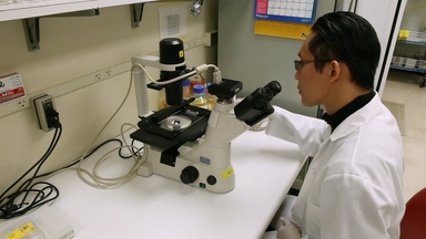
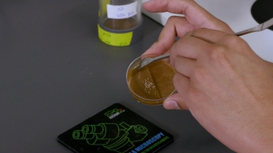
•This protocol describes the dissection and immunostaining of adult Drosophila melanogaster brain tissues. Specifically, this protocol highlights the use of Drosophila mushroom body and photoreceptor neurons as example neuronal subsets that can be accurately used to uncover general principles underlying many aspects of neuronal development.
Related Videos

Assaying Blood Cell Populations of the Drosophila melanogaster Larva

Rearing the Fruit Fly Drosophila melanogaster Under Axenic and Gnotobiotic Conditions

Dissection and Staining of Drosophila Pupal Ovaries

Thawing, Culturing, and Cryopreserving Drosophila Cell Lines

Methods to Test Endocrine Disruption in Drosophila melanogaster

Dissection, Immunohistochemistry and Mounting of Larval and Adult Drosophila Brains for Optic Lobe Visualization

Dissection and Live-Imaging of the Late Embryonic Drosophila Gonad

Investigating Asymmetric Cell Division Dynamics: A Protocol for Live-Imaging of Drosophila Larval Brain Explants (Video) | JoVE

Investigating mRNA Spatial Distribution in Drosophila Muscle Tissue (Video) | JoVE

Staging and Collecting Drosophila melanogaster Third Instar Larvae (Video) | JoVE
Copyright © 2024 MyJoVE Corporation. 판권 소유