
Ferric Chloride-Induced Arterial Thrombosis and Sample Collection for 3D Electron Microscopy Analysis
March 17th, 2023
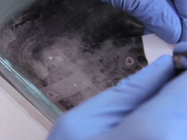
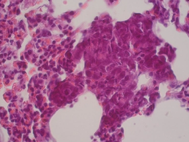
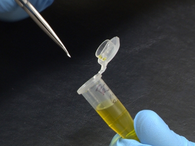
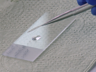
•The present protocol describes how to use a FeCl3-mediated injury to induce arterial thrombosis, and how to collect and prepare arterial injury samples at various stages of thrombosis for electron microscopy analysis.
Related Videos
Quantitative Analysis and Characterization of Atherosclerotic Lesions in the Murine Aortic Sinus
Orthotopic Injection of Breast Cancer Cells into the Mammary Fat Pad of Mice to Study Tumor Growth.
Ferric Chloride-induced Thrombosis Mouse Model on Carotid Artery and Mesentery Vessel

Ferric Chloride-induced Murine Thrombosis Models

Quantitative Visualization of Leukocyte Infiltrate in a Murine Model of Fulminant Myocarditis by Light Sheet Microscopy
Preparation and In Vitro Characterization of Magnetized miR-modified Endothelial Cells

Intravital Microscopy of Monocyte Homing and Tumor-Related Angiogenesis in a Murine Model of Peripheral Arterial Disease

Ferric Chloride-induced Canine Carotid Artery Thrombosis: A Large Animal Model of Vascular Injury

Subclavian Vein Puncture As an Alternative Method of Blood Sample Collection in Rats

Immunofluorescence Labelling of Human and Murine Neutrophil Extracellular Traps in Paraffin-Embedded Tissue
Copyright © 2024 MyJoVE Corporation. 판권 소유