Aby wyświetlić tę treść, wymagana jest subskrypcja JoVE. Zaloguj się lub rozpocznij bezpłatny okres próbny.
Method Article
Collection, Isolation and Enrichment of Naturally Occurring Magnetotactic Bacteria from the Environment
W tym Artykule
Podsumowanie
We demonstrate a method to collect magnetotactic bacteria (MTB) that can be applied to natural waters. MTB can be isolated and enriched from sediment samples using a relatively simple setup that takes advantage of the bacteria's natural magnetism. Isolated MTB can then be examined in detail using both light and electron microscopy.
Streszczenie
Magnetotactic bacteria (MTB) are aquatic microorganisms that were first notably described in 19751 from sediment samples collected in salt marshes of Massachusetts (USA). Since then MTB have been discovered in stratified water- and sediment-columns from all over the world2. One feature common to all MTB is that they contain magnetosomes, which are intracellular, membrane-bound magnetic nanocrystals of magnetite (Fe3O4) and/or greigite (Fe3S4) or both3, 4. In the Northern hemisphere, MTB are typically attracted to the south end of a bar magnet, while in the Southern hemisphere they are usually attracted to the north end of a magnet3,5. This property can be exploited when trying to isolate MTB from environmental samples.
One of the most common ways to enrich MTB is to use a clear plastic container to collect sediment and water from a natural source, such as a freshwater pond. In the Northern hemisphere, the south end of a bar magnet is placed against the outside of the container just above the sediment at the sediment-water interface. After some time, the bacteria can be removed from the inside of the container near the magnet with a pipette and then enriched further by using a capillary racetrack6 and a magnet. Once enriched, the bacteria can be placed on a microscope slide using a hanging drop method and observed in a light microscope or deposited onto a copper grid and observed using transmission electron microscopy (TEM).
Using this method, isolated MTB may be studied microscopically to determine characteristics such as swimming behavior, type and number of flagella, cell morphology of the cells, shape of the magnetic crystals, number of magnetosomes, number of magnetosome chains in each cell, composition of the nanomineral crystals, and presence of intracellular vacuoles.
Protokół
1. MTB Collection
- When deciding on a freshwater site to collect magnetotactic bacteria (MTB), it is often best to start with a pond or slow-moving stream that has a soft muddy sediment layer. In this demonstration we collected a sample at the edge of the Olentangy River on the campus of The Ohio State University (OSU) in Columbus, Ohio (USA). While this was a convenient location for our demonstration, the protocol described here is applicable to any aquatic location. The materials used in this protocol can be found in Table 1. Find a location where the depth of the water is between 10 and 100 cm. At such a location, you should collect the upper-most layer of sediment using a clear, screw-top container. Scoop the sediment and water into the container until it is filled with one-third to one-half sediment and the remaining volume with water. Keep the container submerged until it is filled with water and then tightly cap the container with its screw-top lid. It's not necessary to mix the sediment. Wipe the outside of the container dry with a towel and then take the sample to your laboratory. It's not necessary to rush the sample back to your laboratory. We've left MTB samples in plastic containers in the field for several days before bringing them back to our laboratory. The MTB should be viable for several weeks to months as long as you store the samples in a cool, shaded place in the field.
- Once the sample is in your laboratory, loosen the cap and leave it covering the container to reduce the amount of evaporation. Store the container at room temperature in a dark room, drawer, or completely cover the container with aluminum foil. Allow the sediment and fine particles to completely settle to the bottom of the container by leaving the sample undisturbed for several hours to several days. It is not necessary to mix the sediment, MTB prefer an undisturbed environment. The clear walls of the plastic container will allow you to confirm that the particles have settled to the bottom. Depending on your sample, MTB can remain alive in the sample for many months.
2. MTB Isolation
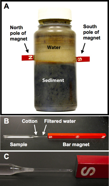
- When you are ready to isolate the MTB, place magnets on the sides of the plastic container approximately 1 cm above the sediment-water interface (Figure 1A). Be careful not to disturb the sediment in the bottom of the container. Place the south pole of a bar magnet on one side of the container and the north side of another bar magnet on the opposite side (Figure 1A). Almost any magnet can be used, such as a magnetic stir bar or large refrigerator magnet. Anything can be used to support the magnets at the correct height above the sediment-water interface. We've found that resting the magnets on the top of a cardboard or plastic box is best, however, the magnets can also be taped to the outside of the plastic container. Wait 30 min to several hours for the bacteria to swim to the magnet.
- Use a sterile pipette to carefully collect the water from inside the container (Figure 1A) near the position of the south-pole bar magnet (for samples collected in the Northern hemisphere). This water should contain the MTB that have been attracted to the south-pole bar magnet. Next, a capillary racetrack should be used to further enrich the MTB.
3. MTB Racetrack
- In order to enrich your sample with magnetotactic bacteria, a capillary racetrack is necessary (Figures 1B and 1C). These need to be made prior to isolating the cells from the clear-plastic container.
- Use a 5.75 inch (146 mm) glass Pasteur pipette to make a racetrack. Use a diamond pen or file to cut off the top of the pipette, the length of the pipette is not crucial, but it should be able to contain approximately 1-2 ml of water. Next, use a Bunsen burner to melt the tip so that it becomes sealed (Figure 1C). The resulting pipette should have an open end and a sealed end.
- Make several of these racetracks and then autoclave. Additionally, you will need to autoclave cotton and several long metal needles.
- Add filtered sample water, collected from near the sediment water interface shown in Figure 1A, to an autoclaved racetrack using a long metal needle attached to a filtered syringe. The pore size of the filter should be 0.22 mm to eliminate debris and contaminants from the water. It is important to be absolutely sure that there are no air bubbles in the glass capillary.
- Plug the bottom of the racetrack with sterile cotton (Figure 1B). Use the metal needle to push the cotton towards the sealed end of the racetrack so it is 0.5 - 1 cm away from the sealed tip (Figure 1C).
- Using a sterile pipette, add the MTB-containing water (from section 2.2) to the sample reservoir (open end) of a prepared MTB racetrack (Figure 1B).
4. MTB Enrichment
- Once the racetrack is filled with sample fluid, lay it on its side on a horizontal surface (e.g., a benchtop) and place the south pole of a bar magnet (in the Northern hemisphere) next to the sealed tip of the racetrack (Figures 1B and 1C).
- Wait 5 to 30 min for the MTB to migrate through the cotton. Then you should collect the fluid near the tip of the racetrack. Waiting too long can introduce contaminants, such as other motile bacteria, to the tip of the capillary. Optionally, you could use a light microscope to view the tip of the racetrack and watch the MTB collect at the racetrack's tip. This will allow you to determine how long it takes the MTB to migrate through the cotton plug.
- Gently use the diamond knife to make a little scratch near cotton plug and snap off the end of the racetrack.
- Use a 1 ml syringe with a narrow needle (25 or 27 gauge) to remove the fluid from the tip of the racetrack. This liquid sample should now contain the enriched MTB.
5. MTB Observation by Light Microscopy
- Place a drop (10-20 μl) of the enriched MTB sample onto a coverslip.
- Quickly flip the coverslip over so the drop is now facing down and hanging from the coverslip.
- Place the coverslip onto an o-ring that is resting on a glass slide. The o-ring should have a slightly smaller diameter then the coverslip (about 1 cm; Figure 2).
- Place this hanging drop onto a light microscope stage and focus on one edge of the drop. A 60X dry objective works very well because most have a high numerical aperture (NA; e.g., 0.93) but do not require oil, which is difficult to use for the hanging drop method (Figure 2).
- Place the south end of a bar magnet close to the hanging drop and MTB will begin to migrate towards the edge of the drop closest to the magnet (Figure 3). Within a few minutes many MTB should be at the edge of the drop (Figure 3). Prove to yourself that the bacteria are magnetic by reversing the pole of the magnet and then observe the bacteria swim in the opposite direction.
6. MTB Observation by Transmission Electron Microscopy (TEM)
- Place a drop (~20 μl) of the enriched MTB onto a copper grid and allow the bacteria to settle for 10 min.
- Wick off excess water with a piece of clean filter paper.
- Optionally, the grid can be negatively stained with 2% uranyl acetate, 2% phosphotungstic acid pH 7.2, or 2.5% sodium molybdate7, 8, 9. This is done by placing the copper grid onto a drop of stain immediately after incubating the grid with the enriched MTB. Incubate the grid with the negative stain, the times will vary depending on the stain used, and then wick off the fluid with a piece of clean filter paper.
- Observe the MTB using transmission electron microscopy (TEM, Figure 4). For the work described here MTB were adsorbed to Formvar stabilized and carbon coated 200 mesh copper grids (Ted Pella #01800). The grids were placed with the carbon side down on a drop of cell suspension for up to 10 min, then immediately washed one time by placing the grid on a drop of water for 30 sec. For staining, the grids were placed on a drop of 2% uranyl acetate (Ted Pella #19481) for 30 sec to 5 min and then dried completely using a piece of filter paper. The grids were analyzed by TEM using either an FEI Tecnai Spirit at 80kV or a FEI Tecnai F20 using high angle annular dark field STEM at 200kV.
Wyniki
A magnet is an effective tool that can be used to isolate magnetotactic bacteria (MTB) contained in environmental samples (Figure 1A). A capillary racetrack (Figure 1B) uses the magnetic properties of MTB to attract them through a cotton plug where they can be separated from non-magnetotactic microorganisms also contained within the environmental sample.

Dyskusje
Magnetotactic bacteria are not necessarily found in every aquatic environment8 but when they do occur, they can be found on the order of 100 - 1,000 cells per milliliter2. In order to observe the MTB using optical microscopy, you will need approximately 50 bacteria/ml in your sample8. If there are no or few MTB in your sample, then you will either need to select a new environmental site to collect your samples or you will need to try one or more of the techniques discussed in the next sec...
Ujawnienia
No conflicts of interest declared.
Podziękowania
This work was supported by grants from the U.S. National Science Foundation (EAR-0920299 and EAR-0745808); U.S. National Science Foundation East Asian and Pacific Summer Institutes; the Geological Society of America Research Grant Program and the Alumni Grants for Graduate Research and Scholarship from The Ohio State University. We would like to thank the editor and two anonymous reviewers for their insightful comments.
Materiały
| Name | Company | Catalog Number | Comments |
| Glass slides | Fisher Scientific | S95933 | |
| Glass Pasteur pipets | Fisher Scientific | 13-678-6A | |
| O-ring | Hardware store | ||
| Cover slips | Fisher Scientific | 12-542B | |
| Bar magnet | Fisher Scientific | S95957 | |
| Container | Any | Any plastic or glass container that can hold at least 0.5 L and can be sealed | |
| Cotton | Any | ||
| Microscope with 60X dry lens | Zeiss | A 60X dry lens is not absolutely necessary, but this gives a high NA without using oil | |
| Diamond pen | Fisher Scientific | 08-675 | |
| 0.22 mm filter | Fisher Scientific | 09-719C | |
| 1 ml syringe | Fisher Scientific | NC9788564 | |
| Microcentrifuge tubes | Fisher Scientific | 02-681-320 | |
| Formvar/Carbon 200 mesh, copper grids | Ted Pella Inc. | 01800 | |
| Uranyl acetate | Ted Pella Inc. | 19481 | |
| Tecnai Spirit TEM | FEI | ||
| Tecnai F20 S/TEM | FEI |
Odniesienia
- Blakemore, R. Magnetotactic bacteria. Science. 190, 377-379 (1975).
- Blakemore, R. P. Magnetotactic bacteria. Annual Reviews in Microbiology. 36, 217-238 (1982).
- Bazylinski, D. A., Frankel, R. B. Controlled Biomineralization of Magnetite (Fe3O4) and Greigite (Fe3S4) in a Magnetotactic Bacterium. Applied and Environmental Microbiology. 61, 3232-3239 (1995).
- Lefevre, C. T., Menguy, N., et al. A Cultured Greigite-Producing Magnetotactic Bacterium in a Novel Group of Sulfate-Reducing Bacteria. Science. 334, 1720-1723 (2011).
- Simmons, S. L., Bazylinski, D. A., et al. South-seeking magnetotactic bacteria in the Northern Hemisphere. Science. 311, 371-374 (2006).
- Wolfe, R., Thauer, R., et al. A 'capillary racetrack' method for isolation of magnetotactic bacteria. FEMS Microbiology Letters. 45, 31-35 (1987).
- Rodgers, F. G., Blakemore, R. P. Intercellular structure in a many-celled magnetotactic prokaryote. Archives of Microbiology. 154, 18-22 (1990).
- Moench, T. T., Konetzka, W., et al. A novel method for the isolation and study of a magnetotactic bacterium. Archives of Microbiology. 119, 203-212 (1978).
- Balkwill, D., Maratea, D. Ultrastructure of a magnetotactic spirillum. Journal of Bacteriology. 141, 1399-1408 (1980).
- Lins, U., Freitas, F., et al. Simple homemade apparatus for harvesting uncultured magnetotactic microorganisms. Brazilian Journal of Microbiology. 34, 111-116 (2003).
- Jogler, C., Lin, W., et al. Toward Cloning of the Magnetotactic Metagenome: Identification of Magnetosome Island Gene Clusters in Uncultivated Magnetotactic Bacteria from Different Aquatic Sediments. Applied and Environmental Microbiology. 75, 3972-3979 (2009).
- Lin, W., Li, J., et al. Newly Isolated but Uncultivated Magnetotactic Bacterium of the Phylum Nitrospirae from Beijing, China. Applied and Environmental Microbiology. 78, 668-675 (2012).
- Li, J., Pan, Y., et al. Biomineralization, crystallography and magnetic properties of bullet-shaped magnetite magnetosomes in giant rod magnetotactic bacteria. Earth and Planetary Science Letters. 293, 368-376 (2010).
- Oestreicher, Z., Valerde-Tercedor, C. Magnetosomes and magnetite crystals produced by magnetotactic bacteria as resolved by atomic force microscopy and transmission electron microscopy. Micron. 43, 1331-1335 (2012).
Przedruki i uprawnienia
Zapytaj o uprawnienia na użycie tekstu lub obrazów z tego artykułu JoVE
Zapytaj o uprawnieniaThis article has been published
Video Coming Soon
Copyright © 2025 MyJoVE Corporation. Wszelkie prawa zastrzeżone