Aby wyświetlić tę treść, wymagana jest subskrypcja JoVE. Zaloguj się lub rozpocznij bezpłatny okres próbny.
Method Article
Quantitative Characterization of Liquid Photosensitive Bioink Properties for Continuous Digital Light Processing Based Printing
* Wspomniani autorzy wnieśli do projektu równy wkład.
W tym Artykule
Podsumowanie
This study uses temperature and material composition to control the yield stress properties of yield stress fluids. The solid-like state of the ink can protect the printing structure, and the liquid-like state can continuously fill the printing position, realizing the digital light processing 3D printing of extremely soft bioinks.
Streszczenie
Precise printing fabrication of bioinks is a prerequisite for tissue engineering; the Jacobs working curve is the tool to determine the precise printing parameters of digital light processing (DLP). However, the acquisition of working curves wastes materials and requires high formability of materials, which are not suitable for biomaterials. In addition, the reduction of cell activity due to multiple exposures and the failure of structural formation due to repeated positioning are both unavoidable problems in conventional DLP bioprinting. This work introduces a new method of obtaining the working curve and the improvement process of continuous DLP printing technology based on such a working curve. This method of obtaining the working curve is based on the absorbance and photorheological properties of the biomaterials, which do not depend on the formability of the biomaterials. The continuous DLP printing process, obtained from improving the printing process by analyzing the working curve, increases the printing efficiency more than tenfold and greatly improves the activity and functionality of cells, which is beneficial to the development of tissue engineering.
Wprowadzenie
Tissue engineering1 is important in the field of organ repair. Due to the lack of organ donation, some diseases, such as liver failure and kidney failure, cannot be cured well, and many patients do not receive timely treatment2. Organoids with the required function of the organs may solve the problem caused by the lack of organ donation. The construction of organoids depends on the progress and development of bioprinting technology3.
Compared with extrusion-type bioprinting4 and inkjet-type bioprinting5, the printing speed and printing accuracy of the digital light processing (DLP) bioprinting method are higher6,7. The printing module of the extrusion-type method is line-by-line, while the printing module of the inkjet-type method is dot-by-dot, which is less efficient than the layer-by-layer printing module of DLP bioprinting. The modulated ultraviolet (UV) light exposure to a whole layer of material to cure a layer in DLP bioprinting and the feature size of the image determines the accuracy of DLP printing. This makes DLP technology very efficient8,9,10. Due to overcuring of the UV light, the precise relationship between the curing time and the printing size is important for high-accuracy DLP bioprinting. Furthermore, continuous DLP printing is a modification of DLP printing method that can greatly improve the printing efficiency11,12,13. For continuous DLP printing, precise printing conditions are the most important factors.
The relationship between the curing time and the printing size is called the Jacobs working curve, which is widely used in DLP printing14,15,16. The traditional method to obtain the relationship is to expose the material for a certain time and measure the curing thickness to obtain a data point about the exposure time and curing thickness. Repeating this operation at least five times and fitting the data points obtains the Jacobs working curve. However, this method has obvious disadvantages; it needs to consume a lot of material to achieve the curing, the results are highly dependent on the printing conditions, the bioinks used in DLP bioprinting are expensive and rare, and the formability of the bioinks is usually not good, which can lead to inaccurate measurements of curing thickness.
This article provides a new method to obtain the curing relationship according to the physical properties of the bioink. Using this theory can optimize continuous DLP printing. This method can be used to obtain the curing relationship more quickly and accurately; the continuous DLP curing can therefore be better determined.
Access restricted. Please log in or start a trial to view this content.
Protokół
1. Theoretical preparation
- Define three parameters: liquid absorbance (Al), solid absorbance (As), and threshold time (tT)17.
- Rewrite the traditional Jacobs working curve using these three parameters17 according to Equation 1:
 (Equation 1)
Here, tH is the curing time of one single layer, and H is the height of one single layer.
2. Parameter acquisition
- Measure the threshold time of the bioink using a rheometer equipped with an element for temperature control.
- Use a 365 nm light source to expose the testing platform of the rheometer and make the light intensity at a certain value.
- Set the rheometer to get the Time-Moduli data during a period of 300 s, and take each data point every 0.3 s through the Time Settings options in the rheometer software.Click the Start Test button of the rheometer to start the test, and at the same time, click the Start Button of the light source.
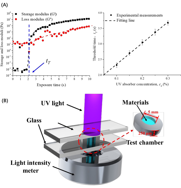
- Counting from the start of exposure, when the storage modulus data is equal to the loss modulus data, the corresponding time is recognized as the threshold time. Record manually.
- Build the absorbance test equipment as shown in the previous work17. Use two upper and lower glass slides to clamp the ring-shaped printed structure (5 mm inner diameter, 10 mm outer diameter) with a thickness of 500µm so that the inner circle of the ring forms a chamber. Place the chamber on the test area of the light intensity meter and set the light source to expose the chamber area.
NOTE: Figure 1 shows the schematic diagram of photorheological test results and data processing results, and the absorbance testing equipment.- Measure the incident light intensity (Ii) when the test chamber is not filled with material from the absorbance test equipment by reading the display of the light intensity meter of the testing equipment.
- Fill the test chamber with 10 µLof bioink.
- Expose the test chamber with bioink to UV light at 365 nm. Obtain the light intensity (Ilh) from the absorbance test equipment by reading the display of the light intensity meter of the testing equipment.
- Obtain the light intensity when the bioink is cured (Ish) from the absorbance test equipment by reading the display of the light intensity meter of the testing equipment when the value no longer changes. This value is the solid absorbance, Ish.
- Calculate the liquid absorbance and solid absorbance using Equations 2 and 3:
 Equation 2
 Equation 3
- Obtain the Jacobs working curve according to the obtained parameters.

Figure 1: Test results and equipment. (A) Schematic diagram of photorheological test results and data processing results. (B) Absorbance testing equipment. This figure has been modified with permission from Li et al.17. Please click here to view a larger version of this figure.
3. Continuous DLP printing parameter settings
- Use DLP software to achieve DLP printing, and the set of printing parameters in the software as follows.
- Set the exposure time of the first single layer as the threshold time (tT) in the software's parameter settings.
- Calculate the exposure time of curing 10 µmthick materials according to Equation 1 and subtract the threshold time to obtain the real exposure time for curing a single layer.
- Set the time interval between adjacent layers to 0 s in the software's parameter settings.
- Start the printer by clicking the Start button in the printing software. When the printing process ends, finish printing by clicking the Stop button in the printing software.
Access restricted. Please log in or start a trial to view this content.
Wyniki
This article shows a new method to obtain curing parameters and introduces a new way to achieve continuous DLP printing, demonstrating the efficiency of this method in determining the working curve.
We used three different materials in DLP printing to verify the accuracy of the theoretical working curve obtained by the method introduced in this article. The materials are 20% (v/v) polyethylene (glycol) diacrylate (PEGDA), 0.5% (w/v) lithium phenyl-2,4,6-trimethylbenzoylphosphinate (LAP) with d...
Access restricted. Please log in or start a trial to view this content.
Dyskusje
The critical steps of this protocol are described in section 2. It is necessary to unify the light intensity used in the photorheology test and the printing light intensity in the actual tests. The absorbance testing equipment is the most important part. The shape of the test chamber should be the same as the photosensitive area of the light intensity meter. Due to the properties of the materials that continuously change during the whole UV light exposure process, the light intensity needs to continue to change
Access restricted. Please log in or start a trial to view this content.
Ujawnienia
The authors have nothing to disclose.
Podziękowania
The authors gratefully acknowledge the support provided by the National Natural Science Foundation of China (Grant Nos. 12125205, 12072316, 12132014), and the China Postdoctoral Science Foundation (Grant No. 2022M712754).
Access restricted. Please log in or start a trial to view this content.
Materiały
| Name | Company | Catalog Number | Comments |
| Brilliant Blue | Aladdin (Shanghai, China). | 6104-59-2 | |
| DLP software | Creation Workshop | N/A | |
| Lithium phenyl-2,4,6-trimethylbenzoylphosphinate | N/A | LAP; synthesized | |
| Light source | OmniCure | https://www.excelitas.com/product-category/omnicure-s-series-lamp-spot-uv-curing-systems | 365 nm |
| Polyethylene (glycol) diacrylate | Sigma-Aldrich | 455008 | PEGDA Mw ~700 |
| Rheometer | Anton Paar, Austria | MCR302 |
Odniesienia
- Berthiaume, F., Maguire, T. J., Yarmush, M. L. Tissue engineering and regenerative medicine: history, progress, and challenges. Annual Review of Chemical and Biomolecular Engineering. 2 (1), 403-430 (2011).
- Ng, W. L., Chua, C. K., Shen, Y. -F. Print me an organ! Why we are not there yet. Progress in Polymer Science. 97, 101145(2019).
- Sun, W., et al. The bioprinting roadmap. Biofabrication. 12 (2), 022002(2020).
- Jiang, T., Munguia-Lopez, J. G., Flores-Torres, S., Kort-Mascort, J., Kinsella, J. M. Extrusion bioprinting of soft materials: An emerging technique for biological model fabrication. Applied Physics Reviews. 6 (1), 011310(2019).
- Ng, W., et al. L.cControlling droplet impact velocity and droplet volume: Key factors to achieving high cell viability in sub-nanoliter droplet-based bioprinting. International Journal of Bioprinting. 8 (1), 424(2021).
- Yu, K., et al. Printability during projection-based 3D bioprinting. Bioactive Materials. 11, 254-267 (2022).
- Zhong, Z., et al. Bioprinting of dual ECM scaffolds encapsulating limbal stem/progenitor cells in active and quiescent statuses. Biofabrication. 13 (4), (2021).
- Huh, J., et al. Combinations of photoinitiator and UV absorber for cell-based digital light processing (DLP) bioprinting. Biofabrication. 13 (3), (2021).
- Saed, A. B., et al. Functionalized poly l-lactic acid synthesis and optimization of process parameters for 3D printing of porous scaffolds via digital light processing (DLP) method. Journal of Manufacturing Processes. 56, 550-561 (2020).
- Ng, W. L., et al. Vat polymerization-based bioprinting-process, materials, applications and regulatory challenges. Biofabrication. 12 (2), 022001(2020).
- Li, Y., et al. High-fidelity and high-efficiency additive manufacturing using tunable pre-curing digital light processing. Additive Manufacturing. 30, 100889(2019).
- Kelly, B. E., et al. Volumetric additive manufacturing via tomographic reconstruction. Science. 363 (6431), 1075-1079 (2019).
- Tumbleston, J. R., et al. Continuous liquid interface production of 3D objects. Science. 347 (6228), 1349-1352 (2015).
- Classens, K., Hafkamp, T., Westbeek, S., Remmers, J. J. C., Weiland, S. Multiphysical modeling and optimal control of material properties for photopolymerization processes. Additive Manufacturing. 38, 101520(2021).
- Gong, H., Beauchamp, M., Perry, S., Woolley, A. T., Nordin, G. P. Optical approach to resin formulation for 3D printed microfluidics. RSC Advances. 5 (129), 106621-106632 (2015).
- Hofstetter, C., Orman, S., Baudis, S., Stampfl, J. Combining cure depth and cure degree, a new way to fully characterize novel photopolymers. Additive Manufacturing. 24, 166-172 (2018).
- Li, Y., et al. Theoretical prediction and experimental validation of the digital light processing (DLP) working curve for photocurable materials. Additive Manufacturing. 37, 101716(2021).
- Wang, M., et al. Molecularly cleavable bioinks facilitate high-performance digital light processing-based bioprinting of functional volumetric soft tissues. Nature Communications. 13 (1), 3317(2022).
Access restricted. Please log in or start a trial to view this content.
Przedruki i uprawnienia
Zapytaj o uprawnienia na użycie tekstu lub obrazów z tego artykułu JoVE
Zapytaj o uprawnieniaThis article has been published
Video Coming Soon
Copyright © 2025 MyJoVE Corporation. Wszelkie prawa zastrzeżone