
In Vitro Canine Neutrophil Extracellular Trap Formation: Dynamic and Quantitative Analysis by Fluorescence Microscopy
August 24th, 2018
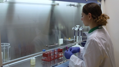
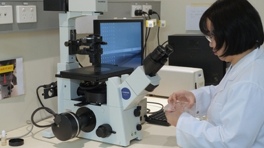
•We describe methods to isolate canine neutrophils from whole blood and visualize NET formation in live neutrophils using fluorescence microscopy. Also described are protocols to quantify NET formation and citrullinated histone H3 (citH3) expression using immunofluorescence microscopy.
Tags
Vídeos Relacionados
Neutrophil Extracellular Traps: How to Generate and Visualize Them

Tracking Neutrophil Intraluminal Crawling, Transendothelial Migration and Chemotaxis in Tissue by Intravital Video Microscopy

Quantitative In vitro Assay to Measure Neutrophil Adhesion to Activated Primary Human Microvascular Endothelial Cells under Static Conditions
High Throughput Measurement of Extracellular DNA Release and Quantitative NET Formation in Human Neutrophils In Vitro
Measuring Phagosome pH by Ratiometric Fluorescence Microscopy

Methods to Study Lipid Alterations in Neutrophils and the Subsequent Formation of Neutrophil Extracellular Traps
A Simple Fluorescence Assay for Quantification of Canine Neutrophil Extracellular Trap Release
A High-throughput Assay to Assess and Quantify Neutrophil Extracellular Trap Formation
In Vitro Stimulation and Visualization of Extracellular Trap Release in Differentiated Human Monocyte-derived Macrophages

Identification of Neutrophil Extracellular Traps in Paraffin-Embedded Feline Arterial Thrombi using Immunofluorescence Microscopy
SOBRE A JoVE
Copyright © 2024 MyJoVE Corporation. Todos os direitos reservados