Войдите в систему

Nose, Sinuses, Oral Cavity and Pharynx Exam

Обзор

Source: Richard Glickman-Simon, MD, Assistant Professor, Department of Public Health and Community Medicine, Tufts University School of Medicine, MA

This video provides an overview of sinus, nose, and throat examinations. The demonstration begins with a brief overview of the anatomy of the region. The upper third of the nose is bony, and the bottom two-thirds are cartilaginous. Air entering the nares passes through the nasal vestibules and into the narrow passageway between the nasal septum medially and the bony turbinates laterally. Beneath each curving turbinate is a groove or meatus. The nasolacrimal duct and most of the air-filled paranasal sinuses drain into the inferior and middle meatuses, respectively. Of the three sets of paranasal sinuses, only the maxillary and frontal can be readily examined. A continuous, highly vascular mucosa lines the entire nasal cavity and sinuses.

Figure 1
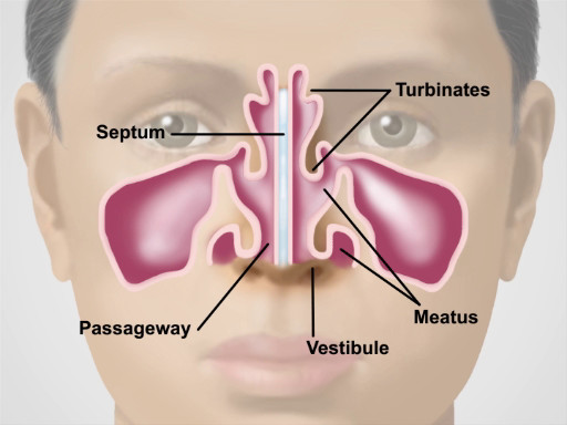
Figure 1. Anatomy of the Nose.

Figure 2
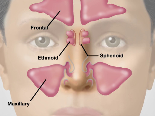
Figure 2. Location of the Major Sinuses.

Muscular folds of the lips mark the entrance to the oral cavity. The pinkish gingiva, or gums, attach firmly to the teeth and adjacent bone into which the teeth embed. The buccal mucosa lines the inner cheeks, where the Stensen duct drains the parotid glands into small papillae near the upper second molars. Small, red papillae cover the dorsal surface of the tongue along with a whitish coat of varying thickness. The midline lingual frenulum connects the undersurface of the tongue to the floor of the mouth, where the Wharton duct drains the submandibular glands into papillae on either side. Arching over the tongue are the anterior and posterior pillars of the soft palate and midline uvula. If present, the tonsils can be seen protruding bilaterally between the pillars. The bony hard palate rises anteriorly above the soft palate. The pharynx lies posteriorly behind the soft palate and tongue.

Figure 3
Figure 3. Anatomy of the Oral Cavity.

Figure 4
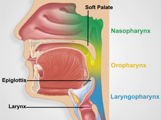
Figure 4. Pharyngeal Regions.

Процедура

1. Nose and Sinus Exam

  1. Inspect the nose for skin changes, nodules, and deformities. Slight deviation of the septum is common, and benign if it does not cause airflow obstruction.
  2. Press gently on the tip of the nose to widen the nares, and inspect the vestibule with the aid of a pen light.
  3. Conduct the nose cavity exam with an otoscope.
    1. Using the largest ear speculum available, inspect the nasal cavity with the otoscope.
    2. Tilt the patient's head back slightly, and holding the otoscope handle perpendicular and away from the patient's chin, gently place the speculum in the nasal vestibule, avoiding contact with the septum.
    3. When looking through the otoscope, gradually move the speculum posteriorly and angle it upward to glimpse the lower and middle turbinates, the nasal septum, and the narrow intervening passage.
    4. Note the color of the mucosa and the presence of any swelling, polyps, ulcerations, bleeding, discharge, blockages, deviations, or perforations.
  4. Palpate the frontal and maxillary sinuses for tenderness.
    1. Bilaterally position your first and second fingers on top of the patient's forehead while firmly pressing your thumbs upward under the bony brows. Avoid placing any pressure on the eyes.
    2. Keeping your fingers in place, move your thumbs down and press firmly on the cheeks, just medial and inferior to the zygomatic bone.
  5. If sinusitis is suspected, consider performing an illumination test.
    1. Darken the room and direct the brightest light of the otoscope upward beneath each brow, close to the nose.
    2. Look for a red glow illuminating the air-filled frontal sinuses.
    3. Have the patient tilt the head back with the mouth open wide.
    4. After removing any upper dentures, place the light source directly over each air-filled maxillary sinus and look for a red glow illuminating the hard palate on each side. Failure to see the red glow suggests mucosal thickening or secretions in the corresponding sinus.

2. Oral Cavity and Pharyngeal Exam

  1. Observe the lips for color, moisture, ulcers, or cracking.
  2. With a fresh tongue depressor and bright light in hand, ask the patient to open the mouth wide. Inspect the gums for bleeding, ulcers, or swelling, and check to see if any teeth are missing, discolored, misshapen, or loose.
  3. Carefully inspect the buccal mucosa for ulcers, nodules, or white patches.
  4. Inspect the tongue.
    1. Ask the patient to protrude the tongue and move it from side to side, assessing for symmetry, and inspect the color and texture of its dorsal surface. Oral cancers most commonly develop on the sides and base of the tongue.
    2. Have the patient touch the tongue to the hard palate, and carefully inspect its undersurface and the floor of the mouth.
    3. Using a gloved hand, gently grasp the tip of the tongue with a square piece of gauze and move it from side to side, inspecting carefully for ulcerations, plaques, masses, or discoloration.
  5. Inspect the pharynx.
    1. With the tongue back inside, have the patient open wide and say "ah" or yawn.
    2. If the pharynx cannot be seen clearly, have the patient repeat this maneuver while you firmly press down on the tongue with the tongue depressor. Take care not to gag the patient.
    3. Watch the soft palate rise, and inspect the uvula, anterior and posterior pillars, tonsils (if present), and pharynx. Check for symmetry, discoloration, ulcerations, swelling, masses, or tonsillar exudate.
Заявка и Краткое содержание

Because of their constant exposure to the environment, the nose, sinuses, oral cavity, and pharynx often suffer from infections and other inflammatory conditions. Viral and allergic rhinitis are by far the most common nasal disorders. Both can produce copious watery discharge, but viral infections tend to produce a deep red mucosa, while allergies produce a pale, bluish one. Other common conditions found on nasal examination include epistaxis, septal deviation, and nasal polyps. Tenderness over the paranasal sinuses suggests acute sinusitis, particularly if accompanied by face pain, fever, and purulent nasal discharge. In such cases, transilluminating the sinuses may be helpful. To avoid missing a lurking carcinoma, it is important to thoroughly inspect the oral mucosa, particularly on the sides and base of the tongue where oral carcinomas are most likely to occur. Other common disorders to look for include gingivitis, gum recession, teeth erosion, aphthous ulcers, exudative tonsillitis, viral pharyngitis, candidiasis, and leukoplakia.

Теги
NoseSinusesOral CavityPharynxExamInterconnected StructuresRespiratory SystemDigestive SystemInfectionsInflammatory ConditionsExamineEssential SkillPhysiciansVideo TutorialAnatomy ReviewPhysical ExaminationSense Of SmellWarming And Moistening AirFiltering AirBony NoseCartilaginous NoseNasal VestibulesNasal SeptumBony TurbinatesMeatusParanasal SinusesMaxillary SinusFrontal SinusSphenoidal SinusEthmoidal Sinus

Перейти к...

0:00

Overview

0:43

Anatomy of the Nose and Sinuses

1:49

Nose and Sinus Examination

4:56

Anatomy of the Oral Cavity and Pharynx

6:31

Oral Cavity and Pharyngeal Exam

8:20

Summary

Видео из этой коллекции:

article

Now Playing

Nose, Sinuses, Oral Cavity and Pharynx Exam

Physical Examinations II

64.6K Просмотры

article

Eye Exam

Physical Examinations II

75.8K Просмотры

article

Ophthalmoscopic Examination

Physical Examinations II

66.7K Просмотры

article

Ear Exam

Physical Examinations II

53.6K Просмотры

article

Thyroid Exam

Physical Examinations II

103.5K Просмотры

article

Lymph Node Exam

Physical Examinations II

380.9K Просмотры

article

Abdominal Exam I: Inspection and Auscultation

Physical Examinations II

200.5K Просмотры

article

Abdominal Exam II: Percussion

Physical Examinations II

245.4K Просмотры

article

Abdominal Exam III: Palpation

Physical Examinations II

137.5K Просмотры

article

Abdominal Exam IV: Acute Abdominal Pain Assessment

Physical Examinations II

66.4K Просмотры

article

Male Rectal Exam

Physical Examinations II

112.4K Просмотры

article

Comprehensive Breast Exam

Physical Examinations II

85.4K Просмотры

article

Pelvic Exam I: Assessment of the External Genitalia

Physical Examinations II

299.4K Просмотры

article

Pelvic Exam II: Speculum Exam

Physical Examinations II

148.1K Просмотры

article

Pelvic Exam III: Bimanual and Rectovaginal Exam

Physical Examinations II

145.3K Просмотры

JoVE Logo

Исследования

Образование

О JoVE

Авторские права © 2025 MyJoVE Corporation. Все права защищены