Method Article
Cell Block Preparation from Cytology Specimen with Predominance of Individually Scattered Cells
Summary
Shidham's method for preparation of cell blocks with AV-marker from cytology specimens containing individually scattered cells and small cell groups.
Abstract
This video demonstrates Shidham's method for preparation of cell blocks from liquid based cervicovaginal cytology specimens containing individually scattered cells and small cell groups. This technique uses HistoGel (Thermo Scientific) with conventional laboratory equipment.
The use of cell block sections is a valuable ancillary tool for evaluation of non-gynecologic cytology. They enable the cytopathologist to study additional morphologic specimen detail including the architecture of the lesion. Most importantly, they allow for the evaluation of ancillary studies such as immunocytochemistry, in-situ hybridization tests (FISH/CISH) and in-situ polymerase chain reaction (PCR). Traditional cell block preparation techniques have mostly been applied to non-gynecologic cytology specimens, typically for body fluid effusions and fine needle aspiration biopsies.
Liquid based cervicovaginal specimens are relatively less cellular than their non-gynecologic counterparts with many individual scattered cells. Because of this, adequate cellularity within the cell block sections is difficult to achieve. In addition, the histotechnologist sectioning the block cannot visualize the level at which the cells are at the highest concentration. Therefore, it is difficult to monitor the appropriate level at which sections can be selected to be transferred to the glass slides for testing. As a result, the area of the cell block with the cells of interest may be missed, either by cutting past or not cutting deep enough. Current protocol for Shidham's method addresses these issues. Although this protocol is standardized and reported for gynecologic liquid based cytology specimens, it can also be applied to non-gynecologic specimens such as effusion fluids, FNA, brushings, cyst contents etc for improved quality of diagnostic material in cell block sections.
Protocol
Introduction:
This is a video describing Shidham’s method for cell block preparation from liquid based cytology (LBC) specimen using HistoGelTM (Thermo Scientific) (HG). As compared to other random approaches, the following are the two critical features of this protocol for preparing cell blocks from relatively hypocellular specimens with singly scattered loose cells (1-5).
- This protocol involves steps to concentrate the cells along the plane parallel to the cutting surface of the cell block.
- It also includes a beacon-like dark inclusion of AV-marker (Figure 1b), which serves two of the following purposes:
- To visualize the level at which the cells are concentrated. The area of the cell block with the cells of interest now could be visualized by the histotechnologist when the dark colored beacon is exposed during cutting. This ability to monitor would prevent one from cutting through the level with most of the cells, or not cutting too superficial into the level with highest concentration of sample cells.
- To serve as a locator reference point in serial cell block section on different slides. This reference point acts as a beacon to help locate particular cells or groups of cells for evaluation of a coordinate immunoreactivity pattern with the SCIP approach (6,7).
PROTOCOL (Figure 2)
Preparation of the sample.
- Transfer the residual LBC cervical cytology specimen to a flat bottom glass tube (15mm diameter x 45mm) (Figure 2.1 through 2.4). Place the glass tube into a larger plastic carrier tube (28 x 85mm) and centrifuge. Remove the glass bottom tube from the carrier tube and pour off the supernatant.
- The glass tube is then capped (to prevent spillage of heating water in the next step) and placed back inside a larger flat bottom carrier plastic tube
- The carrier plastic tube containing the glass tube is then capped, placed in a centrifuge (with swiveling cups and not fixed angle cups so that the cells fall perpendicularly to the flat bottom of the glass tube), and spun at 1805 G (3000 rpms, rotor radius- 17cm) for five minutes (Figure 2.5).
- The tubes are then removed vertically from the centrifuge and the smaller glass tube is removed with forceps from the larger carrier plastic tube without disturbing the sedimented pellet with cells.
- The glass tube with specimen is uncapped and the supernatant is poured off taking care not to disturb the flat layer of sediment cells at the bottom (Figure 2.6).
Inclusion of the reference coordinate AV-marker and addition of gel
- A dark beacon AV-marker (about 2 mm X 2 mm size, flat surfaced, fragment of dark colored, sectionable material) (Figure 3) is added as a signpost to the glass tube (Figure 2.7).
- Liquefy an aliquot of HG by melting it in microwave for 10 seconds at medium power.
- Add 0.5 ml of molten HG to the tube, mix with the sediment quickly, and recap it (Figure 2.8) (Proceed to the next step quickly without allowing the HG to begin solidifying).
- Add about 2.5 ml of warm (45° C) water to the carrier plastic tube (Figure 2.9).
- The smaller capped glass tube is placed inside the plastic tube with warm water. (This step is necessary to keep the HG from solidifying during the next steps) (Figure 2.9).
- The carrier plastic tube is placed in the centrifuge (with swiveling cups and not fixed angle cups so that the cells fall perpendicularly to the flat bottom of the glass tube), and spun at 1805 G (3000 rpms, rotor radius- 17cm) for five minutes. The purpose of this centrifugation step is to push the AV-marker and to concentrate the cells into a layer closer to the cutting surface of the final paraffin embedded cell block (Figure 1d).
- The tubes are then removed gently and vertically from the centrifuge taking care not to disturb the sedimented thin layer with sample cells at the bottom.
- The larger plastic tube is uncapped and the smaller glass tube is removed vertically by a forceps without disturbing the sediment layer of specimen cells.
- The small glass tube is refrigerated in vertical position for 15 minutes to cool and solidify the HG (Figure 2.11).
Removal of the cell block as a button of gel with specimen for final processing
- The solidified HG disk, with the layer of concentrated/sediment specimen at the bottom is dislodged from the flat bottom glass tube by squirting 10% formalin through a 23 gauge needle with the syringe (Figure 2.12).
- The needle is inserted along the side of the tube at the periphery of solidified HG disc with specimen (Figure 2.12).
- The needle is rotated along the side of the tube while formalin is slowly pushed through the syringe. This results in the separation of the HG button along with dark colored beacon AV-marker and the concentrated specimen in it from the flat bottom of the glass tube (Figure 2.12).
- The cell block (gel button with specimen cells) is then placed in a labeled cassette and submitted for tissue processing to prepare paraffin embedded cell blocks (Figure 2.13).
Embedding and cutting of the specimen
- The disk is embedded in paraffin with the dark beacon marker side down as cutting surface (Figure 1).
- The block is sectioned until the dark colored AV-marker as a beacon is exposed and clearly visible.
- Three to four micron sections are cut from this level which should contain most of the singly scattered cells from the specimen.
- The sections are collected on the glass slide for further staining, immunohistochemical staining, or other tests as indicated. The protocols for these tests including the type of slides to use for mounting the sections may vary. Generally for immunostaining, coated slides are used to prevent floating and loss of sections from the slides during the immunostaining steps.
Abbreviations used (in alphabetic order): CISH, chromogenic in-situ hybridization tests; FISH, Fluorescent in-situ hybridization tests; FFPE, formalin-fixed paraffin-embedded; FNA, fine needle aspiration; HG, HistoGel™ (Thermo Scientific); LBC, liquid based cytology; PCR polymerase chain reaction;
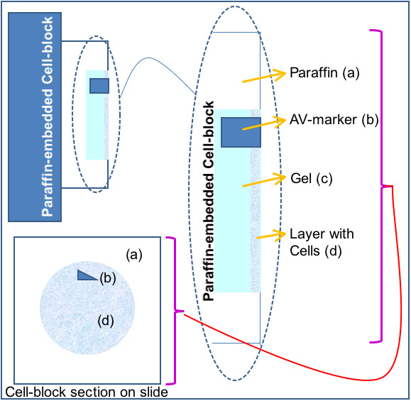
Figure 1. The structure of cell block prepared by Shidham’s protocol.
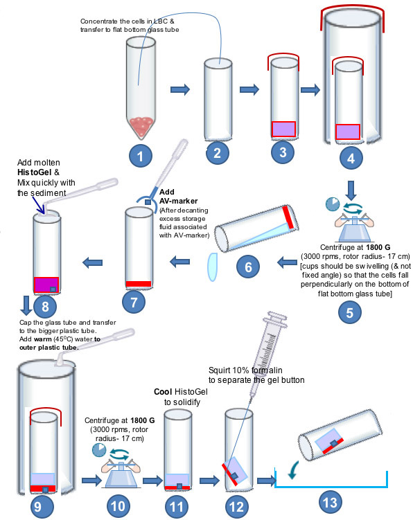
Figure 2. The summary of different steps for preparing cell block from LBC specimen by Shidham’s protocol.
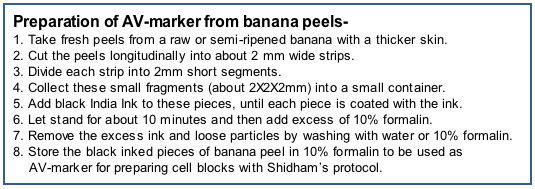
Figure 3. Preparation of AV-marker from banana peels.
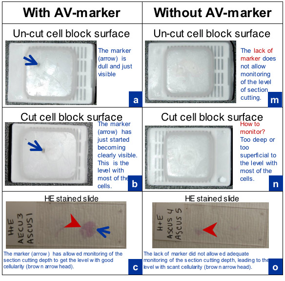
Figure 4. Comparison of cell blocks and sections with and without AV-marker.
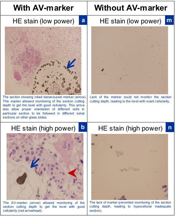
Figure 5. Comparison of cellularity of sections of cell block with and without AV-marker.
Discussion
Cell blocks are a valuable tool for evaluation of various cytology specimens (1). Most importantly, in addition to the architectural details of the specimen, cell blocks allow for evaluation of ancillary studies such as immunocytochemistry, fluorescent/chromogenic in-situ hybridization tests (FISH/CISH) and in-situ PCR. A variety of methods for preparation of cell block are described. However, most of these are suitable for non-gynecologic cytology specimens which contain relatively many cells and with tissue microfragments such as in FNA aspirates and some serous fluids such as effusions (1,3,4,5).
Because cell blocks primarily provide the opportunity for immunocytochemical evaluation, their processing should preferably be similar to that of the formalin-fixed paraffin-embedded (FFPE) tissues. Ultimately the results obtained after immunocytochemical evaluation of cell block sections are compared to those with the published literature performed predominantly on FFPE tissue sections. Any alterations in the protocol potentially compromise and nullify the validity of the results obtained on cell blocks processed through different fixatives and reagent sequences other than that used for routine FFPE. Some commercial techniques may have the drawback of processing through a protocol of exposure to other fixative-reagent exposure.
Various methods of cell block preparation are available for specimens with a significant quantity of sediment and tissue fragments. Principally, the concentrated sediments are supported by some gel or coagulation principle. The maneuverable button is then embedded in paraffin after processing like the surgical pathology specimens/biopsies.
The gels used include gelatin, agar, fibrinogen/plasma-thrombin, and other commercial gels such as HG. The methods of concentration vary from simple pelleting of the sediment by centrifugation to concentration of cells along various types of membranes. Examples include: Milipore, collodin (Celloidin) bags or scraping the cells from the cytology smears on glass slides (1). We also evaluated a variety of gels by trying different combinations of agar and gelatin. None of the combinations achieved the a firm enough consistency to obtain an easily maneuverable disc of solidified gel with embedded cells from the specimen in one piece. HG showed appropriate consistency and in our experience the immunostaining results on HG cell block sections have been excellent.
Liquid based cytology (LBC) specimens for cervicovaginal cytology are generally less cellular than non-gynecologic specimens as mentioned above. In addition, the gynecologic LBC specimens predominantly contain individual scattered exfoliated superficial cells from cervicovaginal mucosa. Due to this, appropriate cellularity within the cell block sections may not be achieved without a special approach. As these singly scattered cellular components in the the block cannot be seen by the histotechnologist during section cutting, the level at which the cells start appearing in the sections cannot be appreciated and may be missed, either by cutting past the level with most cells or not cutting deep enough into the level with highest concentration of sample cells (Figure 4). Shidham’s protocol addresses both of these issues using HG as embedding medium and conventional lab equipments (2) (Figure 2).
This protocol involves following two major features (Figure 1):
- Concentration and alignment of singly scattered cells in a narrow plane adjacent and parallel to the cutting surface of the cell block (Figure 5).
- Inclusion of a beacon-like dark AV-marker as a signpost. This is critical for achieving following features:
- Identifying and monitoring the level at which the cells are concentrated in the cell block. The exposure of the dark colored signpost highlights the level at which most of the singly scattered cells in the cell block are expected to be located (Figure 4). This prevents the overcutting (cutting past the level with most cells) or undercutting (not cutting deep enough into the level with highest concentration of sample cells) in to the cell block and allow selection of the sections from the level in the cell block corresponding with highest concentration of cells.
- The dark colored signpost in the sections also serves as a reference point to survey and identify exactly the same cells in different serial sections of the cell block on different slides (Figure 4). This is critical while interpreting and evaluating the coordinate properties such immunoprofile of particular cells by the SCIP approach to follow the same cells in different sections (6,7).
Although our protocol is standardized and reported for liquid based cervical cytology specimens, it can also be used to enhance diagnostic yield of many other non-gynecologic specimens such as effusion fluids, FNA, brushings, cyst contents etc. In addition the AV marker would facilitate improved application of SCIP approach during immunohistochemical evaluation of cell block sections of these specimens. The embedding medium may be replaced by other reagents with appropriate modifications at relevant steps. HG may be replaced by plasma (Fibrinogen) to be gelled by Thrombin at room temperature (1).
Acknowledgements
The authors thank Chris Chartrand, HT(ASCP) for demonstrating the section cutting of the cell block with AV marker.
References
- Shidham, V. B., Epple, J. Chapter 14, Appendix I: Collection and processing of effusion fluids for cytopathologic evaluation. Cytopathologic Diagnosis of Serous Fluids. Shidham, V. B., Atkinson, B. F. , Elsevier. Forthcoming.
- Varsegi, G., D'Amore, K., Shidham, V. p16INK4a Immunocytochemistry as an Adjunct to Cervical Cytology - Potential Reflex Testing on Specially Prepared Cellblocks from Residual Liquid Based Cytology (LBC) Specimens. Modern Pathology 22: 98th Annual Meeting of United States and Canadian Academy of Pathology. 2009 Mar 7-13, Boston, Ma, , Abstract 424 97a-97a (2009).
- Nigro, K., Tynski, Z., Wasman, J., Abdul-Karim, F., Wang, N. Comparison of cell block preparation methods for nongynecologic ThinPrep specimens. Diagn Cytopathol. 35, 640-643 (2007).
- Saleh, H. A., Hammoud, J., Zakaria, R., Khan, A. Z. Comparison of Thin-Prep and cell block preparation for the evaluation of Thyroid epithelial lesions on fine needle aspiration biopsy. CytoJournal. 5, 3-3 (2008).
- Kyroudi, A., Paefthimiou, M., Symiakaki, H., Mentzelopoulou, P., Voulgaris, Z., Karakitsos, P. Increasing diagnostic accuracy with a cell block preparation from thin-layer endometrial cytology: a feasibility study. Acta Cytol. 50, 63-69 (2006).
- Atkinson, B. F. Chapter 5: Immunocytochemistry of effusion fluids: introduction to SCIP approach (Chapter 5. Cytopathologic Diagnosis of Serous Fluids. Shidham, V. B., Atkinson, B. F. , Elsevier. (2007).
- Shidham, V. B. Chapter 15, Appendix II: Immunocytochemistry of effusions processing and commonly used immunomarkers. Cytopathologic Diagnosis of Serous Fluids. Shidham, V. B., Atkinson, B. F. , Elsevier. (2007).
Reprints and Permissions
Request permission to reuse the text or figures of this JoVE article
Request PermissionExplore More Articles
This article has been published
Video Coming Soon
Copyright © 2025 MyJoVE Corporation. All rights reserved