A subscription to JoVE is required to view this content. Sign in or start your free trial.
Whole-mount Immunofluorescence Imaging: A Fixing and Staining Technique to Evaluate Intact Organoids
In This Article
Overview
This video describes the technique of immunofluorescent staining of intact prostate cancer organoids and their imaging by whole-mount confocal microscopy. This technique aims to evaluate the ex vivo differentiation capacity of basal and luminal prostate epithelial cells.
Protocol
1. Fixing and Staining Prostate Organoids for Immunohistochemical Analysis by Whole-mount Confocal Microscopy
- Collecting prostate organoids from 24-well plates — TIMING: 45-60 min
NOTE: When collecting prostate organoids to process for confocal microscopy, it is critical to handle them with care in order to maintain their structure. The collection protocol below is designed to reduce disruption of the organoid structure during isolation.- Remove the media from each well of the plate containing organoids by tilting the plate at a 45° angle.
- Digest the matrix gel by incubating it with 500 µL of dispase-containing media (Table 1) for 30 min in a 37 °C 5% CO2 incubator.
- Collect digested organoid suspension in a microcentrifuge tube and pellet the organoids by centrifugation at 800 x g for 3 min at RT. Remove the supernatant.
- Whole-mount immunofluorescent staining of prostate organoids — TIMING: 3-4 days (1-5 h/day)
- Add 500 µL of 4% paraformaldehyde in PBS and incubate for 2 h at RT with gentle shaking.
- Pellet the organoids by centrifugation at 800 x g for 3 min at RT, remove the supernatant, and wash the pellet with 1 mL of PBS for 15 min with gentle shaking.
- Wash the pellet as in step 1.2.2 for an additional two times.
- Pellet the organoids by centrifugation at 800 x g for 3 min at RT and remove the supernatant. Add 1 µg/mL DAPI in blocking solution (Table 1). Incubate for 2 h at RT or alternatively overnight at 4 °C with gentle shaking.
- Pellet the organoids by centrifugation at 800 x g for 3 min at RT and remove the supernatant. Add primary antibody (rabbit anti-p63, mouse anti-cytokeratin 8) in blocking solution and incubate overnight at 4 °C with gentle shaking.
- Pellet the organoids by centrifugation at 800 x g for 3 min at RT and remove the supernatant. Wash the pellet with 1 mL of PBS for 15 min with gentle shaking.
- Wash the pellet as in step 1.2.6 for an additional two times.
- Pellet the organoids by centrifugation at 800 x g for 3 min at RT and remove the supernatant. Add secondary antibody (goat anti-rabbit IgG-Alexa Fluor 594, goat anti-mouse IgG-Alexa Fluor 488) in blocking solution and incubate overnight at 4 °C with gentle shaking.
- Pellet the organoids by centrifugation at 800 x g for 3 min at RT, remove the supernatant, and wash the pellet with 1 mL of PBS for 15 min with gentle shaking.
- Wash the pellet as in step 1.2.9 for an additional two times.
2. Tissue Clearing and Mounting of the Stained Prostate Organoids for Whole-mount Confocal Microscopy — TIMING: 7 H
- Pellet the organoids by centrifugation at 800 x g for 3 min at RT and remove the supernatant.
- Add 1 mL of 30% sucrose in PBS with 1% Triton X-100 and incubate for 2 h at RT with gentle shaking.
- Pellet the organoids by centrifugation at 800 x g for 3 min at RT and remove the supernatant.
- Add 1 mL of 45% sucrose in PBS with 1% Triton X-100 and incubate for 2 h at RT with gentle shaking.
- Pellet the organoids by centrifugation at 800 x g for 3 min at RT and remove the supernatant.
- Add 1 mL of 60% sucrose in PBS with 1% Triton X-100 and incubate for 2 h at RT with gentle shaking.
- Pellet the organoids by centrifugation at 800 x g for 3 min at RT and remove 95% of the supernatant.
NOTE: The pellet becomes looser as the concentration of sucrose becomes higher. Observing the DAPI-stained organoids under the UV light to confirm that they were not lost during removal of the supernatant is recommended. - Transfer a 10-20 µL droplet of the remaining suspension to a chambered coverslip and proceed to confocal microscopy.
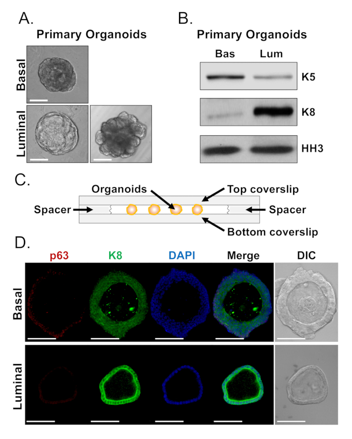
NOTE: Coverslip fragments can be placed on either side of the droplet to be used as spacers (Figure 1C). These prevent organoids from collapsing when a coverslip is placed over the droplet.
Table 1: Instructions for the preparation of key solutions
| Component | Concentration |
| B-27 | 1x (dilute from 50x concentrate) |
| GlutaMAX | 1x (dilute from 100x concentrate) |
| N-acetyl-L-cysteine | 1.25 mM |
| Normocin | 50 µg/mL |
| Recombinant Human EGF, Animal-Free | 50 ng/mL |
| Recombinant Human Noggin | 100 ng/mL |
| R-spondin 1-conditioned media | 10% conditioned media |
| A83-01 | 200 nM |
| DHT | 1 nM |
| Y-27632 dihydrochloride (ROCK inhibitor) | 10 µM |
| Advanced DMEM/F-12 | Base media |
| R-spondin 1-conditioned media is generated as described in Drost, et al. After addition of all components, filter sterilize mouse organoid media using 0.22 µm filter. ROCK inhibitor is only added during establishment of culture and passaging of organoids. | |
Results

Figure 1: Analysis of lineage marker expression in prostate organoids by Western blot and whole-mount confocal microscopy. (A) Representative phase contrast images of basal-derived (top), and luminal-derived (bottom) organoids after 7 days of culture. Scale bar = 100 µm. (B) Western blot analysis of basal-derived (Bas) and luminal-derived (Lum) organoid...
Disclosures
Materials
| Name | Company | Catalog Number | Comments |
| 16% Paraformaldehyde | Thermo Fisher Scientific | 50-980-487 | |
| 4’,6-diamidino-2-phenylindole (DAPI) | Thermo Fisher Scientific | D1306 | |
| Advanced DMEM/F-12 | Thermo Fisher Scientific | 12634010 | |
| Dispase II, Powder | Thermo Fisher Scientific | 17-105-041 | |
| Fetal Bovine Serum (FBS) | Sigma | F8667 | |
| Goat anti-mouse IgG-Alexa Fluor 488 | Invitrogen | A28175 | |
| Goat anti-rabbit IgG-Alexa Fluor 594 | Invitrogen | A11012 | |
| Mouse anti-cytokeratin 8 | BioLegend | 904804 | |
| Penicillin-Streptomycin (10,000 U/ mL) | Thermo Fisher Scientific | 15-140-122 | |
| Rabbit anti-p63 | BioLegend | 619002 | |
| RPMI 1640 Medium, HEPES (cs of 10) | Thermo Fisher Scientific | 22400105 | |
| Sucrose | Sigma | S0389-500G | |
| Triton X-100 | Sigma | X100-5ML |
References
This article has been published
Video Coming Soon
Source: Crowell, P. D. et al. Evaluating the Differentiation Capacity of Mouse Prostate Epithelial Cells Using Organoid Culture. J. Vis. Exp. (2019)
Copyright © 2025 MyJoVE Corporation. All rights reserved