Method Article
How to Detect Amygdala Activity with Magnetoencephalography using Source Imaging
In This Article
Summary
This article describes how to record amygdala activity with magnetoencephalography (MEG). In addition this article will describe how to conduct trace fear conditioning without awareness, a task that activates the amygdala. It will cover 3 topics: 1) Designing a trace conditioning paradigm using backward masking to manipulate awareness. 2) Recording brain activity during the task using magnetoencephalography. 3) Using source imaging to recover signal from subcortical structures.
Abstract
In trace fear conditioning a conditional stimulus (CS) predicts the occurrence of the unconditional stimulus (UCS), which is presented after a brief stimulus free period (trace interval)1. Because the CS and UCS do not co-occur temporally, the subject must maintain a representation of that CS during the trace interval. In humans, this type of learning requires awareness of the stimulus contingencies in order to bridge the trace interval2-4. However when a face is used as a CS, subjects can implicitly learn to fear the face even in the absence of explicit awareness*. This suggests that there may be additional neural mechanisms capable of maintaining certain types of "biologically-relevant" stimuli during a brief trace interval. Given that the amygdala is involved in trace conditioning, and is sensitive to faces, it is possible that this structure can maintain a representation of a face CS during a brief trace interval.
It is challenging to understand how the brain can associate an unperceived face with an aversive outcome, even though the two stimuli are separated in time. Furthermore investigations of this phenomenon are made difficult by two specific challenges. First, it is difficult to manipulate the subject's awareness of the visual stimuli. One common way to manipulate visual awareness is to use backward masking. In backward masking, a target stimulus is briefly presented (< 30 msec) and immediately followed by a presentation of an overlapping masking stimulus5. The presentation of the mask renders the target invisible6-8. Second, masking requires very rapid and precise timing making it difficult to investigate neural responses evoked by masked stimuli using many common approaches. Blood-oxygenation level dependent (BOLD) responses resolve at a timescale too slow for this type of methodology, and real time recording techniques like electroencephalography (EEG) and magnetoencephalography (MEG) have difficulties recovering signal from deep sources.
However, there have been recent advances in the methods used to localize the neural sources of the MEG signal9-11. By collecting high-resolution MRI images of the subject's brain, it is possible to create a source model based on individual neural anatomy. Using this model to "image" the sources of the MEG signal, it is possible to recover signal from deep subcortical structures, like the amygdala and the hippocampus*.
Protocol
Designing a trace conditioning paradigm using backward masking to block awareness
1. Design Stimuli
- Design the stimuli for the two groups.
- Choose 4 neutral expressions from different individuals.
- Align the faces so that the eye region of each face is in the same location.
- Crop the faces using an oval so that the hair, ears, and other peripheral features are no longer visible.
- Use the signal processing toolbox in Matlab (See Table 1 for software necessary to run the experiment) to create high-pass filtered images, by removing all information that is fewer than 5 cycles per degree12.
- Create the mask by merging several neutral expressions together, and adding high spatial frequency noise to the image.
- Normalize all images so that they have equal luminance.
2. Program the Experiment using Presentation
- Program the training and testing phases in Presentation using the parameters described below (See Figure 1).
- In addition, program a separate file (PCC) that will be used by the PSYLAB data acquisition software package during training and testing, in order to deliver the shock triggered by Presentation.
- For the training session program 4 blocks of differential trace fear conditioning with 15 trials per CS, per block.
- On each trial present the CSs for 30 msec.
- On each trial present the mask for 970 msec.
- On each CS+ trial present the shock UCS for 100 msec, so that it coterminates with the mask.
- Vary the location of the CS/mask combination so that it randomly appears in 1 of 4 quadrants.
- Present 1 trial every 6±2 sec using a variable intertrial interval.
- For the testing session program 1 block of reacquisition with 5 trials of each face CS, and 5 trials each of two new face CSs.
- In order to maximize your ability to record skin conductance responses (SCRs) during the testing trials, present the CS for 8 sec.
- On each CS+ trial present the shock UCS for 100 msec, so that it coterminates with the CS+.
- Present 1 trial every 20±4 sec using a variable intertrial interval.
- Instruct subjects to report UCS expectancy during both sessions, and record their responses using an MRI/MEG compatible axis device (joystick, slider, dial; See Section 7).
- Record SCRs during the testing session using electrodes attached to the bottom of the participants left foot (See Section 9).
Recording brain activity during the task using magnetoencephalography
3. Setup Equipment for Training at MEG Suite (See Figure 2)
- Connect the stimulus presentation computer to the MEG acquisition system using a standard DB25 multi connector ribbon cable (See Table 2 for equipment necessary to conduct the experiment.).
- Connect the stimulus presentation computer to the PSYLAB stand-alone monitor (SAM) using the 8-bit to 2-bit isolation adapter and the synchronization cable.
- The transistor -transistor logic (TTL) pulses used to mark the stimulus presentations can cause artifacts in the MEG data if they are sent to the SAM. To avoid these artifacts, mark the onset of the stimuli using only the bits blocked by the isolation adapter.
- Connect the shock stimulator (SHK1) to the SAM using the cable provided with the unit.
- Pass the shielded extension cable through the wave guide and connect it to the shock stimulator.
- Connect the SAM to a computer running the PSYLAB data acquisition software using a standard USB cable.
- Connect the rotary dial to the stimulus presentation computer (USB) and the MEG acquisition system (BNC) using the gameport-to-gameport/BNC splitter and the gameport-to-USB adapter.
- Record two minutes of sensor data without the subject in the room.
4. Setup Equipment for Testing at MRI Suite
- Connect the stimulus presentation computer to the SAM using the synchronization cable.
- Connect the shock stimulator (SHK1) and the skin conductance amplifier (SC5) to the SAM using the cables provided with the unit.
- Pass the shielded extension cables for SCR and shock through the wave guide and connect them to their respective units.
- Connect the SAM to a computer running the PSYLAB data acquisition software using a standard USB cable.
- Connect the rotary dial to the stimulus presentation computer using the gameport to USB adapter.
5. Setup Subject for Training at MEG Suite (See Figure 3)
- Attach electrodes and sensors to the subject using the schematic in Figure 3 as a guide.
- Attach disposable electrodes to monitor eyeblinks above and below the subject's right eye.
- Attach disposable electrodes to monitor heart rate to the subject's left side just below the heart and to the right chest just below the collarbone.
- Attach one disposable electrode as a reference to the back of the subject's left shoulder.
- Attach two cup electrodes to the subject's right leg over the right tibial nerve above the medial malleolus to administer the shock.
- Attach 4 head position indicator (HPI) coils to the subject, one above each eye and one behind each ear.
- Digitize the position of the subject's head relative to the HPI coils using fiducial points.
- Using the Polhemus system, map the position of the subject's nasion, and left and right tragi.
- Align the subject's digital head position relative to the fiducial points, making sure that the points are symmetrical.
- Next map the position of the subject's HPI coils.
- Finally, digitize 50-100 points along the subject's scalp.
- Escort the subject to the MEG system and connect the electrodes and sensors to the appropriate interface.
- Plug the disposable electrode leads into the MEG system amplifier.
- Plug the HPI wiring harness into the MEG system.
- Plug the shock electrode leads into the shielded extension cable.
- Raise the chair so that the subject's head is touching the top of the MEG helmet.
- Position the screen so that the projected image is in focus.
6. Shock Workup
- Set the shock to a level that the subject reports as painful but tolerable.
- Arm the shock stimulator by turning the dial from the 0 mA position the 5 mA position.
- Administer several presentations of the shock using the stimulus control window from the PSYLAB data acquisition software package.
- After each presentation have the subject rate the intensity of the shock on a scale from 0 (not at all painful) to 10 (painful but tolerable).
- Gradually increase the intensity of the shock until the subject rates it as a 10.
- Record the value from the scale in the parameter value box in the subject details window; shocks will be administered during the experiment at the value indicated in this box.
7. Response Device
- Instruct the subject on the proper use of the dial using an example Presentation scenario.
- Instructions: "Move the cursor all the way to the right (100) if you are absolutely sure that you will receive a presentation of the stimulation in the near future. Move the cursor all the way to the left (0) if you are sure that you will not receive a stimulation in the near future. Move the cursor to the middle (50) if you are unsure whether or not you will receive the stimulation in the near future."
8. Record MEG During Training
- Record two minutes of raw data at 2 kHz, while the subject rests with their eyes open.
- Prior to training begin recording event codes and shock delivery using the PSYLAB data acquisition software.
- Be sure that PSYLAB is running the proper PCC code so that it sends the shock when triggered by the computer.
- Record raw data at 2 kHz during each of the four training runs.
- Record online averages as a way to visually inspect the data in realtime for systematic sources of noise.
- Ask the subject to rate the intensity of the shock after each run to assess habituation.
9. Setup Subject for Testing at MRI Suite
- Escort the subject from the MEG suite to the MRI suite.
- Reattach the shock electrodes and recalibrate the intensity of the shock.
- Attach two cup electrodes to the bottom of the subject's left foot to monitor SCRs.
- Make sure that the subject still understands how to use the response device.
- Position the subject on the MRI table, secure their head, and connect the SCR and shock electrode leads to the corresponding shielded cables.
- Position the mirror attached to the head coil so that the participant can see the screen placed behind the head coil.
10. Record fMRI During Testing
- Collect high-resolution anatomical images (SPGR).
- Record blood-oxygenation level dependent responses during the testing session using standard imaging parameters (TR = 2 sec; TE = 25 msec; fleld of view = 24 cm; flip angle = 90 °).
- After testing have the subject complete a post experimental questionnaire.
Using source imaging to recover signal from subcortical structures.
11. Analyze Behavioral and fMRI Data
- Use UCS expectancy to determine whether subjects were able to discriminate between the stimuli.
- Average the UCS expectancy data for the 900 msec trace interval and the preceding 900 msec baseline period for each trial.
- Subtract the value for the baseline period from the value for the trace interval to determine how the subject moved the dial after the stimulus presentation.
- Perform a CS type by trial repeated measures ANOVA across subjects.
- Analyze behavioral and fMRI data from the testing session using previously published standards5,13-15.
12. Preprocess MRI Volume
- Use Freesurfer16 to create a segmented subcortical volume, and surfaces of the cortex, outer skin, and outer skull.
- Convert volumes and surfaces to AFNI readable format.
- Run importsurfaces.csh - the first time you run the program it will copy all the files you need into a new 'MODEL' folder in each subject's segmentation folder. It will also create an 'importsurface.mrml' file that is used to create the surface models of the amygdala and hippocampus.
- Create and convert the amygdala and hippocampus volumes into surfaces using Slicer3 and Paraview.
- Run Slicer3 importsurface.mrml from the subject's 'MODEL' directory. This will load the surfaces and volumes into 3dslicer.
- Generate models of amygdala and hippocampus, save models as {structure}.vtk.
- Import .vtk files into paraview.
- Run filter "generate surface normals."
- Export surface normals for amy and hipp as {structure}.ply (ascii) files.
- Import the surfaces and MRI volume into Brainstorm.
- Run importsurfaces.csh again - this will convert the surfaces into files that can be read by matlab and will copy all of the tess_{structure}.mat files into the Brainstorm database directory.
- Make sure that you have already created the subject in Brainstorm before copying tess_{structure}.mat files to Brainstorm folder (See Step 14.1).
- Once you get the surfaces into Brainstorm be sure to refresh the database.
- Warp the MRI volume into standard space by identifying the fiducial points.
- Manually align scalp surface with MRI, then apply the warp to all other surfaces.
- Merge the two pial surfaces and reduce the total number of vertices to 15,000.
- Merge the two hippocampal surfaces and reduce the total number of vertices to 2,000.
- Merge the two amygdala surfaces and reduce the total number of vertices to 1,000.
- Merge the pial, hippocampal, and amygdala surfaces.
- Create regions of interest (scouts) for the amygdala and hippocampus.
13. Preprocess MEG Recordings using Brainstorm11
- Create new subject in the Brainstorm database.
- Import the MEG recording file for each training session.
- Remove artifacts caused by sources outside the magnetically-shielded room (MSR) using signal space separation17.
- Remove artifacts caused by heart beats and eye movements using signal space projections from events identified on the electrocardiography (ECG) and electrooculography (EOG) channels.
- Inspect the recordings to ensure that Brainstorm correctly identified heart beat and eyeblinks events.
- Inspect the recordings for other possible sources of noise.
- Inspect the evoked data created from the online averages for systematic sources of artifact.
- Note that the TTL pulses used to mark the onset of the stimuli can cause artifacts in the recordings if sent to PSYLAB SAM unit.
- Send only TTL pulses needed to administer the shock to the SAM unit and isolate the unit from the remaining pulses using the 8-bit to 2-bit isolation adapter.
14. Analyze Evoked Responses using Brainstorm
- Use the event channel to identify epochs (-200 msec to 900 msec) corresponding to each of the experimental trials.
- Refine MRI registration using head points.
- Compute noise covariance from recordings.
- Compute head model using overlapping spheres method with cortex as input.
- Compute sources using the minimum-norm estimate method10.
- Continue analysis on sources.
- Band-pass filter sources for individual trials (1 Hz to 20 Hz).
- Take the absolute value of the band-pass filtered sources and convert those values to z-scores based on baseline variability.
- Spatially smooth the sources (sigma = 5 mm).
- Average sources across trials.
- Project the averages onto the default anatomy for the experiment.
- Compute t-tests on the sources across the different conditions.
- Filter significant t-test results using spatial and temporal thresholds to correct for family-wise error.
- Identify significantly activated regions and export the time course of activation for each subject.
- Compute the mean and standard error of the mean across subjects at each time point.
15. Perform Time-frequency Decompositions on ROI using Brainstorm
- Project the raw data from the individual trials onto the default anatomy for the experiment.
- Identify and create regions of interest from the analysis of evoked response or from anatomo-functional a priori hypotheses.
- Compute time-frequency decompositions of the data from your ROI for each trial using standard parameters (central frequency = 1 Hz; time resolution [FWHM] = 3 sec; frequency range = 10:90 Hz; frequency resolution = 1 Hz).
- Convert resulting time-frequency decomposition maps to z-scores.
- Average the resulting maps across trials for each subject.
- Perform t-tests on the maps across the different conditions.
Results
Using the methods described here, our investigations have led to two major findings: 1) It is possible to manipulate awareness of visual CSs during trace conditioning, and still show evidence of learning. 2) It is possible to recover MEG signals from the amygdala using source imaging*.
In Section 2, we described how to manipulate awareness of visual CSs with backward masking. When exposed to a masked stimulus that is displayed for ~30 msec, the subjects are generally unaware of the stimulus presentation5,6,8*. One way to verify the success of this manipulation is to measure the subjects' ability to predict the occurrence of the UCS. If the masking manipulation is successful, subjects should be unable to accurately predict the occurrence of the UCS based on the CS type (See Figure 4).
Although the timing in this type of training makes it difficult to directly measure learning during the training session. It is possible to indirectly measure learning by exposing them to a subsequent unmasked reacquisition testing session with new and old stimuli5*. If subjects are able to learn about the contingencies during the training phase, they should show larger magnitude differential (CS+ > CS-) SCRs to the old stimuli relative to the new stimuli. This effect is apparent in the Unfiltered group when we look at testing phase trials after the subjects have been re-exposed to the CS-UCS contingencies (i.e. Trials 2-5; See Figure 4).
In Section 8, we described how to record MEG during the masked trace conditioning session. Using source imaging to process these recordings, it is possible to recover MEG signal from subcortical structures like the amygdala18*. Subjects shown unfiltered face (N = 9) CSs exhibit larger amygdala responses (Figure 5) and gamma oscillations (Figure 6) than subjects shown high-pass filtered faces (N = 9). In addition, these subjects also show larger responses in a network of face processing regions like the occipital face area (Figure 7 and Supplemental Video).
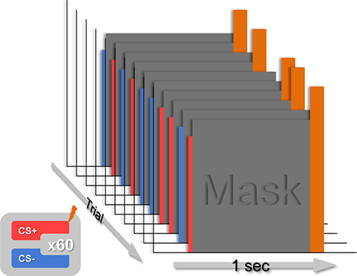
Figure 1. Schematic depicting a typical training session. Present 60 trials of a CS+ and 60 trials of a CS-, in pseudorandom order, such that there are 4 blocks of 15 trials each. Present the CSs for 30 msec, immediately followed by an 970 msec mask that coterminates with the shock UCS on CS+ trials.
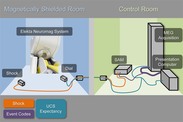
Figure 2. Schematic depicting the equipment used in a typical conditioning experiment. This setup makes it possible to: 1) present visual stimuli via the Presentation software, 2) administer an electrical stimulation UCS via the Psylab hardware (SAM), 3) record UCS expectancy using an axis device (dial) attached to the presentation computer, and 4) synchronize the stimulus presentations and responses with the MEG recordings via the MEG acquisition system interface.
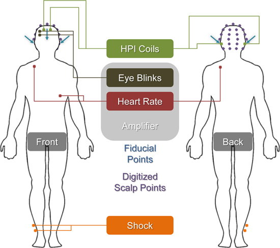
Figure 3. Illustration showing the location of each of the sensors and fiducial points described in Section 5. Dots with attached lines correspond to the labeled sensors and leads. Blue arrows represent the fiducial points used to register the MEG recordings with the MRI anatomical volume. Purple points represent digitized scalp points used to further refine the MEG-MRI coregistration.
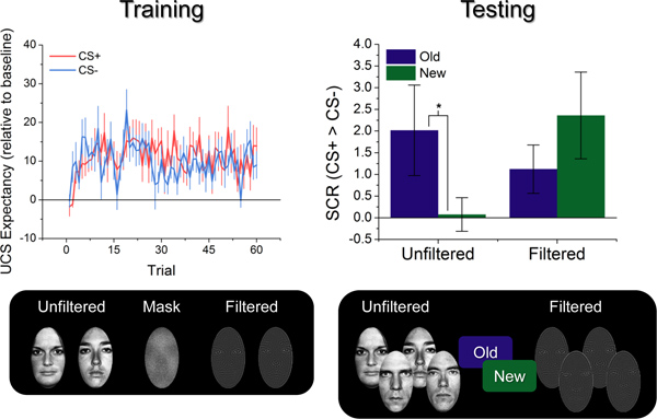
Figure 4. Behavioral results from a typical conditioning study. The graph on the left shows UCS expectancy across the training session, collapsed across the Unfiltered and Filtered groups. Notice that subjects are showing similar levels of UCS expectancy for the CS+ and CS- across the 60 trials, suggesting that the masking procedure blocked their ability to discriminate between the CSs (F(1,17) = 2.19; p = 0.16). The graph on the right shows the differential SCRs during the testing session. Notice that the Unfiltered, but not the Filtered group seems to be showing larger differential SCRs to the Old stimuli than the New stimuli (Unfiltered New/Old x CS+/CS- interaction: F(1,7) = 5.94; p = 0.045; Filtered New/Old x CS+/CS- interaction: F(1,7) = 1.13; p = 0.32), suggesting that the training leads to better reacquisition of the CS-UCS associations for these subjects. (*p < 0.05).
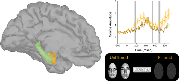
Figure 5. MEG results from a typical conditioning experiment. The figure on the left shows the 3d models of the amygdala (orange), hippocampus (green), and cerebral cortex used to model the sources of the MEG signal. The graph on the right represents activity from an amygdala cluster modeled from the MEG recordings. The light colored line represents the activity evoked by Unfiltered faces, while the dark colored line represents the activity evoked by Filtered faces. Vertical gray shaded sections represent time intervals where Unfiltered faces evoke significantly larger responses than Filtered faces (F(1,17) > 3.44; p < 0.05). Click here to view larger figure.
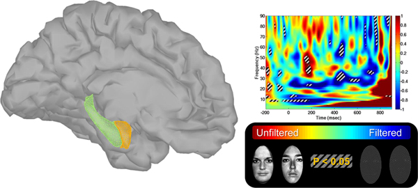
Figure 6. Amygdala time frequency results from a typical conditioning experiment. The figure on the left shows the 3d models of the amygdala (orange), hippocampus (green), and cerebral cortex used to model the sources of the MEG signal. The graph on the right represents the MEG signal recorded from the amygdala broken down by time and frequency. Warm colors represent regions in the spectrograph that show significantly more power for unfiltered faces than for filtered faces. Cool colors represent the opposite. Regions with the striped overlay represent significant differences across the groups. Click here to view larger figure.
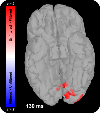
Figure 7. Figure showing occipital face area activation in a typical conditioning experiment. Colors represent the magnitude of the Unfiltered > Filtered t-test at the corresponding dipole. Warm colors represent larger responses to Unfiltered faces than to Filtered faces. Cool colors represent larger responses to Filtered faces than to Unfiltered faces.
Supplemental Video. Video showing cortical responses in a typical conditioning experiment. Colors represent the magnitude of the Unfiltered > Filtered t-test at the corresponding dipole. Warm colors represent larger responses to Unfiltered faces than to Filtered faces. Cool colors represent larger responses to Filtered faces than to Unfiltered faces. Click here to view supplemental movie.
Discussion
In this paper we describe methods 1) to manipulate subjects' awareness of target CSs during a trace fear conditioning paradigm. 2) and to recover MEG signal from the amygdala during trace fear conditioning without awareness. Using these methodologies, we were able to show that trace conditioning without awareness is possible when faces are used to predict the UCS. This result suggests that faces receive special processing even when presented below the perceptual detection threshold*. Consistent with this conclusion we found that broad spectrum faces evoke robust amygdala responses and bursts of gamma oscillations during the trace interval. This result suggests that the amygdala is capable of maintaining a representation of a face CS during a brief trace interval.
Although presented together, these two methods can be used independently as well. For instance it is possible to use backward masking to manipulate target visibility in other paradigms where behavior may be affected by emotional cues processed below the level of conscious awareness5,6,8*. In addition, using the source imaging approach described here it is possible to create 3d models of other subcortical structures, and it may be possible to recover signal from these structures during other region specific tasks. For instance, by using source imaging to model hippocampal activity, it may be possible recover MEG signal from hippocampal sources during tasks like spatial navigation.
The methods described here were designed with two goals in mind: 1) block awareness of the target stimuli, 2) and maximize the ability to detect stimulus evoked amygdala responses using MEG. These design constraints make it difficult to measure the subjects' implicit knowledge of the stimulus contingencies. For instance, SCRs resolve over the course of several seconds5,13; however, the CSs are only presented for ~30 msec during training, and the shock is presented shortly after (~900 msec). Given these time constraints, CR expression will be inevitably confounded by UCR expression during training. Because of this colinearity, it is necessary to test the subjects' knowledge of the stimulus contingencies using a subsequent unmasked testing session. However a testing session at the end of the experiment is not optimal because SCRs tend to habituate over the course of the experiment1. Given the number of trials needed to show reliable evoked responses with MEG, this SCR habituation will decrease considerably the power to detect a behavioral effect of the training. Future studies should focus on finding better ways to index implicit learning during fear conditioning with masked CSs. This could be done by either finding an alternative index of fear during the training (i.e. pupil dilation19,20) or find a more sensitive measure of fear that can be administered after the training session.
Disclosures
The authors have nothing to disclose.
Acknowledgements
This study was supported by the National Institute of Mental Health (MH060668 and MH069558).
Materials
| Name | Company | Catalog Number | Comments |
| Software | |||
| Matlab | Mathworks | mathworks.com/products/matlab | |
| Presentation | Neurobehavioral Systems | neurobs.com | |
| Psylab | Contact Precision Instruments | psychlab.com | |
| AFNI | NIMH - Scientific and Statistical Computing Core | afni.nimh.nih.gov/afni | |
| Freesurfer | Martinos Center for Biomedical Imaging | surfer.nmr.mgh.harvard.edu/fswiki | |
| MNE | Martinos Center for Biomedical Imaging | nmr.mgh.harvard.edu/martinos/userInfo/data/sofMNE.php | |
| Brainstorm | open-source collaboration | neuroimage.usc.edu/brainstorm | |
| 3d Slicer | open-source collaboration | slicer.org | |
| Paraview | Kitware | paraview.org | |
| Table 1. Software used Software used. | |||
| Equipment | |||
| Physiological Monitoring System | |||
| Psylab stand alone monitor (x2) | Contact Precision Instruments | SAM | |
| Skin conductance amplifier | Contact Precision Instruments | SC5 | |
| Shock stimulator (x2) | Contact Precision Instruments | SHK1 | |
| Additional Components | |||
| 8-bit synchronization cable (x2) | Contact Precision Instruments | Included with SAM | |
| 8-bit to 2-bit isolation adapter | N/A | Custom | |
| DB25 ribbon cable (x2) | N/A | Standard | |
| Shielded extension cable (x3) | Contact Precision Instruments | CL41 | |
| Radiotranslucent cup electrodes for SCR and shock (x6) | Biopac | EL258-RT | |
| Signa Gel | Parker Laboratories | 15-250 | |
| Response Device | |||
| Rotary dial with gameport connector (x2) | N/A | Custom | |
| Gameport-to-gameport/BNC splitter | N/A | Custom | |
| BNC cable | N/A | Standard | |
| Gameport-to-USB adapter (x2) | Rockfire | RM203U | |
| Additional Components for MEG Setup | |||
| HPI coils and wiring harness | N/A | Custom | |
| HPI positioning system | Inition | Polhemus Isotrak | |
| Table 2. Equipment used. | |||
References
- Knight, D. C., Cheng, D. T., Smith, C. N., Stein, E. A., Helmstetter, F. J. Neural substrates mediating human delay and trace fear conditioning. Journal of Neuroscience. 24, 218-228 (2004).
- Knight, D. C., Nguyen, H. T., Bandettini, P. A. The role of awareness in delay and trace fear conditioning in humans. Cognitive, Affective, & Behavioral Neuroscience. 6, 157-162 (2006).
- Manns, J. R., Clark, R. E., Squire, L. R. Parallel acquisition of awareness and trace eyeblink classical conditioning. Learning & Memory. 7, 267-272 (2000).
- Weike, A. I. I., Schupp, H. T. T., Hamm, A. O. Fear acquisition requires awareness in trace but not delay conditioning. Psychophysiology. 44, 170-180 (2007).
- Balderston, N. L., Helmstetter, F. J. Conditioning with masked stimuli affects the timecourse of skin conductance responses. Behavioral Neuroscience. 124, 478-489 (2010).
- Flykt, A., Esteves, F., Öhman, A., Ohman, A. Skin conductance responses to masked conditioned stimuli: phylogenetic/ontogenetic factors versus direction of threat. Biological Psychology. 74, 328-336 (2007).
- Öhman, A., Carlsson, K., Lundqvist, D., Ingvar, M. On the unconscious subcortical origin of human fear. Physiology & Behavior. 92, 180-185 (2007).
- Whalen, P. J., et al. Masked presentations of emotional facial expressions modulate amygdala activity without explicit knowledge. Journal of Neuroscience. 18, 411(1998).
- Huang, M. X., Mosher, J. C., Leahy, R. M. A sensor-weighted overlapping-sphere head model and exhaustive head model comparison for MEG. Physics in Medicine and Biology. 44, 423-440 (1999).
- Hämäläinen, M. S., Ilmoniemi, R. J. Interpreting magnetic fields of the brain: minimum norm estimates. Medical & Biological Engineering & Computing. 32, 35-42 (1994).
- Tadel, F., Baillet, S., Mosher, J. C., Pantazis, D., Leahy, R. M. Brainstorm: A User-Friendly Application for MEG/EEG Analysis. Computational Intelligence and Neuroscience. 2011, 879716-87 (2011).
- Rotshtein, P., Vuilleumier, P., Winston, J., Driver, J., Dolan, R. J. Distinct and convergent visual processing of high and low spatial frequency information in faces. Cerebral Cortex. 17, 2713-2724 (2007).
- Cheng, D. T., Richards, J., Helmstetter, F. J. Activity in the human amygdala corresponds to early, rather than late period autonomic responses to a signal for shock. Learning & Memory. 14, 485-490 (2007).
- Knight, D. C., Smith, C. N., Stein, E. A., Helmstetter, F. J. Functional MRI of human Pavlovian fear conditioning: patterns of activation as a function of learning. Neuroreport. 10, 3665-3670 (1999).
- Schultz, D. H., Helmstetter, F. J. Classical conditioning of autonomic fear responses is independent of contingency awareness. Journal of Experimental Psychology Animal Behavior Processes. 36, 495-500 (2010).
- Fischl, B., et al. Sequence-independent segmentation of magnetic resonance images. NeuroImage. 23, 69-84 (2004).
- Taulu, S., Kajola, M., Simola, J. Suppression of interference and artifacts by the Signal Space Separation Method. Brain Topography. 16, 269-275 (2004).
- Dumas, T., et al. MEG study of amygdala responses during the perception of emotional faces and gaze. 17th International Conference on Biomagnetism Advances in Biomagnetism-Biomag, , (2010).
- Reinhard, G., Lachnit, H., König, S. Tracking stimulus processing in Pavlovian pupillary conditioning. Psychophysiology. 43, 73-83 (2006).
- Reinhard, G., Lachnit, H. Differential conditioning of anticipatory pupillary dilation responses in humans. Biological Psychology. 60, 51-68 (2002).
Reprints and Permissions
Request permission to reuse the text or figures of this JoVE article
Request PermissionThis article has been published
Video Coming Soon
Copyright © 2025 MyJoVE Corporation. All rights reserved