Method Article
Three-Dimensional (3D) Tumor Spheroid Invasion Assay
In This Article
Summary
Invasion of surrounding normal tissues is a defining characteristic of malignant tumors. We provide here a simple, semi-automated micro-plate assay of invasion into a natural 3D biomatrix that has been exemplified with a number of models of advanced human cancers.
Abstract
Invasion of surrounding normal tissues is generally considered to be a key hallmark of malignant (as opposed to benign) tumors. For some cancers in particular (e.g., brain tumors such as glioblastoma multiforme and squamous cell carcinoma of the head and neck – SCCHN) it is a cause of severe morbidity and can be life-threatening even in the absence of distant metastases. In addition, cancers which have relapsed following treatment unfortunately often present with a more aggressive phenotype. Therefore, there is an opportunity to target the process of invasion to provide novel therapies that could be complementary to standard anti-proliferative agents. Until now, this strategy has been hampered by the lack of robust, reproducible assays suitable for a detailed analysis of invasion and for drug screening. Here we provide a simple micro-plate method (based on uniform, self-assembling 3D tumor spheroids) which has great potential for such studies. We exemplify the assay platform using a human glioblastoma cell line and also an SCCHN model where the development of resistance against targeted epidermal growth factor receptor (EGFR) inhibitors is associated with enhanced matrix-invasive potential. We also provide two alternative methods of semi-automated quantification: one using an imaging cytometer and a second which simply requires standard microscopy and image capture with digital image analysis.
Introduction
Classical anticancer drug development is largely focused on the use of in vitro cell-based assays to screen for cytotoxic agents that inhibit tumor cell proliferation and/or promote programmed cell death (apoptosis). More recently, efforts have moved towards targeted therapies with the development of inhibitors of the underlying molecular causes of malignant cell proliferation. In spite of some spectacular results, such agents are often associated with the rapid development of drug resistance. Given that it is the invasive and metastatic potential of tumor cells that is responsible for the majority of cancer deaths, and that relapsed disease often presents a more aggressive phenotype, it is logical also to consider complementary in vitro experimental models1,2 to identify novel agents that will inhibit these additional key ‘hallmarks’ of cancer.
During malignant progression, tumor cells acquire the ability to invade the surrounding tissue and/or spread into distant organs (metastasis). Cancer cells penetrate the basement membrane by the formation of invadopodia3,4. These structures are enriched with actin filaments, specific adhesion proteins and proteinases and are collectively responsible for tumor cell motility and degradation of the extracellular matrix (ECM)5. Invadopodia extend into the ECM and are believed to be important for tumor cell invasion and also extravasation into vascular channels, facilitating hematogenous (or lymphatic) dissemination and metastasis.
Current standard methods to assess tumor cell invasion in vitro include the following. Transwell-based or Boyden chamber assays2,6 where single cell suspensions are seeded on top of a filter coated with a thick layer of ECM-derived proteins. Cells then invade and move into the lower chamber in response to a chemo-attractant. Commonly used ECM proteins are type I collagen, or a basement membrane-like matrix (BMM, e.g., Matrigel, derived from the EHS tumor7) that mimics the natural composition of basement membranes.
As an alternative to the filter-based Boyden chamber type assays8, cells can be seeded on top of an ECM gel (to which fibroblasts may be added) where they form a monolayer and then individually or collectively invade into the gel. Invasion can be measured in terms of number of invading cells and/or distance travelled from the gel surface9. Tumor cells can also be completely embedded into a matrix either as a single cell suspension or as spheroids - usually placed between two layers of ECM gel- allowing cells to invade out of the tumor mass into the surrounding matrix2,6,10,11.
The three-dimensional (3D) tumor spheroid invasion assay described here provides a rapid, automatable invasion system using a highly reproducible, standardized method12 in a 96-well plate format with one spheroid per well. BMM is added directly to each well to provide a semi-solid matrix into which tumor cells invade from the spheroid body. The extent of tumor cell invasion is monitored at intervals over a period of 72 - 96 hr. This method provides the following advantages over the current in vitro invasion assays mentioned above: the tumor cells are organized into a 3D structure mimicking a tumor micro-region or a micro-metastasis; the tumor spheroids are highly reproducible in size; the invasion assay is performed in situ in the same plate as tumor spheroid development, without the need to move them to secondary plates; the method, combined with the latest technologies of automated image analysis, enables both high content and high throughput analyses of tumor cell invasion.
The image analysis is performed using an imaging cytometer, which scans a 96-well plate within 10 min. By using the confluence application, the extent and rate of invasion achieved either by single cells or by cell clusters spreading out from the tumor spheroids and invading into the matrix can be measured in a dynamic fashion. For lower throughput, an alternative method for image analysis is presented, based on the use of an inverted microscope and standard imaging software.
Protocol
1. Generation of Reproducibly Sized Tumor Spheroids
- Wash tumor cell monolayers with phosphate buffered saline (PBS; 5 ml for a 25 cm2 or 8 - 10 ml for a 75 cm2 flask), add cell dissociation enzyme (1 ml for a 25 cm2 or 2 ml for a 75 cm2 flask) and incubate cells at 37 °C for 2 - 5 min.
- Check cell detachment under a microscope and neutralize cell dissociation enzyme with complete growth medium (5 ml for a 25 cm2 or 8 ml for a 75 cm2 flask).
- Centrifuge cell suspension at 500 x g for 5 min.
- Remove supernatant, tap the tube and re-suspend cell pellet in 1 ml of complete growth medium using a P1000 pipette. This should yield a single cell suspension without cell clusters.
- Count cells using a hemocytometer and dilute the cell suspension to obtain 0.5 - 2 x 10⁴ cells/ml (optimal cell density needs to be determined for each cell line in order to obtain tumor spheroids of 300 - 500 µm diameter 4 days after cell seeding12,13).
- Transfer the cell suspension to a sterile reservoir and, using a multichannel pipette, dispense 200 µl/well into ultra-low attachment (ULA) 96-well round bottom plates12.
- Transfer the plates to an incubator (37 °C, 5% CO2, 95% humidity). Four days later, visually confirm tumor spheroid formation and proceed with the 3D invasion assay.
2. 3D Tumor Spheroid Invasion Assay
- Thaw BMM on ice.
- Keep a set of sterile filter tips for P10, P200 and P1000 pipettes and sterile tubes (1.5 ml volume or larger depending on total volume required) at -20 °C.
- Place the ULA 96-well plates containing 4-day old spheroids on ice.
- Using a multichannel pipette, gently remove 100 µl/well of growth medium from the spheroid plates. For this step angle the tips towards the inside wall of the U-bottom wells, avoiding contact with the bottom of the well and the location of the spheroids; minimize disturbance of the spheroids.
- Using ice-cold tips, transfer BMM to ice-cold tubes. For cytokine-induced invasion or for drug evaluation studies, add reagents (at 2x the final concentration) to the BMM using ice-cold tips. For example, use epidermal growth factor (EGF) to stimulate the invasion of CALS and CALR squamous carcinoma cells. Mix well by swirling gently, avoiding the formation of bubbles.
- Gently dispense 100 µl of BMM into the U-bottom well, aiming the tip towards the inside wall of the well. This step is the most critical as, for optimal image analysis, spheroids must remain in the center of the well12.
- Repeat step 2.6 for all the required wells, allowing 5 - 6 replicates/condition. If necessary, change to a freshly-chilled tip.
NOTE: When more experienced, dispense BMM using a multichannel pipette. - Using a sterile needle, remove bubbles, if present.
- Using a microscope, visually check that spheroids are in a central position. If not, centrifuge the plate at 300 x g for 3 min at 4 °C. This will ensure that spheroids are centrally located in each well.
- Transfer the plate to an incubator at 37 °C and allow the BMM to solidify.
- 1 hr later, using a multichannel pipette, gently add 100 µl/well of complete growth medium. For cytokine-induced invasion or drug evaluation studies, include cytokines or inhibitors in the medium (1x final concentration). Alternatively, if reagents were not added to the BMM in step 2.5, add them to the medium at this point (3x the desired final concentration).
3. Automated Image Acquisition
- Scan plates on the cytometer (for specifications see Table of Equipment and Materials) at intervals starting from time zero (t0 = day of the invasion assay set up, immediately after step 2.10 up to 72 hr, or 96 hr for slower invading tumor cell lines.
- Select the ‘Confluence’ application.
- Check that the spheroid is in focus and, if needed, adjust manually and fix the ‘Focus off-set’.
- Under ‘Sampling settings’, select to scan one central field of view (FOV). Following BMM addition, tumor spheroids should have remained in a central position, thus there is no need to scan the entire well.
- In the software, define the wells to be scanned by highlighting them on the plate map. Click on ‘Start scan’.
NOTE: The scan is automatically saved and can be reviewed later off-line.
4. Automated Image Analysis
- Select ‘Confluence’ application.
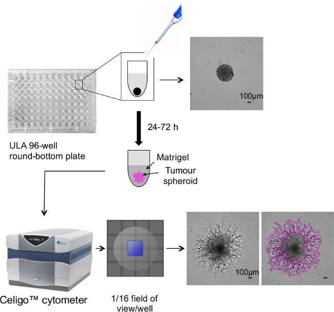
- In the ‘Analysis’ tab, adjust application settings (e.g., “precision”: low, normal, high; “intensity threshold”: 1 - 15) to produce a precise segmentation around the spheroid, including cell processes and invadopodia extending into the BMM (see Figure 1). Adjust the settings for each tumor cell line and/or at various time points. The ‘Confluence’ application measures the % of the well area covered by the invading cells, in the selected FOV.
- Check that the analysis settings are appropriate (i.e., segmentation is accurate) for other spheroids in the plate by clicking on a few different wells. If the segmentation does not accurately outline a spheroid, adjust the application settings further.
- Click on ‘Start analysis’.
- On the ‘Results’ tab, check multiple wells to verify analysis quality. Export ‘Well level data’ to a spreadsheet program.
- Repeat analysis for all the time points. Calculate the mean % confluence for replicate wells and plot invasion (% confluence) against time in a bar chart, using scientific graphing and statistical software of choice.
5. Manual Image Acquisition
- Record an image for each tumor spheroid at intervals starting from t = 0 up to 72 - 96 hr, depending on the speed of invasion of the cell line in question, using an inverted microscope (for a 96-well plate this takes approximately 20 - 30 min). Use a 10X objective. Use a 4X objective when an invaded area is too large to be completely captured within the 10X field of view.
- Save each individual image acquired.
- For calibration, take images of a stage graticule (1 mm) using both 10X and 4X objectives (and save images).
6. Manual Image Analysis
- Open the stage graticule images into the image analysis software of choice.
- Enter the graticule measurements, the units (µm), the objectives used (10X or 4X) and perform the calibration. This step is required only once. For subsequent image analysis, simply reload the required calibration settings.
- Open the invasion assay images and select the calibration settings depending on the microscope objective (10X or 4X) used to obtain the images.
- Alternatively, import images obtained on an imaging cytometer (step 3) and analyze manually, as described for images obtained using an inverted microscope (see results shown in Figure 3 and Figure 4).
- Measure the area covered by the spheroids.
- In the software used here for the Representative Results (for specifications see Table of Equipment and Materials), go to ‘Measure’ and select ‘Count/size’. Chose ‘File’ then ‘Load settings’ and select pre-determined assay settings. Then choose ‘Count’. The spheroid should then be accurately segmented.
- Alternatively go to ‘Edit’ then ‘Draw/Merge Objects’ to open the ‘Magic Wand/Trace’ window. Use the wand cursor to click on the spheroid image and obtain segmentation. Select ‘Trace’ (in the ‘Magic Wand’ window) to manually outline the spheroid using a mouse.
- Export measurements of different parameters (area, diameter, perimeter etc.) to a spreadsheet. Record on the spreadsheet the relevant image information (e.g., well number, relative time point).
- Plot the mean area of replicate spheroids (invasion) versus time using scientific graphing and statistical software. Alternatively, calculate the change in spheroid area at each time point, relative to the area at t = 0. Then plot invasion (% t0) versus time as a linear graph.
Results
3D tumor spheroid invasion is assessed using a simple, reproducible, automated method schematically illustrated in Figure 1: reproducibly sized tumor spheroids are obtained by plating cells into ULA 96-well round bottom plates. 4 days post-initiation spheroids are embedded into BMM. This provides a semi-solid structure into which tumor cells invade and spread out of the spheroid. Spheroids are located centrally at the base of each well and invasion into the BMM is easily monitored at intervals starting from t = 0 up to 72 - 96 hr using an imaging cytometer. This can provide fully automated image analysis.
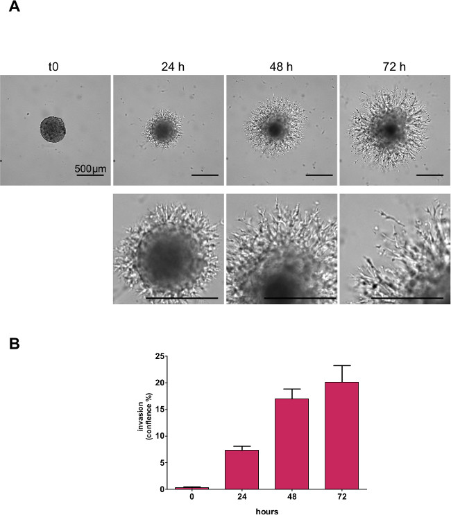
Examples of different types of cancer cell invasion are illustrated in Figures 2 - 4. The highly malignant human glioblastoma multiforme (GBM) cell line U-87 MG, forms a tight, spherical 3D structure and is used here to exemplify brain tumors in which local invasion is a key life-threatening feature. Once embedded in BMM, cells from U-87 MG spheroids spread with a typical “starburst” invasion pattern12. Tumor cells radially extend into the matrix in which they maintain a 3-dimensional shape. The process is followed over a period of 72 hr as shown in Figure 2 for U-87 MG spheroids, when the extent of tumor cell invasion, with details of the cellular processes and invadopodia extending into the BMM is clearly evident. Fully automated image analysis using an image cytometer enables precise quantitation of the tumor cell invasion over time. The quantification in Figure 2 is obtained as illustrated in Figure 1, where the automated analysis produces segmentation around the cell protrusions extending into the BMM from the U-87 MG spheroid body. Note that for this cell line, the tumor spheroid body is excluded from the measurement of the invaded area.
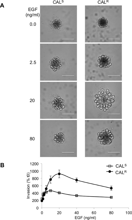
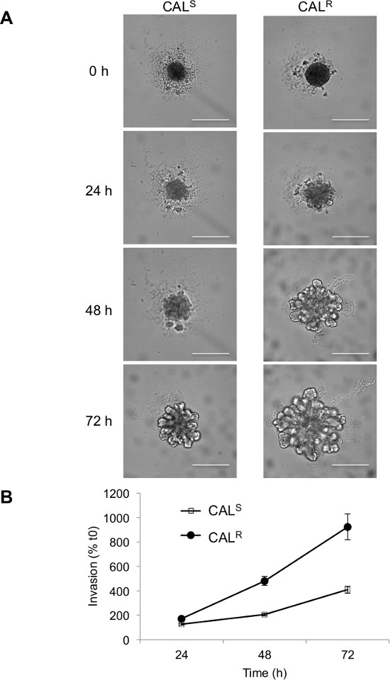
A different pattern of invasion was displayed by human squamous head and neck cancer cell lines. CALS and CALR are isogenic but CALS is sensitive to, and CALR resistant to EGFR tyrosine kinase inhibitors14. In the absence of EGF, neither cell line invaded into the BMM (Figure 3) however, as the concentration of EGF was increased (to 2.5 ng/ml), the spheroids appear to ‘bud’. At the higher concentrations of EGF (40 and 80 ng/ml) the degree of invasion was reduced. This is consistent with the classical bell-shaped dose response to EGF in terms of invasion that has been previously reported15. In 20 ng/ml EGF, finger-like protrusions extruded from the main body of CALR spheroids whilst CALS cells were less invasive after 72 hr (Figure 3). This differential invasion was evident 48 hr after spheroids were placed into BMM (containing 20 ng/ml EGF for maximal distinction between cell lines) and greatest after 72 hr (Figure 4). These images were acquired rapidly using an imaging cytometer, as for U-87 MG. However, in this case, to exemplify an alternative method, the degree of invasion was quantified manually using stand-alone image analysis software and is presented graphically (Figures 3 and 4). These results illustrate a phenotypic difference between drug-sensitive and -resistant cells that could be utilised to screen for compounds that will specifically antagonize the drug resistant, invasive phenotype.

Figure 1. Schematic overview of 3D tumor spheroid invasion assay. The workflow shows the steps the method involves including representative images of U-87 MG glioblastoma spheroids just after embedding into BMM (top panel; t = 0) and after invasion into BMM (bottom panel; t = 72 hr) with and without the segmentation that measures the area covered by the invading cells. Bar = 100 μm. Please click here to view a larger version of this figure.

Figure 2. Time-course of U-87 MG glioblastoma spheroid invasion into BMM. U-87 MG spheroid 3D invasion into BMM was monitored and quantified up to 72 hr using an imaging cytometer. (A) Representative images and (B) quantification of tumor cell invasion are shown. Bar = 500 μm. Please click here to view a larger version of this figure.

Figure 3. CALR (resistant) and CALS (sensitive) tumor spheroid invasion. (A) Representative images and (B) manual quantification of EGF dose-dependent CALR and CALS tumor spheroid invasion are shown. Bar = 500 µm. Please click here to view a larger version of this figure.

Figure 4. Cells fromCALR (resistant) spheroids are more invasive than CALS (sensitive) tumor spheroids when placed in BMM. Upon EGF stimulation CALR tumor spheroids show a more pronounced invasion than CALS. (A) Representative images and (B) manual quantification of invasion are shown. Bar = 500 µm. Please click here to view a larger version of this figure.
Discussion
The two tumor models described here (glioblastoma and SCCHN) were specifically selected to exemplify our 3D assay as they are clinically relevant locally invasive cancers with an unmet need for effective therapies12. Following drug treatment, assessment of in vitro pharmacodynamic (PD) biomarker changes can be performed easily by western blot or immunoassays of lysates following the use of specific commercially available reagents to allow cell recovery from BMM and/or immunofluorescent analysis of invading tumor spheroids by whole mount staining.
This invasion assay is a useful technique for rapid and reproducible assessment of tumor cell invasion into a semisolid medium and therefore particularly appropriate for future in vitro drug screening. Cancer cells invade the matrix in a 3D manner as they spread out from a “micro-tumor”, represented by the tumor spheroid, and extend into an extracellular matrix-like environment. The true three-dimensionality of the assay is evident in the cell morphology, which is different from the flat, adherent morphology cells assume when moving on a solid substrate. The degree of invasion is easily quantified using either an imaging cytometer, allowing an automated read-out, or by using a standard microscope in combination with imaging software. Moreover, this method is suitable for fluorescent imaging13 (for example with cell lines expressing fluorescent proteins or pre-labelled with fluorescent dyes).
The method is simple to perform with the only critical step represented by the addition of the BMM, when there is a risk of disturbing the central position of the spheroid in the U-shaped well. This can result in suboptimal image analysis, with spheroids in different focal planes. It is therefore critical to add the BMM carefully and slowly to each well. After BMM addition it is advisable to check the plate under a microscope and if the localization is considered unacceptable, this can be remedied by gentle centrifugation. With experience, this is rarely necessary. Whilst this method is robust and very reproducible, inter-experimental variation could occur with different batches of BMM. To avoid this, it is advisable to purchase sufficient BMM from a single batch to complete a series of studies.
A limitation of the method (as for any such assays) is the difficulty in distinguishing between invasion and proliferation, which the tumor cells likely undergo during the assay time frame. Although the doubling time of cells can be taken into account, or cell cycle inhibitors such as cytochalasin D introduced, it is not easy to control or clearly distinguish between these two different aspects of tumor progression, especially for fast growing tumor cells and for those that have a more “expansive”, rather than infiltrative, invasion pattern. For this reason it is suggested that a parallel 3D growth assay is performed to evaluate specific effects of any inhibitory or stimulatory agents. If careful dose response studies are performed, it may be possible to select concentrations that minimize effects on proliferation. For example we have shown that the HSP90 inhibitor 17-AAG inhibits U-87 MG 3D tumor spheroid invasion already at 24 hr and at concentrations below the concentration inhibiting 3D growth by 50% (GI50)12.
On the other hand, the significance of this assay compared to other standard invasion assays (e.g., filter based assays or invasion of single cells into 3D matrices1), is that tumor cells invade into the surrounding matrix from the spheroid, that resembles a “micro-tumor” or a “micro-metastasis”, and therefore takes into account important aspects of the pathophysiology of a tumor mass. The method we present provides information on the collective behavior of tumor cells, when initially leaving the spheroids, and also individually, when single cells reach more distant regions in the BMM. Additionally, cells within tumor spheroids may experience hypoxia and nutrient deprivation which, through changes in gene expression, can promote migration and invasion; such features are absent in 2D assays. Moreover, all of this is available in a high-throughput format through the use of specific 96-well plates and the latest imaging technology, allowing a more complex 3D assay to be easily used in target validation and drug discovery.
The method we present can accommodate further applications to address additional aspects of tumor cell invasion. In particular, extra complexity can be envisaged by the addition of host cells, such as fibroblasts and/or endothelial cells, into the spheroid itself or the surrounding matrix. This can also be modified, not only in terms of stiffness (by the use of different concentrations of BMM), but also in terms of composition (by the addition of other components dependent on the specific tissue/organ of the adopted tumor model: e.g., tenascin for brain cancers16 and collagen for breast carcinoma17).
A similar set-up (generation of spheroids and imaging) can also be adapted to assess tissue invasion in 3D, where tumor spheroids are co-cultured with embryoid bodies resembling a complex tissue12 or with other cell-specific organoids (e.g., astrocytes for glioma18 or crypt cultures for gastrointestinal cancers) but further work is required to enable automated image analysis.
Disclosures
Open Access fees were supported by Nexcelom Bioscience, LLC.
Acknowledgements
This work was funded by The National Centre for the Replacement, Refinement and Reduction of Animals in Research (G1000121 ID no.94513) and the Oracle Cancer Trust. SE is supported by The Institute of Cancer Research and Cancer Research UK (grant number C309/A8274). We acknowledge NHS funding to the NIHR Biomedical Research Centre.
Materials
| Name | Company | Catalog Number | Comments |
| Corning Matrigel basement membrane matrix |  Corning Corning | 354234 | Keep at 4 °C to prevent solidification and dispense using cooled pipette tips |
| Cultrex basement membrane extract, PathClear |  Trevigen Trevigen | 3432-005-01 | Keep at 4 °C to prevent solidification and dispense using cooled pipette tips |
| Ultra-low attachment 96-well plate |  Corning Corning | 7007 | |
| EGF |  Miltenyi Biotec Miltenyi Biotec | 130-093-825 | Add 100 μl 1% BSA (w/v) in water to 100 μg EGF. To 25 μl of this, add 4,975 μl RHB-A medium to give a 5 μg/ml working stock. Aliquot and store at -20 °C |
| RHB-A medium |  Stem Cells Inc. Stem Cells Inc. | SCS-SF-NB-01 | For diluting EGF |
| DMEM |  GIBCO GIBCO | 31966-021 | Warm in 37 °C water bath before use |
| Fetal bovine serum |  Biosera Biosera | 1001/500 | Heat at 56 °C to inactivate complement before use |
| TrypLE |  GIBCO GIBCO | 12605 | Cell dissociation solution |
| Celigo cytometer |  Nexcelom Bioscience Nexcelom Bioscience | n/a | Specialist equipment for image capture and analysis. Manual image capture by microscopy and independent image analysis software is an alternative |
| IX70 inverted microscope |  Olympus Olympus | n/a | Used with camera for manual image capture |
| Retiga Exi Fast 1394 digital camera |  Qimaging Qimaging | n/a | Attached to microscope for manual image capture |
| Image-Pro Plus 7.0 |  Media Cybernetics Media Cybernetics | n/a | Software used to control camera and microscope for manual image capture |
| Image-Pro Analyzer 7.0 |  Media Cybernetics Media Cybernetics | n/a | Stand-alone image analysis software for manual measurment of spheroid size |
| Excel 2010 |  Microsoft Microsoft | n/a | Scientific graphing and statistical software |
| Prism 6.0 |  GraphPad GraphPad | n/a | Scientific graphing and statistical software |
References
- Astashkina, A., Mann, B., Grainger, D. W. A critical evaluation of in vitro cell culture models for high-throughput drug screening and toxicity. Pharmacology & therapeutics. 134 (1), 82-106 (2012).
- Zimmermann, M., Box, C., Eccles, S. Two-Dimensional vs. Three-Dimensional In Vitro Tumor Migration and Invasion Assays. Methods and Protocols. 986, 227-252 (2013).
- Hwang, Y. S., Park, K. K., Chung, W. Y. Invadopodia formation in oral squamous cell carcinoma: the role of epidermal growth factor receptor signalling. Arch. Oral. Biol. 57 (4), 335-343 (2012).
- Wang, S., et al. Study on invadopodia formation for lung carcinoma invasion with a microfluidic 3D culture device. PLoS One. 8 (2), e56448 (2013).
- Friedl, P., Alexander, S. Cancer invasion and the microenvironment: plasticity and reciprocity. Cell. 147 (5), 992-1009 (2011).
- Brekhman, V., Neufeld, G. A novel asymmetric 3D in-vitro assay for the study of tumor cell invasion. BMC Cancer. 9, 415 (2009).
- Kleinman, H. K., Martin, G. R. Matrigel: basement membrane matrix with biological activity. Semin. Cancer Biol. 15 (5), 378-386 (2005).
- Boyden, S. The chemotactic effect of mixtures of antibody and antigen on polymorphonuclear leucocytes. J. Exp. Med. 115, 453-466 (1962).
- Nystrom, M. L., et al. Development of a quantitative method to analyse tumour cell invasion in organotypic culture. The Journal of pathology. 205 (4), 468-475 (2005).
- Deisboeck, T. S., et al. Pattern of self-organization in tumour systems: complex growth dynamics in a novel brain tumour spheroid model. Cell Prolif. 34 (2), 115-134 (2001).
- Harma, V., et al. A comprehensive panel of three-dimensional models for studies of prostate cancer growth, invasion and drug responses. PLoS One. 5 (5), e10431 (2010).
- Vinci, M., et al. Advances in establishment and analysis of three-dimensional tumor spheroid-based functional assays for target validation and drug evaluation. BMC Biol. 10, 29 (2012).
- Vinci, M., Zimmermann, M., Box, C., Eccles, S. Tumor spheroid-based migration assays for evaluation of therapeutic agents. Methods and Protocols. , 253-266 (2013).
- Box, C., et al. A novel serum protein signature associated with resistance to epidermal growth factor receptor tyrosine kinase inhibitors in head and neck squamous cell carcinoma. Eur. J. Cancer. 49 (11), 2512-2521 (2013).
- Khajah, M. A., Al Saleh, S., Mathew, S., M, P., Luqmani, Y. A. Differential effect of growth factors on invasion and proliferation of endocrine resistant breast cancer cells. PLoS One. 7 (7), e41847 (2012).
- Brosicke, N., van Landeghem, F. K., Scheffler, B., Faissner, A. Tenascin-C is expressed by human glioma in vivo and shows a strong association with tumor blood vessels. Cell and tissue research. 354 (2), 409-430 (2013).
- Cheung, K. J., Gabrielson, E., Werb, Z., Ewald, A. J. Collective invasion in breast cancer requires a conserved basal epithelial program. Cell. 155 (7), 1639-1651 (2013).
- Molina, J. R., Hayashi, Y., Stephens, C., Georgescu, M. M. Invasive glioblastoma cells acquire stemness and increased Akt activation. Neoplasia. 12 (6), 453-463 (2010).
Reprints and Permissions
Request permission to reuse the text or figures of this JoVE article
Request PermissionThis article has been published
Video Coming Soon
Copyright © 2025 MyJoVE Corporation. All rights reserved