Color Afterimages
Source: Laboratory of Jonathan Flombaum—Johns Hopkins University
Human color vision is impressive. People with normal color vision can tell apart millions of individual hues. Most amazingly, this ability is achieved with fairly simple hardware.
Part of the power of human color vision comes from a clever bit of engineering in the human brain. There, color perception relies on what is known as an 'opponent system.' This means that the presence of one kind of stimulus is treated as evidence for the absence of another, and vice versa; absence of one kind of stimulus is taken as evidence for the presence of the other. In particular, in the human brain there are cells that fire both when they receive signals to suggest that blue light is present, or when they do not receive signals suggesting yellow light. Similarly, there are cells that fire in the presence of yellow or the absence of blue. Blue and yellow are thus treated as opponent values in one dimension, and can be thought of as negative versus positive values on one axis of a Cartesian plane. If a stimulus is characterized as having a negative value on that axis, it can't also have a positive value. So, if it is characterized as yellow, it can't also be characterized as blue. Similarly, green and red (or really, magenta), occupy another opponent dimension. There are cells in the human brain that respond to the presence of one or the absence of the other. Figures 1 and 2 explain color opponency in Cartesian terms.
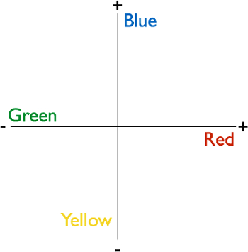
Figure 1. Opponent color dimensions. The human brain processes color using an opponent dimensions system. This is a two-dimensional plane with blue and yellow occupying one axis, which can be thought of as simply positive or negative, and red and green occupying the other axis. The consequence of the system is that the brain processes the presence of some colors as indicating the absence of others, and vice-versa. All perceivable colors occupy a point in the opponent space.
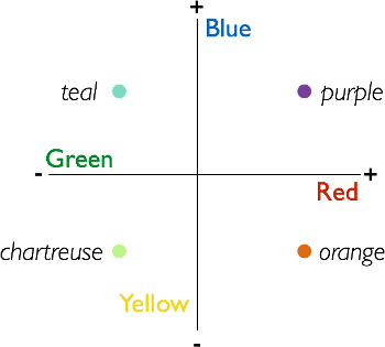
Figure 2. All perceivable colors occupy a point in the opponent space. Shown here are examples of colors that have nonzero values in each of two dimensions of the opponent space.
One way that color opponency was discovered-in 1878 by Ewald Hering, even before scientists had access to techniques for imaging the brain itself-is through an illusion known as a color afterimage. Afterimages are still used today both to demonstrate the opponent properties of human color perception and to study them.
This video demonstrates how to create a color afterimage illusion, and a simple way to collect subjective perceptual responses from human observers.
1. Stimuli
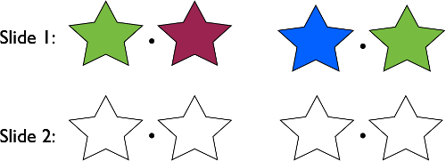
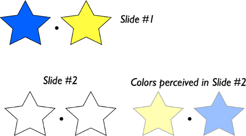
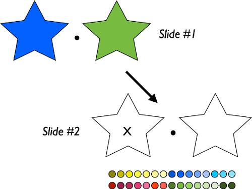
- Open a blank white slide in a slide editor (software such as PowerPoint or Keynote will suffice).
- Use the shape tool to make two equally sized stars with no color fill, only a thin black outline. Center them vertically on the slide, and place one on each side (left and right).
- Place a small black disc in the center of the slide between the stars. This is the fixation point.
- Now, make a copy of this black and white slide. The second copy of it will be the second slide i
For each of the inducer colors in the experiment, identify the most frequently selected after-color. Make a table that visualizes the results, like the one in Figure 8.
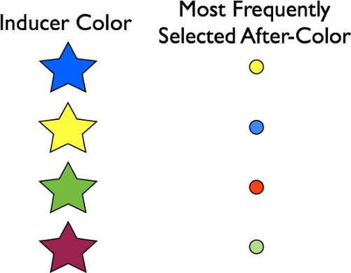
Figure 8. Representative result. Most frequently selected after-colors as a function of inducer colors. The most frequently perceived after-colors will be opponent values of the respective
Color opponency is among the great demonstrations of the scientific method. Researchers in the 1800s were able to infer the nature of color representation in the human brain without any ability to observe brain activity. Today, in fact, color afterimages have become a useful tool for identifying the brain regions involved in processing color. In monkeys, scientists have recorded neurons that fire as though color is present, when it is not, after showing the monkeys sequences of slides that produce afterimages in human ob
- Zeki, S. Colour coding in the cerebral cortex: the reaction of cells in monkey visual cortex to wavelengths and colours. Neuroscience, 9(4), 741-765 (1983).
- Conway, B. R., & Tsao, D. Y. Color architecture in alert macaque cortex revealed by FMRI. Cerebral Cortex, 16(11), 1604-1613 (2006).


Skip to...
Videos from this collection:

Now Playing
Color Afterimages
Sensation and Perception
11.0K Views

Finding Your Blind Spot and Perceptual Filling-in
Sensation and Perception
17.2K Views

Perspectives on Sensation and Perception
Sensation and Perception
11.7K Views

Motion-induced Blindness
Sensation and Perception
6.8K Views

The Rubber Hand Illusion
Sensation and Perception
18.2K Views

The Ames Room
Sensation and Perception
17.2K Views

Inattentional Blindness
Sensation and Perception
13.2K Views

Spatial Cueing
Sensation and Perception
14.8K Views

The Attentional Blink
Sensation and Perception
15.8K Views

Crowding
Sensation and Perception
5.7K Views

The Inverted-face Effect
Sensation and Perception
15.4K Views

The McGurk Effect
Sensation and Perception
15.9K Views

Just-noticeable Differences
Sensation and Perception
15.3K Views

The Staircase Procedure for Finding a Perceptual Threshold
Sensation and Perception
24.2K Views

Object Substitution Masking
Sensation and Perception
6.4K Views
Copyright © 2025 MyJoVE Corporation. All rights reserved