Visual Attention: fMRI Investigation of Object-based Attentional Control
Overview
Source: Laboratories of Jonas T. Kaplan and Sarah I. Gimbel— University of Southern California
The human visual system is incredibly sophisticated and capable of processing large amounts of information very quickly. However, the brain's capacity to process information is not an unlimited resource. Attention, the ability to selectively process information that is relevant to current goals and to ignore information that is not, is therefore an essential part of visual perception. Some aspects of attention are automatic, while others are subject to voluntary, conscious control. In this experiment we explore the mechanisms of voluntary, or "top-down" attentional control on visual processing.
This experiment leverages the orderly organization of visual cortex to examine how top-down attention can selectively modulate the processing of visual stimuli. Certain regions of the visual cortex appear to be specialized for processing specific visual items. Specifically, work by Kanwisher et al.1 has identified an area in the fusiform gyrus of the inferior temporal lobe that is significantly more active when subjects view faces compared to when they observe other common objects. This area has come to be known as the Fusiform Face Area (FFA). Another brain region, known as the Parahippocampal Place Area (PPA), responds strongly to houses and places, but not to faces.2 Given that we know how these regions respond to specific types of stimuli, their activity can be further explored to identify a key component of vision-visual attention.
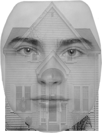
This video shows how to use fMRI to localize the FFA and PPA in the brain, and then examines how object-based attentional control modulates activity in these areas. The use of a functional localizer to restrict subsequent hypothesis testing is a powerful technique in functional imaging. Participants will undergo functional MRI while being presented with an overlaid image of a face and a house. Even though both a face and a house are presented in each stimulus, we predict that patterns of activity in their FFA and PPA will change based on which item is being attended to.3
Procedure
1. Participant recruitment
- Recruit 20 participants.
- Participants should be right-handed and have no history of neurological or psychological disorders.
- Participants should have normal or corrected-to-normal vision to ensure that they will be able to see the visual cues properly.
- Participants should not have metal in their body. This is an important safety requirement due to the high magnetic field involved in fMRI.
- Participants should not suffer from claustrophobia
Results
In the localizer scans, bilateral FFA were more active when subjects were viewing faces than when they were viewing houses. Conversely, the PPA was more active when subjects were viewing houses than when they were viewing faces (Figure 2). These regions, localized via the block-design scans, were later used as regions of interest to extract signal related to shifting attention to faces and to houses during the functional runs.
Application and Summary
The use of localizer scans is a powerful tool for cognitive neuroimaging and has some distinct advantages over whole-brain imaging. By focusing a hypothesis on a small number of specific locations that have known response properties, we can generate very specific predictions with high statistical power. Whole-brain voxel-wise neuroimaging studies must control for the tens of thousands of statistical tests performed at every location in the brain, a process that reduces statistical power. Also, defining these regions base
Tags
Skip to...
Videos from this collection:

Now Playing
Visual Attention: fMRI Investigation of Object-based Attentional Control
Neuropsychology
42.3K Views

The Split Brain
Neuropsychology
68.6K Views

Motor Maps
Neuropsychology
27.7K Views

Perspectives on Neuropsychology
Neuropsychology
12.2K Views

Decision-making and the Iowa Gambling Task
Neuropsychology
33.1K Views

Executive Function in Autism Spectrum Disorder
Neuropsychology
18.0K Views

Anterograde Amnesia
Neuropsychology
30.5K Views

Physiological Correlates of Emotion Recognition
Neuropsychology
16.4K Views

Event-related Potentials and the Oddball Task
Neuropsychology
27.7K Views

Language: The N400 in Semantic Incongruity
Neuropsychology
19.7K Views

Learning and Memory: The Remember-Know Task
Neuropsychology
17.3K Views

Measuring Grey Matter Differences with Voxel-based Morphometry: The Musical Brain
Neuropsychology
17.4K Views

Decoding Auditory Imagery with Multivoxel Pattern Analysis
Neuropsychology
6.5K Views

Using Diffusion Tensor Imaging in Traumatic Brain Injury
Neuropsychology
16.9K Views

Using TMS to Measure Motor Excitability During Action Observation
Neuropsychology
10.3K Views
Copyright © 2025 MyJoVE Corporation. All rights reserved
