
In Vitro Stimulation and Visualization of Macrophage Extracellular Traps
Transcript
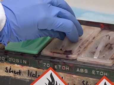
Begin with an adherent macrophage culture.
Add pro-inflammatory cytokines.
The cytokine binds to the macrophage surface receptors, initiating a series of inflammatory events.
First, the cytokine signaling triggers the production of antimicrobial peptides.
Simultaneously, the chromatin within the macrophage nucleus undergoes decondensation.
This process, coupled with changes in membrane composition, disrupts the nuclear membrane, releasing decondensed chromatin into the cytoplasm.
In the cytoplasm, the antimicrobial peptide interacts with the released chromatin.
Cytokine signaling-mediated cell membrane permeabilization causes the decondensed chromatin to be released into the extracellular space, forming a Macrophage Extracellular Trap, or MET.
These web-like structures consist of DNA, histones, antimicrobial molecules, and enzymes.
Add SYTOX green, a fluorescent nucleic acid dye that selectively binds to the exposed DNA in the extracellular traps.
Under a fluorescence microscope, the MET appears as green streaks of extracellular DNA released from the stimulated macrophage.

ABOUT JoVE
Copyright © 2024 MyJoVE Corporation. All rights reserved