
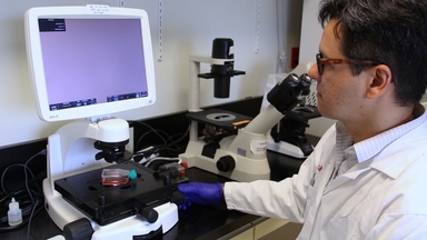
Monitoring B Cell Activation with Antigenic Liposomes using a Calcium-Flux Assay
Transcript
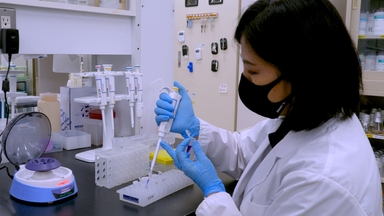
Take red blood cell-depleted splenocytes in a buffer containing a cell-permeant calcium -sensitive fluorescent dye, Indo-1 AM.
Intracellular esterases cleave Indo-1 AM into membrane-impermeable Indo-1, which binds to calcium ions, changing Indo-1's fluorescence signal from blue to violet.
Introduce fluorophore-conjugated antigen-specific antibodies that bind specifically to the respective antigens on cells.
Using flow cytometry, identify fluorophore-labeled B cells and measure their Indo-1 violet-to-blue fluorescence ratio.
Now, treat the cells with antigenic liposomes comprising IgM Fab fragments conjugated to liposome bilayers.
The B cell receptors bind to the liposome-coupled antigens, activating the associated cytoplasmic tyrosine kinases and triggering phospholipase-C gamma-2 activation.
Activated phospholipase-C cleaves the membrane phospholipid, phosphatidylinositol 4,5-bisphosphate, generating second messengers, including inositol 1,4,5- trisphosphate, or IP3.
IP3 binds to IP3-gated calcium channels on the endoplasmic reticulum, releasing calcium ions into the cytoplasm.
These calcium ions bind the unbound Indo-1 molecules, increasing violet fluorescence.
A higher Indo-1 violet-to-blue fluorescence ratio in labeled B cells confirms their successful activation by antigenic liposomes.
Related Videos
Identification of Inositol Phosphate or Phosphoinositide Interacting Proteins by Affinity Chromatography Coupled to Western Blot or Mass Spectrometry
Cell-Free Production of Proteoliposomes for Functional Analysis and Antibody Development Targeting Membrane Proteins

Targeting Drugs to Larval Zebrafish Macrophages by Injecting Drug-Loaded Liposomes
ABOUT JoVE
Copyright © 2024 MyJoVE Corporation. All rights reserved