Fluorescence is a phenomenon that takes place when a substance absorbs light at a given wavelength and emits light at another wavelength. Fluorescence occurs as an electron, which has been excited to a higher, and more unstable energy state, relaxes to its ground state and gives off a photon of light. The light that is responsible for excitation, or moving the electron to a higher energy state, is of shorter wavelength and higher energy than the fluorescence emission, which has a longer wavelength, lower energy, and different color.
Fluorescence microscopy combines the magnifying properties of the light microscope with fluorescence technology that allows the excitation of- and detection of emissions from- fluorophores - fluorescent chemical compounds. With fluorescence microscopy, scientists can observe the location of specific cell types within tissues or molecules within cells.
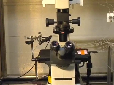
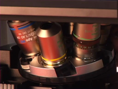
The main components of the fluorescent microscope overlap greatly with the traditional light microscope. However the 2 main differences are the type of light source and the use of the specialized filter elements.
Fluorescence microscopy requires a very powerful light source such as a xenon or mercury arch lamp like the one shown here. The light emitted from the mercury arc lamp is 10-100 times brighter than most incandescent lamps and provides light in a wide range of wavelengths, from ultra-violet to the infrared. This high-powered light source is the most dangerous part of the fluorescence microscope setup as looking directly into unfiltered light can seriously damage your retinas and mishandling the bulbs can cause them to explode.
The principle behind fluorescence microscopy is simple. As light leaves the arc lamp it is directed through an exciter filter, which selects the excitation wavelength.
This light is reflected toward the sample by a special mirror called a dichroic mirror, which is designed to reflect light only at the excitation wavelength. The reflected light passes through the objective where it is focused onto the fluorescent specimen. The emissions from the specimen are in turn, passed back up through the objective – where magnification of the image occurs –and now through the dichroic mirror.
This light is filtered by the barrier filter, which selects for the emission wavelength and filters out contaminating light from the arc lamp or other sources that are reflected off of the microscope components. Finally, the filtered fluorescent emission is sent to a detector where the image can be digitized, or it’s transmitted to the eyepiece for optical viewing.
The exciter filter, dichroic mirror, and barrier filter can be assembled together into a component known as the filter cube. Different filter cubes can be changed during specimen viewing to change the excitation wavelength, and a series of diaphrams can be used to modify the intensity of excitation.
When it comes to performing fluorescence microscopy, the fluorophore can be just as important as the microscope itself, and the type of fluorophore being imaged dictates the excitation wavelength used and emission wavelength that’s detected. The excitation wavelengths contain a small range of energies that can be absorbed by the fluorophore and cause it to transition into an excited state. Once excited, a wide range of emissions, or transitions back to the lower energy state, are possible resulting in an emission spectrum.
The difference between the peak of the absorption, or excitation curve and the peak of the emission curve is known as Stoke’s Shift. The greater the distance in this shift, the easier it is to separate the two different wavelengths. Additionally, any overlapping spectrum needs to be removed by the components of the filter cube for reduced background and improved image quality.
Exposure of the fluorophore to prolonged excitation will cause it to photobleach, which is a weakening or loss of fluorescence. To reduce photobleaching, you can add an anti-fade mounting medium to the slide and seal the edges with nail polish. The slide should also be kept in the dark when not being imaged.
To begin fluorescence imaging, turn on the xenon or mercury light source and allow it to warm up for as long as 15 minutes in order for it to reach constant illumination.
Next, place your sample on the stage and secure it in place. Then, turn on the white light source of your microscope. Focus on your sample using the lowest powered objective by adjusting the coarse and fine focus knobs. Then, use the stage adjustment knobs to find your area of interest.
Next, turn off the white light source, as well as any unnecessary room lights to reduce background.
Select the correct filter cube for the dye you are imaging and open the shutter to illuminate your sample.
Finally, make fine focus adjustments and direct the output light to the imaging camera. You will likely need to make adjustments to the exposure time for each different fluorophore or fluorescent dye used. However, it is important to keep the exposure time constant when comparing features with the same dye on different samples.
To image multiple dyes on the same sample, change the filter cube to match each fluorophore and record the new image.
After each dye in the sample has been imaged, individual images can be overlaid and merged.
Many different types of experiments can make use of fluorescent microscopy and involve different types of fluorophores One of the most common applications of fluorescent microscopy is the imaging of proteins that have been labeled with antibodies that are attached to, or “conjugated” to fluorescent compounds.. Here, an antibody towards leptospiral surface proteins was detected using a secondary antibody conjugated to alexafluor-488, which fluoresces green when excited.
Another way to highlight a specific feature with fluorescence is to integrate the code for a fluorescent protein such as green fluorescent protein, or GFP, into the DNA of an organism. The gene for GFP was originally isolated from jellyfish and can be expressed, or produced, by cultured cells in response to specific triggers or as part of a specific cell type like the tumor cells shown glowing in this image
Another application of fluorescence imaging is Fluorescence Speckle Microscopy which is a technology that uses fluorescently labeled macromolecular assemblies such as the F-actin network seen here, to study movement and turnover kinetics of this important cytoskeletal protein.
An advanced technique known as Fluorescence recovery after photobleaching, or FRAP, is performed by intentionally photobleaching a small region of a sample in order to monitor the diffusion rate of fluorescently labeled molecules back into the photobleached region.
You’ve just watched JoVE’s introduction to Fluorescence Microscopy.
In this video we learned about the concept of fluorescence, how fluorescence microscopy differs from light microscopy, and how to take a fluorescence image through the scope. We also learned about some basic and advanced applications that use fluorescence. Thanks for watching and don’t forget while photobleaching looks great on your teeth it’s not so good for your samples.