
Workflow for High-content, Individual Cell Quantification of Fluorescent Markers from Universal Microscope Data, Supported by Open Source Software
December 16th, 2014
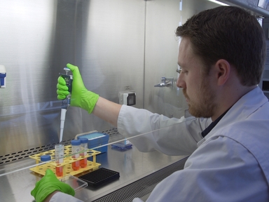
•Presented is a flexible informatics workflow enabling multiplexed image-based analysis of fluorescently labeled cells. The workflow quantifies nuclear and cytoplasmic markers and computes marker translocation between these compartments. Procedures are provided for perturbation of cells using siRNA and reliable methodology for marker detection by indirect immunofluorescence in 96-well formats.
Tags
Related Videos
A Neuronal and Astrocyte Co-Culture Assay for High Content Analysis of Neurotoxicity

Imaging C. elegans Embryos using an Epifluorescent Microscope and Open Source Software
A Quantitative Fitness Analysis Workflow
A High-content Imaging Workflow to Study Grb2 Signaling Complexes by Expression Cloning
Profiling Individual Human Embryonic Stem Cells by Quantitative RT-PCR

Tracking and Quantifying Developmental Processes in C. elegans Using Open-source Tools

Open Source High Content Analysis Utilizing Automated Fluorescence Lifetime Imaging Microscopy
Cellular Redox Profiling Using High-content Microscopy

Open-source Single-particle Analysis for Super-resolution Microscopy with VirusMapper

IR-TEx: An Open Source Data Integration Tool for Big Data Transcriptomics Designed for the Malaria Vector Anopheles gambiae
ABOUT JoVE
Copyright © 2024 MyJoVE Corporation. All rights reserved