
Prolonged Incubation of Acute Neuronal Tissue for Electrophysiology and Calcium-imaging
February 15th, 2017
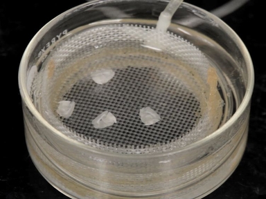
•Once removed from the body, neuronal tissue is greatly affected by environmental conditions, leading to eventual degradation of the tissue after 6 - 8 h. Using a unique incubation method, which closely monitors and regulates the extracellular environment of the tissue, tissue viability can be significantly extended for >24 h.
Related Videos

Cryopreservation of Cortical Tissue Blocks for the Generation of Highly Enriched Neuronal Cultures
Preparation of Acute Subventricular Zone Slices for Calcium Imaging

Dual Electrophysiological Recordings of Synaptically-evoked Astroglial and Neuronal Responses in Acute Hippocampal Slices
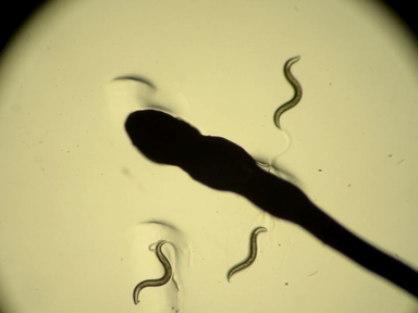
In vivo Neuronal Calcium Imaging in C. elegans

Simultaneous Electrophysiological Recording and Calcium Imaging of Suprachiasmatic Nucleus Neurons

Real-time Electrophysiology: Using Closed-loop Protocols to Probe Neuronal Dynamics and Beyond

Two-photon Calcium Imaging in Neuronal Dendrites in Brain Slices

Ablation of a Neuronal Population Using a Two-photon Laser and Its Assessment Using Calcium Imaging and Behavioral Recording in Zebrafish Larvae

In vivo Calcium Imaging in Mouse Inferior Olive
In Vivo Calcium Imaging of Neuronal Ensembles in Networks of Primary Sensory Neurons in Intact Dorsal Root Ganglia
ABOUT JoVE
Copyright © 2024 MyJoVE Corporation. All rights reserved