
An In Vivo Duo-color Method for Imaging Vascular Dynamics Following Contusive Spinal Cord Injury
December 31st, 2017
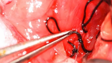
•We introduce an in vivo imaging method using two different fluorescent dyes to track dynamic spinal vascular changes following a contusive spinal cord injury in adult Sprague-Dawley rats.
Related Videos

In vivo Imaging of the Mouse Spinal Cord Using Two-photon Microscopy

Lateral Fluid Percussion: Model of Traumatic Brain Injury in Mice

An Ex Vivo Laser-induced Spinal Cord Injury Model to Assess Mechanisms of Axonal Degeneration in Real-time

A Procedure for Implanting a Spinal Chamber for Longitudinal In Vivo Imaging of the Mouse Spinal Cord

Diffusion Imaging in the Rat Cervical Spinal Cord

Two-photon Imaging of Cellular Dynamics in the Mouse Spinal Cord

Imaging Neural Activity in the Primary Somatosensory Cortex Using Thy1-GCaMP6s Transgenic Mice

Activity-based Training on a Treadmill with Spinal Cord Injured Wistar Rats
Intra-Arterial Delivery of Neural Stem Cells to the Rat and Mouse Brain: Application to Cerebral Ischemia

Induction of Complete Transection-Type Spinal Cord Injury in Mice
ABOUT JoVE
Copyright © 2024 MyJoVE Corporation. All rights reserved