
Whole-mount Confocal Microscopy for Adult Ear Skin: A Model System to Study Neuro-vascular Branching Morphogenesis and Immune Cell Distribution
March 29th, 2018
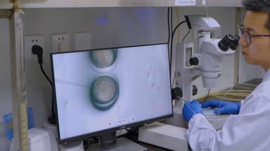
•Here, we describe a high resolution whole-mount imaging method in the entire adult mouse ear skin, which enables us to visualize branching morphogenesis and patterning of peripheral nerves and blood vessels, as well as immune cell distribution.
Tags
Related Videos
Live Imaging of Innate Immune and Preneoplastic Cell Interactions Using an Inducible Gal4/UAS Expression System in Larval Zebrafish Skin
Epigenetic Conversion as a Safe and Simple Method to Obtain Insulin-secreting Cells from Adult Skin Fibroblasts

Horizontal Whole Mount: A Novel Processing and Imaging Protocol for Thick, Three-dimensional Tissue Cross-sections of Skin

Quantitative Whole-mount Immunofluorescence Analysis of Cardiac Progenitor Populations in Mouse Embryos

A Simplified and Efficient Method to Isolate Primary Human Keratinocytes from Adult Skin Tissue
Visualizing the Node and Notochordal Plate In Gastrulating Mouse Embryos Using Scanning Electron Microscopy and Whole Mount Immunofluorescence
An In Vitro Model of a Parallel-Plate Perfusion System to Study Bacterial Adherence to Graft Tissues

Analysis of Cardiac Chamber Development During Mouse Embryogenesis Using Whole Mount Epifluorescence

A Layered Mounting Method for Extended Time-Lapse Confocal Microscopy of Whole Zebrafish Embryos
Whole-Mount In Situ Hybridization in Zebrafish Embryos and Tube Formation Assay in iPSC-ECs to Study the Role of Endoglin in Vascular Development
ABOUT JoVE
Copyright © 2024 MyJoVE Corporation. All rights reserved