
Examination of Mitotic and Meiotic Fission Yeast Nuclear Dynamics by Fluorescence Live-cell Microscopy
June 24th, 2019
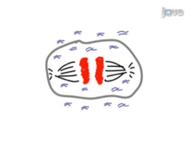
•Here, we present live-cell imaging which is a non-toxic microscopy method that allows researchers to study protein behavior and nuclear dynamics in living fission yeast cells during mitosis and meiosis.
Tags
Related Videos

Live Cell Imaging of F-actin Dynamics via Fluorescent Speckle Microscopy (FSM)
Time-lapse Imaging of Mitosis After siRNA Transfection
Quantitative Live Cell Fluorescence-microscopy Analysis of Fission Yeast

Acquiring Fluorescence Time-lapse Movies of Budding Yeast and Analyzing Single-cell Dynamics using GRAFTS

Examination of Drosophila Larval Tracheal Terminal Cells by Light Microscopy

In Vivo 4-Dimensional Tracking of Hematopoietic Stem and Progenitor Cells in Adult Mouse Calvarial Bone Marrow

Microscopy of Fission Yeast Sexual Lifecycle

Spatiotemporal Analysis of Cytokinetic Events in Fission Yeast
Live Cell Fluorescence Microscopy to Observe Essential Processes During Microbial Cell Growth

Live Cell Imaging to Assess the Dynamics of Metaphase Timing and Cell Fate Following Mitotic Spindle Perturbations
ABOUT JoVE
Copyright © 2024 MyJoVE Corporation. All rights reserved