
Labeling Stem Cells with Fluorescent Dyes for non-invasive Detection with Optical Imaging
April 2nd, 2008
•This video shows techniques for labeling of human embryonic stem cells and mesenchymal stem cells with fluorescent dyes. This technique can be used for an in vivo tracking of transplanted stem cells with optical imaging and for histopathological correlations with fluorescence microscopy.
Tags
Related Videos
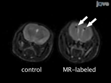
Labeling hESCs and hMSCs with Iron Oxide Nanoparticles for Non-Invasive in vivo Tracking with MR Imaging

Non-invasive 3D-Visualization with Sub-micron Resolution Using Synchrotron-X-ray-tomography

In vitro Labeling of Human Embryonic Stem Cells for Magnetic Resonance Imaging

Proper Care and Cleaning of the Microscope

Phase Contrast and Differential Interference Contrast (DIC) Microscopy

Visualizing the Live Drosophila Glial-neuromuscular Junction with Fluorescent Dyes

Fluorescent Labeling of Drosophila Heart Structures

Lineage Labeling of Zebrafish Cells with Laser Uncagable Fluorescein Dextran

Fluorescence Imaging with One-nanometer Accuracy (FIONA)

Detection of Modified Forms of Cytosine Using Sensitive Immunohistochemistry
ABOUT JoVE
Copyright © 2024 MyJoVE Corporation. All rights reserved